
Like
Share
멍실장
반려동물 엑스레이 판독 기초 — 보호자가 알아야 할 영상 검사 가이드

Basics of Reading Pet X-rays — An Imaging Test Guide for Pet Owners

ImmunityQ&AMeongsiljang Veterinary Advisory Board

We’ve put together an easy-to-understand guide for pet owners, covering the basics of reading veterinary X-rays, the differences between chest and abdominal imaging, and the systematic approach veterinarians use to interpret them.

What Is Reading a Pet’s X-ray?

A veterinarian analyzing an X-ray image of a pet.
Reading a pet’s X-ray is a diagnostic imaging process that systematically evaluates the bones, organs, and soft tissues for abnormalities. What truly matters is that diagnostic accuracy can vary significantly depending on the veterinarian’s experience and their systematic approach, even when looking at the same X-ray image. When pet owners understand the basic principles of X-ray interpretation, they can better comprehend their veterinarian’s explanations and ask more relevant questions.

Situations Requiring an X-ray

X-rays are the most fundamental imaging test in veterinary diagnostics. Your veterinarian may recommend an X-ray in the following situations:
Suspected fractures or dislocations: To assess bone integrity when your pet is limping or shows severe pain upon touching a specific area.
Coughing or difficulty breathing: To evaluate heart size, lung condition, and tracheal structure to identify the underlying cause.
Persistent vomiting or loss of appetite: To determine if there is foreign body ingestion, intestinal obstruction, or abnormalities in abdominal organs.
Routine health screenings: For senior pets, regular chest and abdominal X-rays during check-ups can help detect hidden diseases at an early stage.

Chest vs. Abdominal X-rays: A Comparison

ItemChest X-rayAbdominal X-ray
Main areas observedHeart, lungs, trachea, esophagusStomach, intestines, liver, spleen, kidneys, bladder
Main conditions diagnosedCardiomegaly, pneumonia, tracheal collapse, pleural effusionForeign body ingestion, intestinal obstruction, bladder stones, tumors
Basic imaging positionsLateral + frontal (at least 2 views)Lateral + supine (at least 2 views)
Imaging tipAdjust timing to the respiratory cycleFasting before imaging improves reading accuracy

Additional imaging may be needed depending on the region and purpose

What to Look for in Chest X-rays
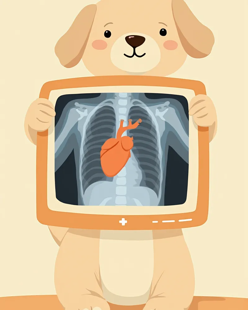
Chest X-rays are primarily interpreted by focusing on the heart and lungs. According to veterinary radiology textbooks, the key is to systematically evaluate each structure one by one.
Heart: The size and shape are assessed systematically. If the heart is abnormally enlarged beyond the normal range, cardiomegaly may be suspected. Accurate assessment of heart size is determined by a trained veterinarian reviewing the entire image.
Lungs: Normal lungs appear dark on an X-ray. If there are areas that appear white, conditions such as pneumonia, hemorrhage, or tumors should be considered.
Trachea: If the trachea appears narrowed or compressed, tracheal collapse or compression by a tumor may be suspected.
Pleural effusion: If fluid accumulates around the lungs, it may be caused by heart disease or infection.
A chest X-ray of a dog showing the heart and lungs

What to Look for in Abdominal X-rays

Abdominal X-rays allow us to evaluate a wide range of structures, from the digestive tract to the urinary system. Veterinary radiology textbooks emphasize the importance of systematically assessing the entire area, keeping in mind even organs that may not be directly visible.
Stomach and Intestines: We check the gas pattern and the diameter of the intestines. Abnormally distended intestines may indicate an obstruction.
Liver and Spleen: We verify whether their size and position are normal. An enlarged liver may suggest liver disease, while an enlarged spleen could indicate a tumor or infection.
Kidneys and Bladder: If a white structure is visible inside the bladder, there is a high likelihood of bladder stones.
Foreign Bodies: Ingested foreign objects such as bones or toy fragments are often visible on the X-ray.

Cases Where Diagnosis Is Difficult with X-rays Alone

X-rays are excellent for visualizing bones and large organs, but they may not clearly show soft tissue details or early-stage diseases. If your veterinarian recommends additional tests such as ultrasound, CT, or MRI, it means that X-rays alone may not provide sufficient information. In such cases, it’s a good idea to actively consider these additional diagnostic options.

The Veterinarian’s Systematic Interpretation Order

Veterinarians follow a strict, systematic order when reviewing X-rays to ensure no anatomical structure is overlooked. Here is a simplified overview of the systematic approach emphasized in veterinary radiology textbooks:
Step 1 — Assess Image Quality: First, evaluate whether the image brightness, patient positioning, and field of view are appropriate.
Step 2 — Review Major Structures: Examine large structures in a consistent order, such as bones, the heart, lungs, or abdominal organs.
Step 3 — Detailed Observation: Compare the size, shape, position, and radiographic density of each organ against normal reference ranges.
Step 4 — Comprehensive Assessment: Synthesize all findings to narrow down the possible diagnoses.
A veterinarian systematically analyzing multiple X-ray images.

Interpretation Terms Pet Owners Should Know
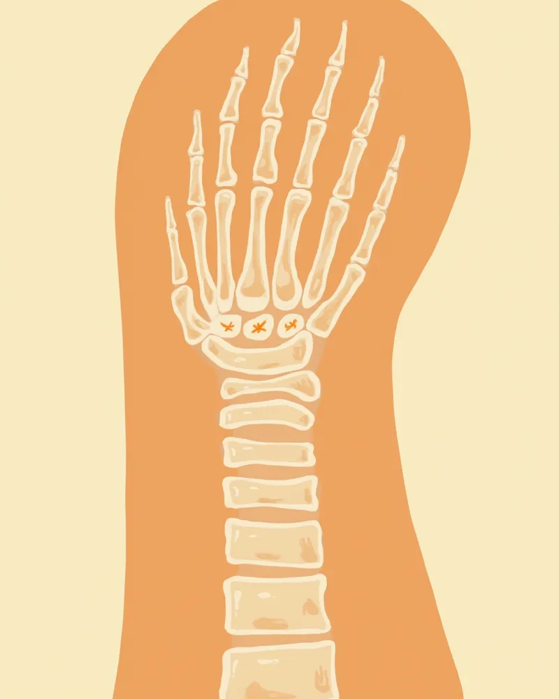
Here are some common terms veterinarians use when explaining X-ray results. Familiarizing yourself with them beforehand will make it much easier to understand the explanation.
Opacity: This refers to how white or black an area appears on an X-ray. Bones appear white, while air appears black.
Soft tissue opacity: This describes the gray areas that are darker than bone but lighter than air, representing structures like muscles and organs.
Radiopaque: This term refers to structures that appear white on an X-ray. Common examples include stones and metallic foreign bodies.
Distension/Dilation: This indicates that the intestines or stomach are swollen beyond their normal size, which can be caused by gas accumulation or obstruction.
The contrast between bone and soft tissue visible on an X-ray image

Sedation or Anesthesia May Be Required During Imaging

For accurate X-ray imaging, your pet needs to remain still. If they’re in significant pain or highly anxious, your veterinarian may recommend administering a mild sedative. This is especially true for areas like the hips or spine, where precise positioning is crucial and clear images are often difficult to obtain without sedation. A thorough health check before using sedatives is essential.

Reviewed by a veterinarian

Dr. Tony — Punnawat Phongkittirak

Dr. Tony — Punnawat Phongkittirak

Veterinarian

A veterinarian who majored in veterinary medicine at Khon Kaen University, Thailand, and completed the IVSA program at North Carolina State University in the United States. Drawing on clinical experience at animal hospitals, he works in the pet healthcare field and is dedicated to building a digital care environment that connects pet parents with veterinarians.

Frequently Asked Questions

How much does an X-ray usually cost?
Costs vary widely depending on the clinic and the specific area being imaged. For the most accurate pricing, it’s best to contact the clinic directly before your visit.
Is frequent X-ray exposure dangerous due to radiation?
Veterinarians recommend X-rays only when they are essential for diagnosis. They weigh the diagnostic benefits against the frequency of imaging to determine whether an X-ray is necessary, so they do not advise unnecessary repeat exposures. If you are concerned about radiation exposure, it is helpful to discuss with your veterinarian why the current X-ray is needed and how often future imaging may be required. You can also ask whether other imaging methods can be used as alternatives or supplements, which can help you feel more at ease with the examination.
Can I interpret X-ray results at home?
Only veterinarians with specialized training can accurately interpret X-rays. Relying on internet information to make your own diagnosis carries a high risk of error, so be sure to listen to your veterinarian’s explanation.
What is the difference between X-rays and ultrasounds?
X-rays provide a clear overview of the overall contours of bones and organs, while ultrasound allows for detailed, real-time visualization of internal organ structures. Since these two diagnostic methods complement each other, they are often used together.
Is fasting required before the imaging procedure?
For abdominal X-rays, fasting before the procedure reduces food in the stomach and intestines, leading to more accurate interpretation. Unless it’s an emergency, it’s best to follow your veterinarian’s instructions and fast appropriately before the scan. The required fasting period can vary depending on your pet’s condition and the purpose of the exam, so be sure to confirm the details with your clinic beforehand.

Share

Related Guides

References

[1] Schaer M, Gaschen F. Clinical Medicine of the Dog and Cat, 4th Ed. CRC Press, 2022.

[2] Thrall DE. Textbook of Veterinary Diagnostic Radiology, 7th Ed. Saunders, 2018.

[3] Dennis R, Kirberger RM, Wrigley RH, Barr FJ. Handbook of Small Animal Radiological Differential Diagnosis. Saunders, 2001.

This information is based on veterinary literature and does not replace diagnosis or treatment. Please consult a veterinarian for specific health concerns.

Metapet Co., Ltd. | CEO: Park Sung-yong | Business Reg. No. 417-88-02562 | Seoul, South Korea | Customer Center

How to Read Pet X-Rays: An Imaging Guide for Owners | Meongsiljang