

Canine Pemphigus (Autoimmune Skin Disease) Guide
Canine pemphigus is an autoimmune disease in which the immune system attacks skin cells, leading to blisters, crusts, and ulcers. Early diagnosis and lifelong management are key.
What is Canine Pemphigus?
What Causes Pemphigus?
At-a-Glance Comparison of Pemphigus Types
Comparison of Pemphigus Types
| Item | Pemphigus foliaceus | Pemphigus erythematosus | Pemphigus vulgaris |
|---|---|---|---|
| Main lesion sites | Nose·face·paw pads·trunk | Mainly limited to the face·nose only | Mouth·lips·mucosa·mucocutaneous junctions |
| Main symptoms | Pustules·crusts·alopecia | Erythema·pustules·crusts | Ulcers·erosions·pain |
| Severity | Moderate | Mild | Severe |
| Frequency | Most common | Rare | Very rare |
General classification based on the veterinary dermatology textbook.
Suspect Pemphigus If You See These Symptoms

When to Seek Immediate Veterinary Care
If your pet develops ulcers on the mouth, gums, or tongue, or shows fever, loss of appetite, and lethargy, it may be suffering from pemphigus vulgaris or a secondary bacterial infection. Deep ulcers in the mouth, mucous membranes, or at the junction of skin and mucous membranes can be a sign of severe pemphigus vulgaris, and if there is a strong foul odor or pus from scabs, a secondary bacterial infection may be spreading. If your pet is undergoing immunosuppressive therapy, they are more vulnerable to infections, so it is safest to visit the vet within 24 hours for a skin biopsy and blood tests.
How is Diagnosis Performed?
How is Treatment Conducted?
Key Facts About Long-Term Steroid Use
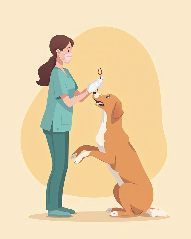
Long-term use of immunosuppressive doses of steroids commonly leads to increased thirst, urination, and appetite. It can also cause gastrointestinal ulcers, thinning of the skin and coat, and delayed wound healing. Prolonged use increases the risk of diabetes. Additionally, because the immune system is suppressed, your pet becomes more vulnerable to secondary infections, including bacterial infections, demodicosis, and dermatophytosis. Never stop the medication abruptly on your own, as this can worsen pemphigus. It is essential to have regular blood and urine tests every three to six months and to keep track of your pet’s weight, water intake, and urine output to share with your veterinarian.
How to Manage at Home

Reviewed by a veterinarian

Dr. Tony — Punnawat Phongkittirak
VeterinarianA veterinarian who majored in veterinary medicine at Khon Kaen University, Thailand, and completed the IVSA program at North Carolina State University in the United States. Drawing on clinical experience at animal hospitals, he works in the pet healthcare field and is dedicated to building a digital care environment that connects pet parents with veterinarians.
Reviewed 2026-04-23
Frequently Asked Questions
Is canine pemphigus curable?
Can it be transmitted to humans? Can it spread to other dogs?
Can vaccines or medications trigger pemphigus?
Can it be improved through diet or supplements?
My dog's nose has turned white—could this also be pemphigus?
Share
Related Guides

Top 5 Medicated Dog Shampoos — For Pyoderma and Bacterial Infections

Veterinarian-Recommended Top 5 Medicated Shampoos for Canine Yeast Dermatitis — Relieves Itching and Greasiness

Top 5 Medicated Cat Shampoos Recommended by Veterinarians — Fungal Dermatitis and Fungal Care

Dog Uric Acid Uroliths FAQ — Key Information for Pet Owners

Frequently Asked Questions About Chronic Recurrent Urinary Tract Infections in Dogs — Key Points Every Pet Owner Should Know
Dog Neurogenic Bladder FAQ — Key Information for Pet Owners

Frequently Asked Questions About the Relationship Between High Blood Pressure and Proteinuria in Dogs — Key Points for Pet Owners

Dog Hematuria FAQ — Key Information Every Pet Owner Should Know

Dog Cystine Urolithiasis FAQ — Key Information for Pet Owners
Dog Bladder Ultrasound Exam FAQ — Key Information Every Pet Owner Should Know
References
[1] Miller WH, Griffin CE, Campbell KL. Muller and Kirk's Small Animal Dermatology, 7th Edition, Chapter 9: Autoimmune and Immune-Mediated Skin Disorders, 2013
[2] Olivry T. A review of autoimmune skin diseases in domestic animals: I - Superficial pemphigus. Veterinary Dermatology, 2006;17(5):291-305
[3] Nishifuji K, Tamura K, Konno H, et al. Development of an enzyme-linked immunosorbent assay for detection of circulating IgG autoantibodies against canine desmoglein 3 in dogs with pemphigus. Veterinary Dermatology, 2009;20(5-6):331-337
[4] Gomez SM, Morris DO, Rosenbaum MR, Goldschmidt MH. Outcome and complications associated with treatment of pemphigus foliaceus in dogs: 43 cases (1994-2000). JAVMA, 2004;224(8):1312-1316