Like
Share
멍실장
강아지·고양이 기관삽관 시술과 모니터링 완벽 가이드

The Complete Guide to Endotracheal Intubation and Monitoring in Dogs and Cats

RespiratoryQ&AMeongsiljang Veterinary Advisory Board

Endotracheal intubation is a procedure that secures the airway with a tube during general anesthesia or emergency respiratory support. We’ve outlined the procedure, equipment preparation, monitoring parameters, and potential complications.

What Is Endotracheal Intubation?

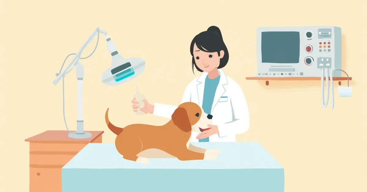
The surgical prep table with the endotracheal tube and laryngoscope in place
Endotracheal intubation is a procedure in which a tube (endotracheal tube) is inserted into the trachea through the mouth to secure an airway, typically for general anesthesia or emergency respiratory support. The most critical aspects are securing the airway and ensuring oxygen supply. This procedure is immediately necessary when an animal cannot breathe on its own during anesthesia or when there is a risk of vomit entering the airway. Although this is not a procedure that pet owners perform themselves, it is almost always carried out when pets undergo surgery or receive emergency treatment.

When Is Endotracheal Intubation Necessary?

There are three main situations in which endotracheal intubation is necessary: - General anesthesia surgery: All procedures under general anesthesia, including spaying/neutering, dental work, and orthopedic surgery - Emergency respiratory failure: Airway obstruction, severe respiratory distress, or cardiopulmonary resuscitation (CPR) - Airway protection: When there is a risk of vomit or blood entering the lungs Brachycephalic breeds such as Pugs and French Bulldogs have excess soft tissue in the upper airway and a narrower anatomical structure, making it difficult to visualize the larynx. As a result, intubation and extubation can be challenging. Even for short procedures, using a laryngoscope to secure a stable airway is the safest approach. Additionally, once anesthesia takes effect, the gag reflex is suppressed, increasing the risk of saliva or secretions entering the lungs and causing aspiration pneumonia. Therefore, it is standard practice to protect the airway with intubation during general anesthesia.

Endotracheal Tube Size Guidelines by Weight

ItemApproximate starting size estimate (inner diameter mm, for reference)Notes
Cats/very small dogs under 2 kg3.0–4.0An uncuffed tube may be used, and if cuffed, inflate carefully
Small dogs 2–5 kg4.0–5.5Along with the estimated size, prepare one size smaller and larger tubes in advance
Medium dogs 5–15 kg5.5–8.0Adjust the length to fit the body type as well
Large dogs 15–30 kg8.0–10.0Check cuff pressure to prevent mucosal injury
Giant dogs over 30 kg10.0–14.0Prepare tubes suited to the body type with some spare

The figures above are only an approximate starting point. In practice, methods such as matching the tube's outer diameter to the width of the narrowest area between the nostrils (nasal septum) are also used, and in addition to the estimated size, a tube one size smaller and one size larger are prepared together and selected while directly viewing the larynx. Especially for brachycephalic breeds or animals that are excessively thin or obese, estimating size by body weight alone may not fit well, so the final choice is determined by the veterinarian's direct assessment. The guardian does not decide the size.

What Is the Sequence of Intubation?
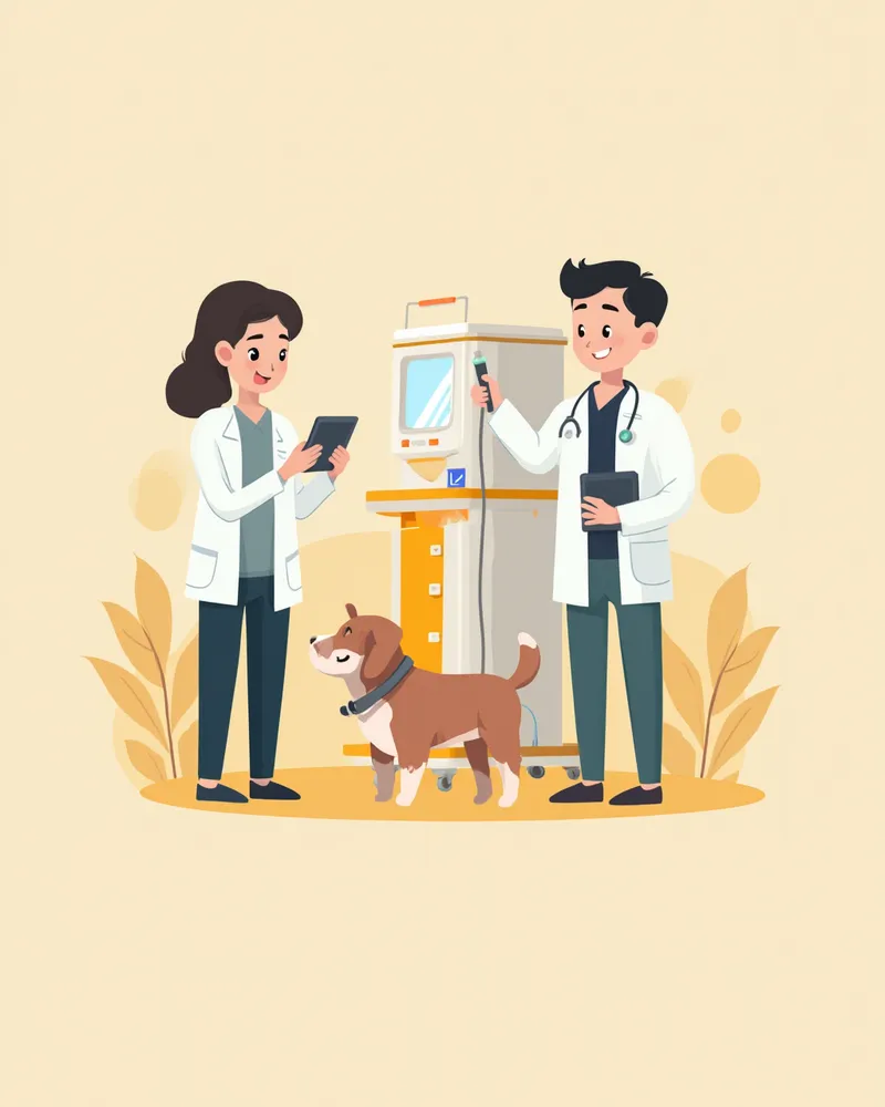
Intubation typically follows this sequence: pre-treatment → induction of anesthesia → intubation → cuff inflation → securing the tube. 1. Secure intravenous access and administer sedatives and analgesics. 2. Induce unconsciousness using an induction agent such as propofol. 3. Open the mouth and visualize the glottis using a laryngoscope. 4. Gently insert the tube into the trachea. 5. Inflate the cuff (balloon) to the appropriate pressure to prevent gas leakage. 6. Secure the tube outside the mouth to the upper or lower jaw with ties. The veterinarian and veterinary nurse work in close coordination, proceeding quickly and accurately while directly visualizing the larynx under adequate anesthetic depth. Pre-oxygenation ensures sufficient oxygen reserves, allowing more time for intubation without risking hypoxia, thereby enhancing safety.
A veterinarian checking the endotracheal tube connected to the anesthesia machine

Warning Signs of Intubation Failure and Complications

If intubation is performed incorrectly, it can lead to esophageal intubation, damage to the tracheal mucosa, or tracheal necrosis and rupture due to cuff overinflation. Cats, in particular, have very sensitive larynges and are prone to complications even from minor irritation, so it is recommended to apply one or two drops of 2% lidocaine to the vocal cords or spray it, then wait long enough for the anesthetic to take full effect before proceeding with intubation. Additionally, forcing a tube that is too large for the trachea can damage the mucosa and tracheal cartilage, potentially leading to necrosis or rupture, making it crucial to select the appropriate tube size. If coughing persists for more than three days after the procedure or if blood-tinged sputum is produced, contact your veterinarian immediately.

What Indicators Are Monitored During Anesthesia?
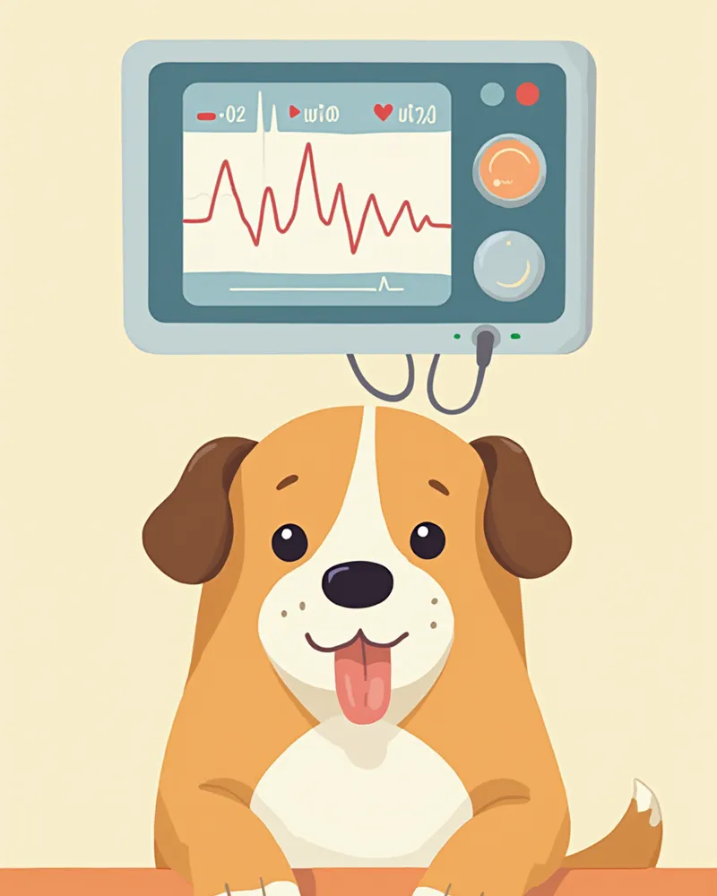
Intubation is just the beginning. Throughout the entire anesthesia process, we monitor the following indicators in real time: - Oxygen saturation (SpO2): Ensure it remains sufficiently high using a pulse oximeter. - End-tidal carbon dioxide (EtCO2): Confirm adequate ventilation through capnography. - Heart rate, electrocardiogram (ECG), and blood pressure: Maintain within normal ranges specific to the breed and condition. - Body temperature: Monitor closely and provide warming to prevent hypothermia. - Respiratory rate and pattern: Check for spontaneous breathing and respiratory effort. - Mucous membrane color, capillary refill time (CRT), and pulse: Assess circulatory status. According to veterinary emergency medicine textbooks, it is crucial to record these indicators in real time on a standardized anesthesia chart to detect changes early. In particular, capnography (EtCO2) is a useful indicator for confirming that the tube is properly placed in the trachea and that ventilation is occurring. Specific normal ranges and target values are determined and managed by the veterinarian based on the pet’s breed, age, and condition.
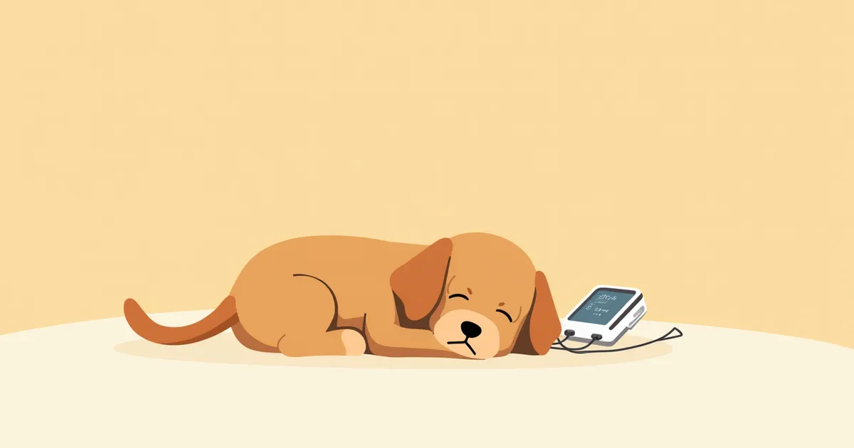
Anesthesia monitoring screen for dogs and oxygen saturation sensor

When and How to Perform Extubation?

Extubation is performed once spontaneous breathing and the swallowing reflex have returned. Removing the tube too early can lead to respiratory failure, while leaving it in too long may cause the animal to bite the tube or develop laryngeal spasms due to irritation. - Cats: The tube should ideally be removed just before the swallowing reflex returns to prevent laryngeal spasms. - Dogs: The tube is removed after the swallowing reflex has clearly returned. - Brachycephalic breeds: It is recommended to keep the tube in place until full consciousness is regained. If oxygen saturation drops after extubation, re-intubation or oxygen therapy via a mask may be necessary.

Key Points for Owners to Check After the Procedure

After your pet recovers from anesthesia and intubation and returns home, mild coughing or throat irritation may occur for a few days. These symptoms typically resolve within 2–3 days. - Normal reactions: Mild coughing, hoarse voice, temporary decreased appetite - Warning signs: Coughing lasting more than 24 hours, difficulty breathing, cyanosis (bluish discoloration of the gums), or coughing up blood Pets with pre-existing tracheal collapse or chronic bronchitis may experience worsened symptoms after the procedure, so it’s a good idea to review the Tracheal Collapse Management Guide as well.

Reviewed by a veterinarian

Dr. Tony — Punnawat Phongkittirak

Dr. Tony — Punnawat Phongkittirak

Veterinarian

A veterinarian who majored in veterinary medicine at Khon Kaen University, Thailand, and completed the IVSA program at North Carolina State University in the United States. Drawing on clinical experience at animal hospitals, he works in the pet healthcare field and is dedicated to building a digital care environment that connects pet parents with veterinarians.

Frequently Asked Questions

Even for a brief procedure, is endotracheal intubation really necessary?
Yes, with general anesthesia, intubation is standard even for brief procedures. Anesthetic agents weaken the respiratory muscles and suppress airway reflexes, which creates a risk of saliva or vomit entering the lungs. Intubation is the most reliable way to prevent this and ensure a stable oxygen supply.
Why is catheterization more challenging in cats?
Cats have sensitive larynges, so even the mild irritation from an endotracheal tube can easily trigger laryngospasms. To prevent this, we apply a small amount of lidocaine to the vocal cords and wait for it to take effect before intubating. We also remove the tube more quickly than in dogs. For safety, it’s best to have this procedure performed at a clinic with experienced veterinarians and veterinary nurses.
My pet’s voice sounds hoarse after the procedure. Is that normal?
Your pet may experience a temporarily hoarse voice or mild cough due to tube irritation. These symptoms typically resolve within 2 to 3 days. However, if they persist for more than 3 days, or are accompanied by blood-tinged mucus or difficulty breathing, it could indicate damage to the tracheal mucosa, and you should seek veterinary care immediately.
I’ve heard that brachycephalic breeds (Pugs, French Bulldogs) are at higher risk during intubation. Is that true?
Brachycephalic breeds have narrower airways and elongated soft palates, making both intubation and extubation challenging. Pre-anesthetic airway assessment is crucial, and oxygen support along with continuous monitoring is necessary until full consciousness returns after extubation. It’s advisable to choose a clinic experienced in handling brachycephalic breeds.
Can I check my pet’s oxygen saturation at home?
It’s difficult to get an accurate reading with home devices. Instead, it’s more practical to monitor whether your pet’s gums stay pink, whether their breathing rate or pattern seems faster or labored compared to normal, and whether they’re showing any unusual signs of respiratory effort. If you notice warning signs such as pale or bluish gums and rapid breathing, it’s best to head to the vet right away.

Share

Related Guides

References

[1] Johnson LR, Lower Airway Disease in Dogs, Small Animal Critical Care Medicine, 3rd Ed

[2] Canine and Feline Respiratory Medicine, 3rd Edition, Tracheal Collapse & Airway Management Chapter

[3] Textbook of Respiratory Disease in Dogs and Cats, Chapter 46 Trachea and Bronchi

[4] The Veterinary Workbook of Small Animal Clinical Cases, Case 42

This information is based on veterinary literature and does not replace diagnosis or treatment. Please consult a veterinarian for specific health concerns.

Metapet Co., Ltd. | CEO: Park Sung-yong | Business Reg. No. 417-88-02562 | Seoul, South Korea | Customer Center

Endotracheal Intubation in Dogs & Cats: Steps & Monitoring | Meongsiljang