Like
Share
멍실장
면역 형광 항체 검사(IFA), 어떤 검사이고 언제 필요해요?

Immunofluorescence Antibody Test (IFA): What Is It and When Is It Needed?

Skin & CoatQ&AMeongsiljang Veterinary Advisory Board

The immunofluorescence antibody test (IFA) is a precise diagnostic tool used to confirm autoimmune skin diseases and certain infectious diseases in dogs and cats. We’ve compiled everything pet owners need to know, from how the test works to interpreting results and important precautions.

What Is the Immunofluorescence Antibody Test (IFA)?
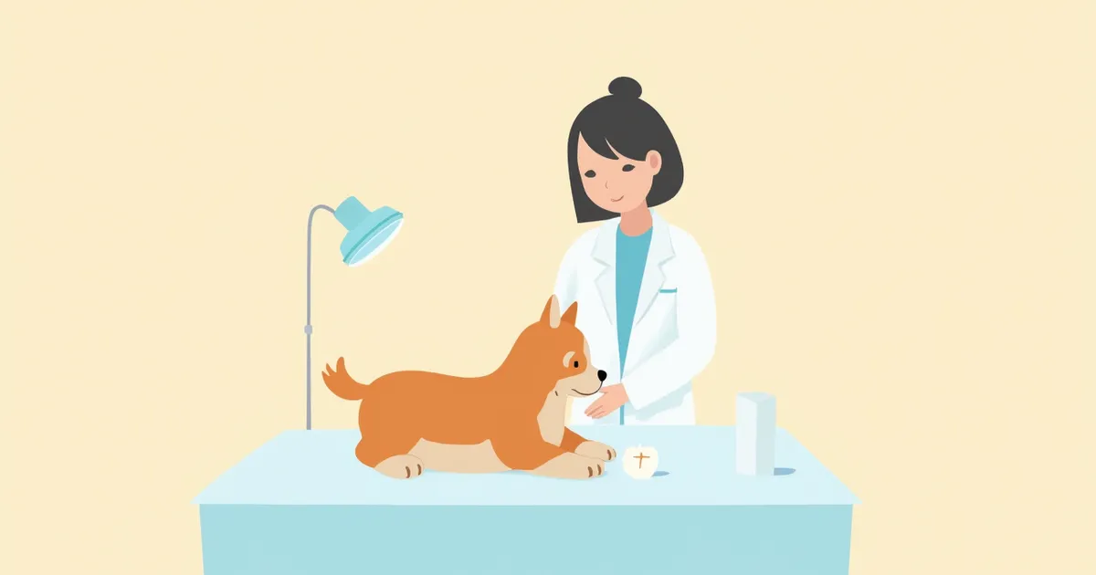
A veterinarian examining a puppy's skin.
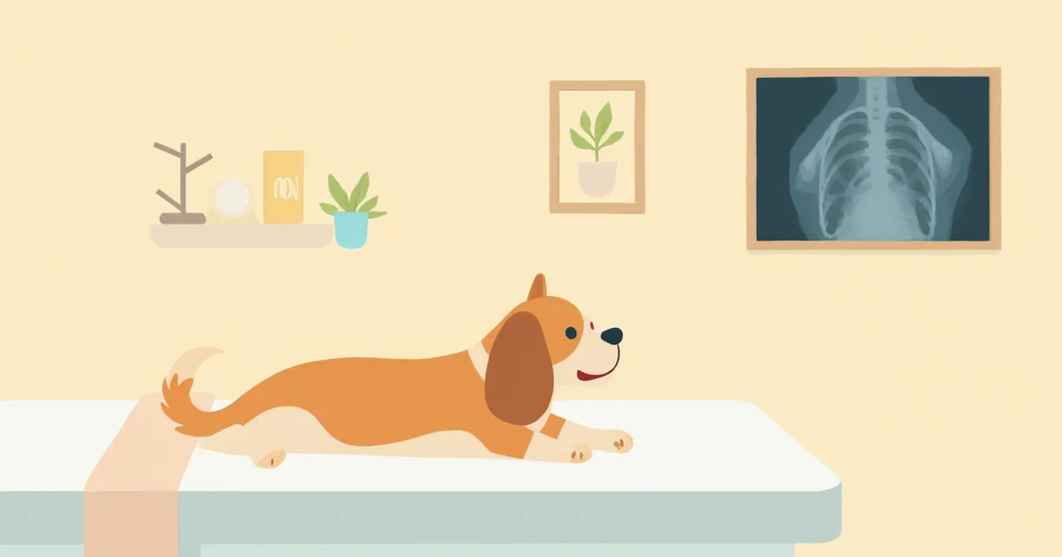
Immunofluorescence antibody testing (IFA) is a precise diagnostic method that uses fluorescently labeled antibodies to directly visualize specific antibodies, proteins, or immune responses within tissues or cells under a fluorescence microscope. What makes this test truly important is its role as a definitive diagnostic tool for confirming skin autoimmune diseases and certain infectious diseases in dogs and cats. It is primarily used to diagnose conditions that are difficult to distinguish based on clinical appearance alone, such as pemphigus and lupus erythematosus.

When Is This Test Performed?

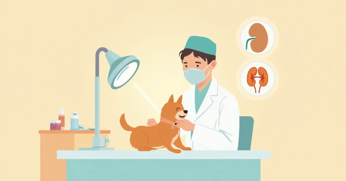
IFA is recommended when skin or mucosal diseases recur or do not respond clearly to standard treatment, making it difficult to identify the cause through routine histopathology. It is primarily used in the following situations: - Suspected autoimmune skin diseases: Pemphigus, lupus erythematosus, bullous pemphigoid - Recurrent skin ulcers or blisters: Lesions extending to the mucous membranes of the lips, nose, or footpads - Confirmation of certain infectious diseases: When direct detection of intracellular viruses such as rabies virus or feline leukemia virus (FeLV) is required, or when serological antibodies need to be confirmed for blood parasite infections like Babesia - Confirmation of immune complex deposition: Evaluation of systemic conditions such as vasculitis or autoimmune kidney disease

Direct vs. Indirect Methods: What’s the Difference?

IFA is broadly divided into two methods. Direct IFA involves directly reacting fluorescent antibodies with a sample of the pet’s tissue to check for the deposition of autoantibodies. It is primarily used to confirm autoimmune diseases in skin biopsy specimens. Indirect IFA involves reacting the pet’s blood (serum) with a reference tissue to indirectly measure the presence and quantity of autoantibodies. Since it can be performed using only a blood sample, it is useful as a screening test.

Direct vs. Indirect Methods at a Glance

ItemDirect IFAIndirect IFA
Test sampleSkin/mucosal tissue biopsyBlood (serum)
Primary useDirect detection of autoantibodiesDetection/titer measurement of autoantibodies
Representative diseasesPemphigus, lupus erythematosusScreening for autoimmune diseases
Collection burdenBiopsy under local anesthesiaPossible with blood draw only
Time to resultAbout 7–14 daysAbout 5–10 days

Turnaround time may vary depending on the diagnostic laboratory and hospital circumstances.

How Is the Test Performed?
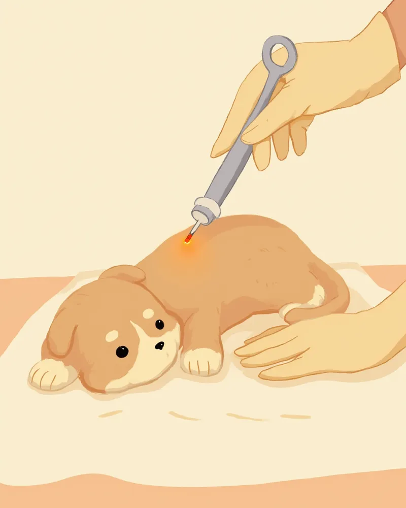
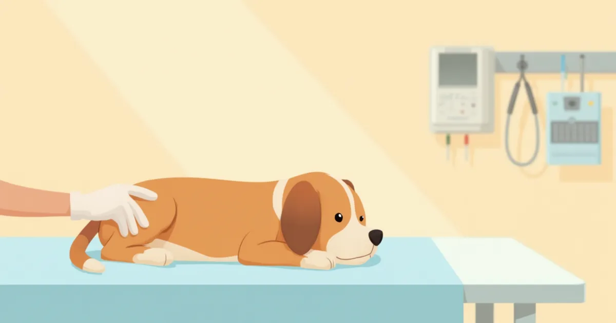
The direct immunofluorescence test follows these steps: 1. Biopsy site selection: We choose the edge of a newly formed blister or rash. We avoid areas with old scabs. 2. Biopsy after local anesthesia: A small biopsy punch is used to collect tissue. The exact size is determined by the veterinarian based on the lesion’s location and your pet’s condition. For sensitive pets, mild sedation may be used alongside the procedure. 3. Storage in specialized transport solution: The tissue is either frozen or placed in a special solution and sent to the laboratory. 4. Fluorescent antibody reaction and interpretation: In the lab, fluorescently labeled antibodies are applied, and the sample is examined under a fluorescence microscope. The time required to receive results can vary from several days to several weeks, depending on the laboratory’s workflow. Please confirm the expected timeline with your veterinarian in advance.
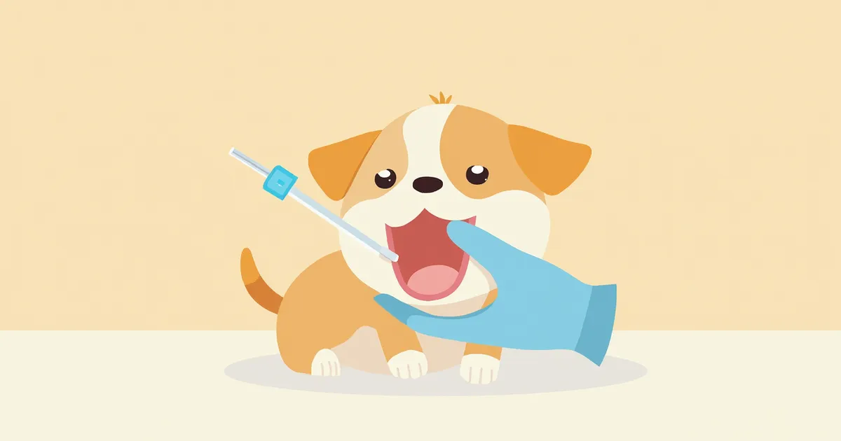
A skin punch biopsy being performed

Key Points to Check Before the Test

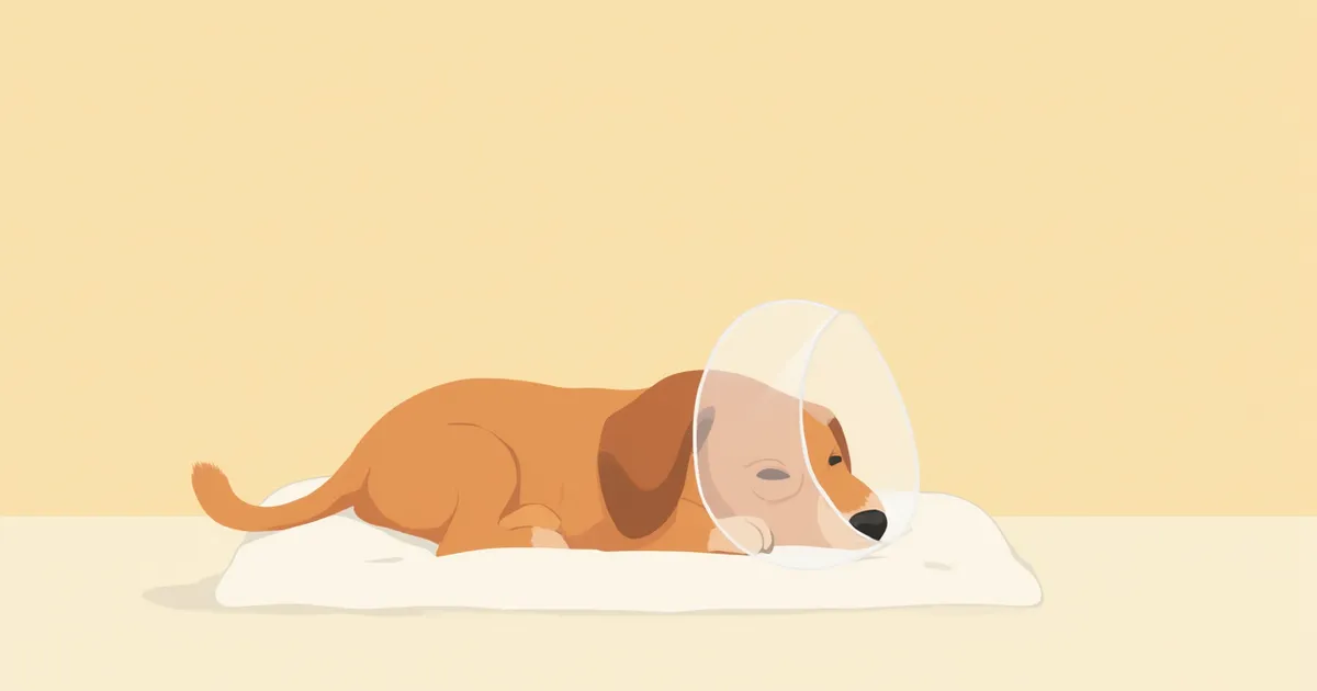
To help ensure accurate biopsy results, it is best to reduce the dose of high-dose steroids or immunosuppressants as much as possible before the procedure. The duration and method of tapering depend on the type of medication, how long it has been administered, and your pet’s current condition, so the specific adjustment plan must be determined in consultation with your veterinarian. If your pet has been on these medications for a long time, the vet will create a tailored tapering schedule. Never stop the medication abruptly on your own, as this can cause the underlying condition to worsen. After the biopsy site is sutured, keep an e-collar on for 1–2 weeks to prevent licking or scratching, and administer any prescribed antibiotics and anti-inflammatory medications on schedule.

How to Interpret the Results

When viewed under a fluorescence microscope, the key diagnostic criterion is where the fluorescence is located and in what pattern it accumulates. The patterns below are generally interpreted in association with specific diseases; however, final interpretation must always be reviewed by a veterinary pathologist and considered alongside clinical findings. - Reticular fluorescence between epidermal cells: Suspect pemphigus-type autoimmune disease - Linear fluorescence at the dermo-epidermal junction: Suspect bullous pemphigoid or lupus erythematosus - Fluorescence along blood vessel walls: Suspect immune complex-mediated vasculitis - Negative (no fluorescence): Low likelihood of autoimmune lesions Results must always be interpreted in conjunction with clinical signs and histopathology. It is rare for immunofluorescence assay (IFA) alone to provide a definitive diagnosis.
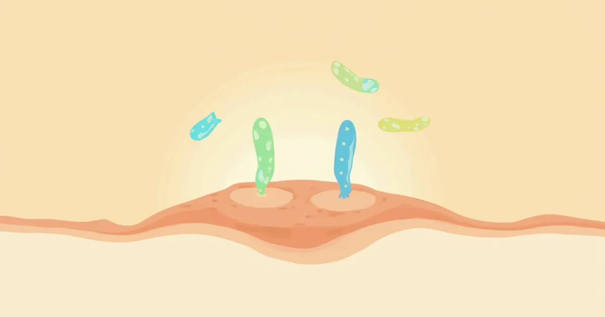
Image of autoantibody deposition observed under a fluorescence microscope

Costs and Limitations to Keep in Mind

IFA is a specialized test conducted exclusively in veterinary diagnostic laboratories, so it costs more and takes longer than routine histopathology. If the specimen isn’t stored properly, the fluorescence may weaken, leading to a false-negative result. Conversely, recent treatments or severe tissue damage can increase the risk of a false-positive. Therefore, veterinarians don’t rely on IFA results alone; they combine them with histopathology, blood tests, and clinical signs to reach a final diagnosis.

Post-Test Care Points

Keep the biopsy site clean and dry to prevent infection. Even if symptoms improve, complete the full course of prescribed antibiotics and anti-inflammatory medications to reduce the risk of resistance. If a skin autoimmune disease is confirmed, long-term management is often necessary, so it is most important to consistently follow the veterinarian’s recommended schedule for follow-up examinations and medication.

Reviewed by a veterinarian

Dr. Tony — Punnawat Phongkittirak

Dr. Tony — Punnawat Phongkittirak

Veterinarian

A veterinarian who majored in veterinary medicine at Khon Kaen University, Thailand, and completed the IVSA program at North Carolina State University in the United States. Drawing on clinical experience at animal hospitals, he works in the pet healthcare field and is dedicated to building a digital care environment that connects pet parents with veterinarians.

Frequently Asked Questions

Is an immunofluorescence antibody test necessary for all skin diseases?
No, if the cause is identified through routine histopathology or bacterial and fungal cultures, an IFA test isn't necessary. It's an additional test we consider when we suspect an autoimmune disease or when dealing with chronic skin conditions of unknown origin.
Does a negative result mean it’s not an autoimmune disease?
Not necessarily. False-negative results can occur depending on the animal’s medication history, how the tissue sample was preserved, and where the sample was collected. If clinical signs are clear, we recommend considering a retest or using an alternative type of immunological test.
Is general anesthesia absolutely necessary?
Most procedures are performed under local anesthesia with mild sedation. General anesthesia is only used briefly when the vet determines it’s necessary, such as if your pet is particularly sensitive or the lesion is in a delicate area.
Is the process the same for cats?
Yes, the basic principle is the same. However, since cats have thinner skin and are more sensitive to stress, we need to be extra careful when selecting the biopsy site and managing sedation. Pemphigus is not uncommon in cats either.
When can my pet return to normal activities after the examination?
Sutures are typically removed 10 to 14 days after surgery. Until then, keep your pet wearing an Elizabethan collar during walks, and postpone bathing until the incision has fully healed, as directed by your veterinarian.

Share

Related Guides

References

[1] Tizard, I.R., Veterinary Immunology, 11th Edition, Elsevier, 2022

[2] Miller, W.H., Griffin, C.E., Campbell, K.L., Muller & Kirk's Small Animal Dermatology, 7th Edition, 2013

[3] Olivry, T. et al., A review of autoimmune skin diseases in domestic animals, Veterinary Dermatology, 2018

This information is based on veterinary literature and does not replace diagnosis or treatment. Please consult a veterinarian for specific health concerns.

Metapet Co., Ltd. | CEO: Park Sung-yong | Business Reg. No. 417-88-02562 | Seoul, South Korea | Customer Center

Immunofluorescence Antibody Test (IFA) in Dogs & Cats | Meongsiljang