Like
Share
멍실장
비강 내시경 검사는 어떻게 진행되나요?

How is nasal endoscopy performed?

RespiratoryQ&AMeongsiljang Veterinary Advisory Board

We’ve put together a comprehensive guide on nasal endoscopy for dogs and cats, covering what it is, when it’s needed, the procedure, and important precautions.

What is nasal endoscopy?
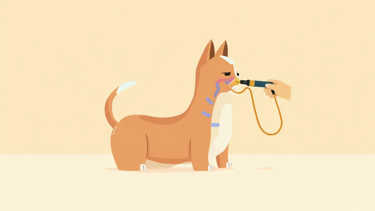
A medical illustration showing the principle of nasal endoscopy in dogs
Nasal endoscopy is a precise diagnostic procedure that involves inserting a thin camera (endoscope) directly into the nasal cavity to visualize the interior in real time. Its greatest advantage is the ability to directly observe the mucosa and perform a biopsy on the spot. While X-rays and CT scans effectively reveal bone structure and the extent of lesions—with CT being particularly valuable for tumor assessment—only endoscopy allows for direct visualization of the mucosal condition, fungal plaques, or deeply embedded foreign bodies, as well as sample collection from those specific sites. Therefore, rather than relying solely on endoscopic findings, it is more accurate to diagnose the cause of chronic sneezing or epistaxis by integrating results from imaging and histopathological examinations. Additionally, to prevent image distortion caused by bleeding, it is standard practice to perform imaging studies such as X-rays or CT scans before conducting the endoscopy. This approach is highly beneficial for precisely identifying the underlying cause when chronic sneezing or nosebleeds persist for more than two weeks.

When is nasal endoscopy needed?

This procedure is performed when nasal symptoms persist or do not respond to standard medical treatment. It is particularly indicated when symptoms are confined to one nostril or when recurrent epistaxis (nosebleeds) occurs, as these signs may suggest a tumor, foreign body, or fungal infection. According to veterinary internal medicine textbooks, nasal endoscopy is a key diagnostic tool that allows direct visualization of mucosal lesions, foreign bodies, tumors, and fungal plaques that are difficult to confirm with imaging studies alone (such as X-rays or CT scans), while also enabling simultaneous biopsy. For differentiating the causes of chronic nasal disease, it is recommended to use imaging and endoscopy in a complementary manner.

Comparison of diagnostic tests for nasal diseases

ItemX-rayCTNasal endoscopy
Anesthesia requiredNot requiredRequiredRequired
Bone structure inspectionPossibleExcellentNot possible
Direct observation of mucosaFalseFalseTrue
Simultaneous biopsyFalseFalseTrue
Foreign body removalFalseFalseTrue
Exam time10 minutes20–30 minutes30–60 minutes

Each exam is complementary, and combined diagnosis is common.

How is the procedure performed?
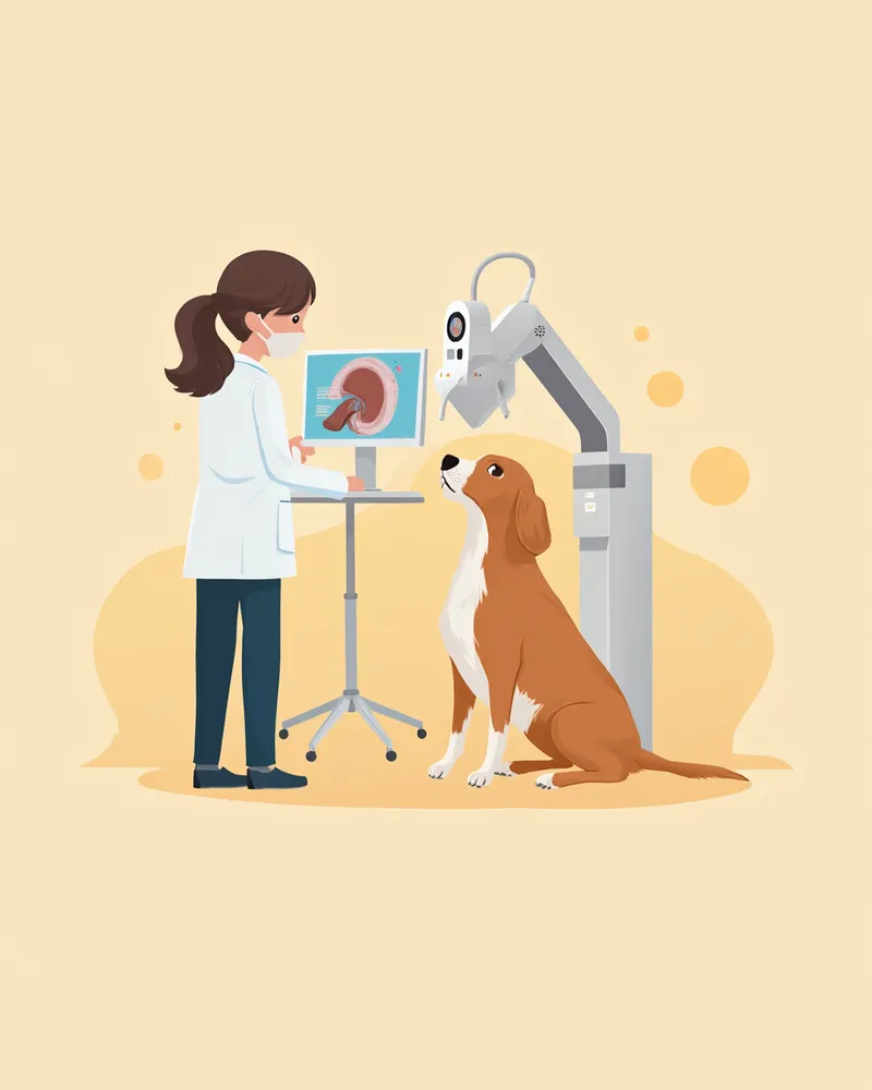
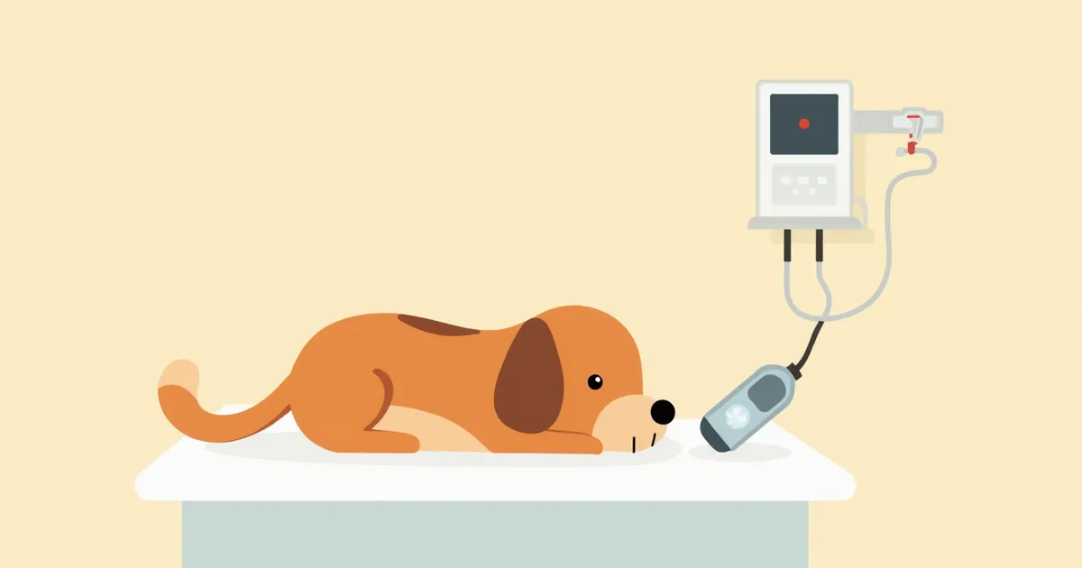
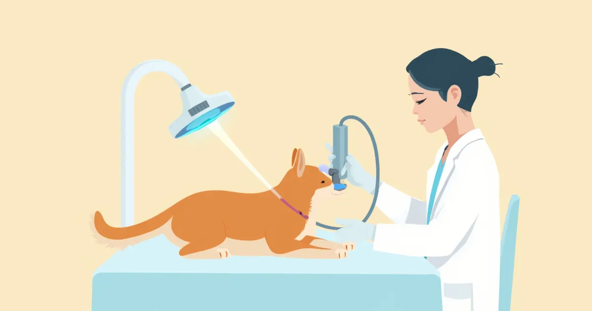
Nasal endoscopy is performed under general anesthesia. Before anesthesia, blood tests and cardiac evaluations are conducted to ensure safety. The front part of the nose is examined using a rigid endoscope, while the back part, including the nasopharynx, is inspected with a flexible endoscope. If necessary, biopsies (tissue sampling), foreign body removal, and flushing can all be done in one session. The entire procedure typically takes about 30 minutes to an hour. Most pets can go home the same day, but older animals may need to stay overnight.
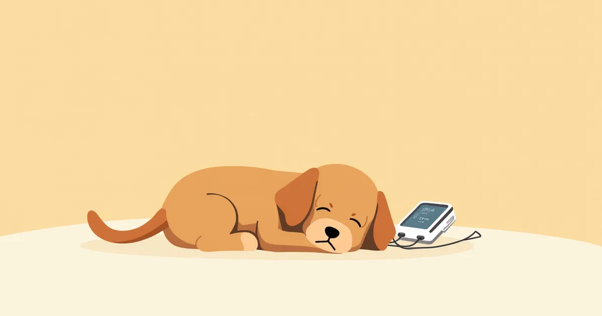
A veterinarian examines a dog while viewing the nasal endoscope monitor.

What to check before the procedure

Because nasal endoscopy requires general anesthesia, it’s essential to follow your veterinarian’s instructions for fasting before the procedure. Older pets or those with heart or kidney disease are at higher risk during anesthesia, so more thorough pre-anesthetic testing will be performed. If your pet is taking blood thinners or has low platelet counts, there may be an increased risk of bleeding, which could necessitate postponing the exam. Be sure to inform your veterinarian about any medications your pet is currently taking.

Conditions that can be identified through nasal endoscopy
The following are the most common conditions that can be identified through nasal endoscopy:
Nasal tumors: We can directly visualize the location and size of both benign and malignant tumors, and confirm the diagnosis through a biopsy. However, since CT scans provide a more accurate assessment of the tumor's invasive extent, we often use imaging tests in conjunction with endoscopy.
Aspergillosis: We can visually identify the characteristic white plaques caused by fungal infection and improve diagnostic accuracy by collecting samples from the affected area.
Foreign bodies: If objects such as grass blades or seeds are lodged deep within the nasal cavity, we can remove them directly using the endoscope.
Chronic rhinitis: We evaluate the state of inflammation, ulceration, and bleeding in the mucous membranes.
Nasal polyps: We can directly observe benign growths in the nasal cavity and nasopharynx and assess them through a biopsy.
Nasal endoscopy equipment and examination monitor

How much does the procedure cost?

The cost of a nasal endoscopy varies significantly depending on the size and location of the veterinary clinic. Generally, including anesthesia, basic tests, and tissue analysis, it ranges from $300 to $700. At secondary care facilities (university hospitals or specialty clinics), costs often exceed $600. If performed alongside a CT scan, additional fees will apply. For an accurate estimate, it’s best to contact the clinic directly. Requesting a cost estimate before the procedure can help you prepare for the expected expenses.

Post-procedure care points

A small amount of nasal bleeding or sneezing may persist for a few days after the examination, but this is usually a normal reaction that resolves on its own. It takes some time for your pet to fully wake up from anesthesia, so during this period, offer small amounts of water and food gradually. Avoid vigorous exercise for several days, and contact the clinic immediately if the bleeding does not stop and continues for an extended period, or if high fever or difficulty breathing occurs. If you notice any unusual symptoms during the recovery process, it is safest to consult your veterinarian rather than making your own judgment.

Reviewed by a veterinarian

Dr. Tony — Punnawat Phongkittirak

Dr. Tony — Punnawat Phongkittirak

Veterinarian

A veterinarian who majored in veterinary medicine at Khon Kaen University, Thailand, and completed the IVSA program at North Carolina State University in the United States. Drawing on clinical experience at animal hospitals, he works in the pet healthcare field and is dedicated to building a digital care environment that connects pet parents with veterinarians.

Frequently asked questions

When will the results of the nasal endoscopy be available?
You can review the gross examination findings right away on the day of the test. If a histopathological examination (biopsy) was also performed, the pathology results typically take about 5 to 10 days.
Is this available at all veterinary clinics?
No, nasal endoscopy requires specialized equipment and a skilled veterinarian, so it’s typically performed at secondary care facilities or internal medicine specialty clinics. Most cases are referred to these centers after an initial evaluation at a primary care clinic.
Can cats also undergo nasal endoscopy?
Yes, it’s possible. Because cats have smaller nasal passages than dogs, we use a finer endoscope. It’s especially useful for confirming nasal polyps and performing mucosal biopsies to identify the underlying cause of chronic rhinitis. However, endoscopic findings in feline chronic rhinitis can be nonspecific, so it’s important to interpret them in conjunction with histopathology and imaging studies.
Is it possible to perform the examination without anesthesia?
Unfortunately, it's not possible. If the animal is conscious, it may move around and damage the nasal mucosa, making accurate examination difficult. General anesthesia is essential.
Is a follow-up test needed after the initial examination?
It depends on the diagnosed condition and how your pet responds to treatment. For fungal infections or during cancer therapy, we may schedule a follow-up exam in two to three months to monitor progress.

Share

Related Guides

References

[1] Clinical Medicine of the Dog and Cat, 4th Edition - Respiratory Endoscopy Chapter

[2] Veterinary Surgical Oncology, 2nd Edition - Nasal Cavity Imaging

[3] McCarthy, T.C., Veterinary Endoscopy for the Small Animal Practitioner, 2005

This information is based on veterinary literature and does not replace diagnosis or treatment. Please consult a veterinarian for specific health concerns.

Metapet Co., Ltd. | CEO: Park Sung-yong | Business Reg. No. 417-88-02562 | Seoul, South Korea | Customer Center

Nasal Endoscopy for Dogs and Cats: Procedure, Cost & Care | Meongsiljang