Like
Share
멍실장
유방 낭종 vs 종양 구별과 절제

Distinguishing Mammary Cysts from Tumors and Surgical Removal

ImmunityQ&AMeongsiljang Veterinary Advisory Board

Dog mammary cysts are benign lumps, but they can be difficult to distinguish from mammary tumors based on appearance alone. Accurate diagnosis through fine-needle aspiration and ultrasound is essential to determine whether surgical removal is necessary.

What Are Canine Mammary Cysts?

A medical illustration showing the cross-sectional structure of a dog’s mammary cyst
Canine mammary cysts are benign, fluid-filled sacs that form within the mammary gland tissue. The most important thing to remember is that it is difficult to distinguish them from tumors by touch alone. Even if a lump is small and soft, you must have your veterinarian confirm whether it is a tumor through fine-needle aspiration and ultrasound if it does not disappear within three weeks or if it grows. Unspayed female dogs are at high risk for mammary tumors, so you should never assume a lump is harmless just because it looks like a cyst.

Causes and Predisposed Dogs

Mammary cysts are closely linked to hormonal fluctuations. Repeated heat cycles stimulate the mammary tissue, and when some of the ducts become blocked, secretions accumulate, leading to cyst formation. They are particularly common in female dogs that are either unspayed or spayed later in life, as well as in those with a history of recurrent pseudopregnancy or prior mammary stimulation. Multiple cysts are often palpable simultaneously rather than a single one, and they can develop in any mammary gland on either side.

Key Points for Distinguishing Mammary Cysts from Mammary Tumors

ItemCystBenign tumorMalignant tumor
TextureSoft·springyElasticFirm·fixed
BorderDistinctGenerally distinctIndistinct
Growth rateSlow or fluctuatingSlowFast
Skin adhesionNoneAlmost noneFrequently present
Ulceration·bleedingRareRareCommonly occurs
Fine-needle aspiration findingsClear fluidUniform cellsAtypical cells

It is difficult to distinguish accurately by palpation alone, so fine-needle aspiration·ultrasound is essential

Accurate Diagnosis — Fine-Needle Aspiration and Ultrasound

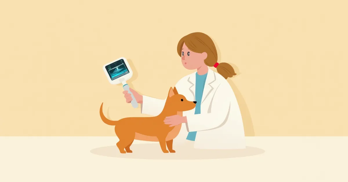
When a lump is felt in the mammary glands, the first test performed is usually a fine-needle aspiration (FNA). A thin needle is used to extract material from inside the lump, which is then examined under a microscope. If it’s a cyst, only clear or yellowish fluid and a few cells will be visible, whereas a tumor will show clusters of atypical cells. Ultrasound can help determine whether the lump contains fluid (appearing black) or solid tissue (appearing gray). However, if the FNA results are inconclusive, surgical removal followed by a histopathological examination is required for a definitive diagnosis.
An ultrasound examination of a dog's mammary gland to assess the internal fluid structure of a cyst.

Signs That Require Immediate Veterinary Attention

If a lump is larger than 2 cm, growing rapidly, or accompanied by redness, ulceration, or bleeding, it may not be a simple cyst. A firm, fixed mass that causes pain when touched is also concerning. Sudden weight loss or a decreased appetite in your dog are additional warning signs. Malignant mammary tumors can metastasize to the lymph nodes, lungs, and other organs, so if you notice any of these symptoms, please seek veterinary care immediately.

When and How to Perform Surgical Removal

Even for simple cysts, surgical removal is recommended in the following cases: - Size: When the cyst is 1 cm or larger in diameter and continues to grow. - Discomfort: When it is painful to touch or repeatedly ruptures. - Uncertain diagnosis: When it is difficult to rule out a tumor through fine-needle aspiration. Surgical options include excision of the lesion alone, partial mastectomy (removal of the affected mammary gland), or unilateral mastectomy (removal of all mammary glands on one side). These procedures are often performed concurrently with spaying or neutering.
A scene of the operating room preparing for a dog’s mastectomy surgery

Post-Surgical Care Precautions

For 7 to 10 days after surgery, your pet must wear an Elizabethan collar to prevent them from licking the incision site. If you notice swelling or discharge at the surgical site, these are signs of infection, so please contact your veterinarian immediately. Limit walks to short trips for bathroom breaks only, and restrict jumping and stair climbing for two weeks. Even if the removed tissue appears benign, a histopathological examination is essential to confirm the diagnosis and assess the risk of recurrence or metastasis.

Prevention and Recurrence Management Points

- Early Spaying: The more frequently a female dog goes into heat and is exposed to hormones, the higher her risk of developing mammary tumors. Tumors are more commonly observed in females that are not spayed or are spayed later in life. Spaying at an early age reduces hormonal exposure, which helps lower the risk of tumors. - Regular Palpation: For unspayed females, owners should gently feel the mammary glands once a month to check for lumps. - Weight Management: Since hormonal exposure plays a role in the development of mammary tumors, maintaining an appropriate body weight is beneficial for overall health and hormonal balance. - Caution with Hormonal Medications: Hormonal medications used to suppress heat cycles can increase exposure to hormones that stimulate the mammary glands, so consult your veterinarian before use. Even if spaying is not feasible, early detection is possible through regular ultrasound examinations.

Reviewed by a veterinarian

Dr. Tony — Punnawat Phongkittirak

Dr. Tony — Punnawat Phongkittirak

Veterinarian

A veterinarian who majored in veterinary medicine at Khon Kaen University, Thailand, and completed the IVSA program at North Carolina State University in the United States. Drawing on clinical experience at animal hospitals, he works in the pet healthcare field and is dedicated to building a digital care environment that connects pet parents with veterinarians.

Frequently Asked Questions

Will mammary cysts go away on their own if left untreated?
Small cysts may shrink in response to hormonal changes, but they rarely disappear completely. More often, they grow larger or fill with fluid over time, so if a cyst persists after three weeks of observation, it should be evaluated by a veterinarian.
If a fine-needle aspiration confirms that it’s a cyst, is surgery not necessary?
If it’s a simple cyst under 1 cm in size and causing no discomfort, regular monitoring every 3 to 6 months is sufficient. However, if fluid repeatedly accumulates in the same spot or the size changes, surgical removal should be considered.
My senior dog hasn’t been spayed or neutered. Is surgery too risky at this age?
We first assess anesthesia risk through blood tests, an echocardiogram, and chest X-rays. If a lump is felt in the mammary gland, surgical removal is generally recommended. However, for very senior dogs where the burden of anesthesia and surgery is significant, we avoid pushing too hard; instead, the veterinarian comprehensively evaluates the dog’s overall condition and risks to determine whether observation or surgery is the better course of action.
If a cyst is removed from one side, can it develop on the other side as well?
Yes, because hormones circulate throughout the body, new growths can develop in the opposite mammary glands as well. Even after surgery, it’s important to regularly palpate and monitor all mammary glands.
Can male dogs also develop mammary cysts?
Although rare, hormonal imbalances can cause male dogs to develop enlarged mammary tissue and lumps. If you feel a lump on a male dog’s mammary gland, it is essential to have it examined to determine whether it is benign or malignant.

Share

Related Guides

References

[1] Withrow & MacEwen's Small Animal Clinical Oncology, 6th Ed, Chapter 28: Tumors of the Mammary Gland

[2] Veterinary Surgical Oncology, 2nd Ed, Chapter on Mammary Tumors

[3] Sorenmo KU et al., Canine mammary gland tumors, Vet Clin North Am Small Anim Pract, 2011

This information is based on veterinary literature and does not replace diagnosis or treatment. Please consult a veterinarian for specific health concerns.

Metapet Co., Ltd. | CEO: Park Sung-yong | Business Reg. No. 417-88-02562 | Seoul, South Korea | Customer Center

Dog Mammary Cysts vs. Tumors: How to Tell & Surgery Guide | Meongsiljang