Like
Share
멍실장
고양이 흉부 X선 판독 자주 묻는 질문 Q&A — 보호자가 꼭 알아야 할 핵심

Frequently Asked Questions on Feline Thoracic X-ray Interpretation — Key Points for Pet Owners

RespiratoryQ&AMeongsiljang Veterinary Advisory Board

Feline thoracic X-rays are a key diagnostic tool for respiratory diseases. Here’s a summary of essential questions and answers for pet owners to understand.

Why is feline thoracic X-ray interpretation important?

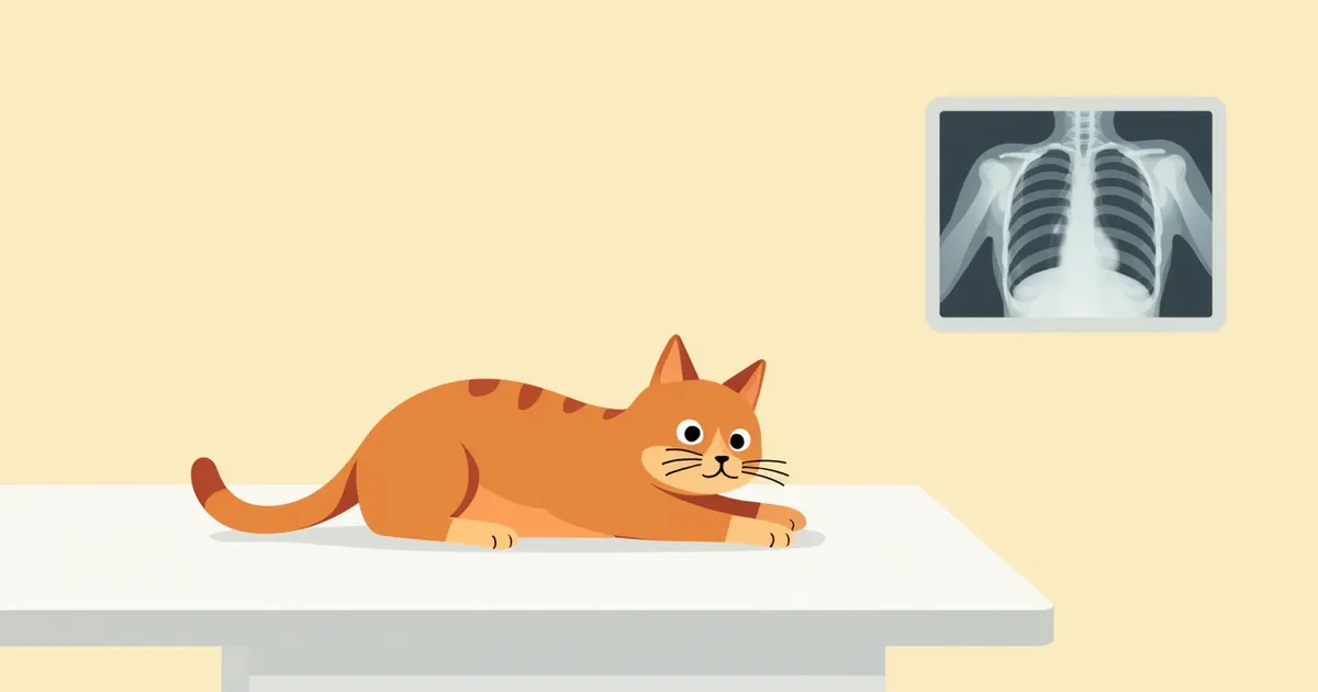
A cat undergoing a chest X-ray, with the technician adjusting its position.
Feline thoracic radiography is a key diagnostic tool for non-invasively identifying structural abnormalities in the lungs and heart. The more precise the imaging technique, combined with knowledge of thoracic anatomy and skilled interpretation, the more valuable information can be obtained. - Pneumonia or pulmonary edema: A hazy appearance of the lungs may suggest inflammation or fluid accumulation. Pulmonary edema, in particular, often presents as patchy opacities within the lung lobes. - Pneumothorax: Air in the thoracic cavity can compress the lungs, leading to respiratory distress. This can be an emergency situation. - Cardiomegaly: An enlarged heart may indicate heart disease, but radiography has limited sensitivity for mild to moderate enlargement, with clear changes typically visible only when the condition is more advanced. Therefore, additional tests and comprehensive interpretation are always necessary. - Pulmonary nodules or pleural effusion: A fixed shadow in a specific area may suggest a tumor, infectious lesion, or fluid accumulation. Accurate diagnosis requires a veterinarian’s comprehensive assessment, and early detection significantly aids treatment.

What are the main abnormalities seen on X-rays?
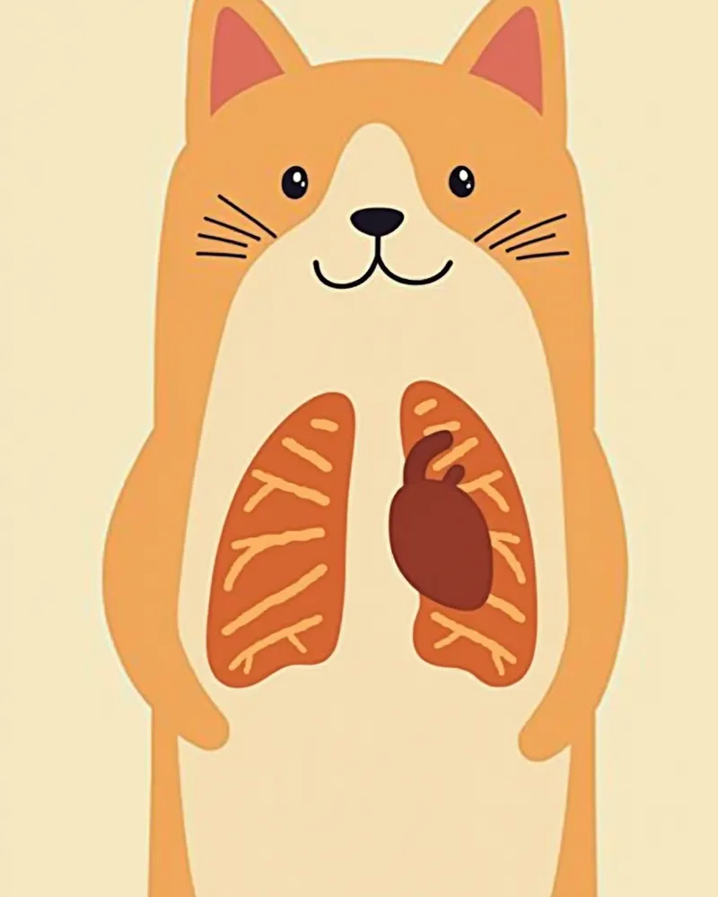
Common abnormalities seen on feline thoracic radiographs include pulmonary opacities, pneumothorax, changes in heart size, pulmonary nodules, and pleural effusion. - Pulmonary opacities: These often indicate pneumonia or pulmonary edema, reflecting the accumulation of fluid or inflammation within lung tissue. The pattern of opacity may be localized or diffuse. - Pneumothorax: Air in the pleural space compresses the lungs, leading to severe respiratory distress. While commonly associated with trauma, it can rarely occur secondary to chronic airway disease, warranting careful attention. - Cardiomegaly: An enlarged heart silhouette may signal heart disease; however, radiography has limited sensitivity for mild to moderate atrial or ventricular enlargement, with distinct enlargement typically visible only when the disease is more advanced. - Pulmonary nodules: Fixed radiographic densities can arise from various causes, including tumors, infections, or fibrosis. Further diagnostic testing is required based on the characteristics of the lesion. - Pleural effusion: Fluid accumulation in the pleural space compromises breathing. It is identified by findings such as interlobar fissures, rounded lung margins, and separation of the lung from the chest wall. Comprehensive interpretation by a veterinarian is essential for accurate diagnosis and treatment.
Areas on a cat’s chest X-ray showing lung haziness and heart enlargement

Is concern necessary even if X-ray results are normal?
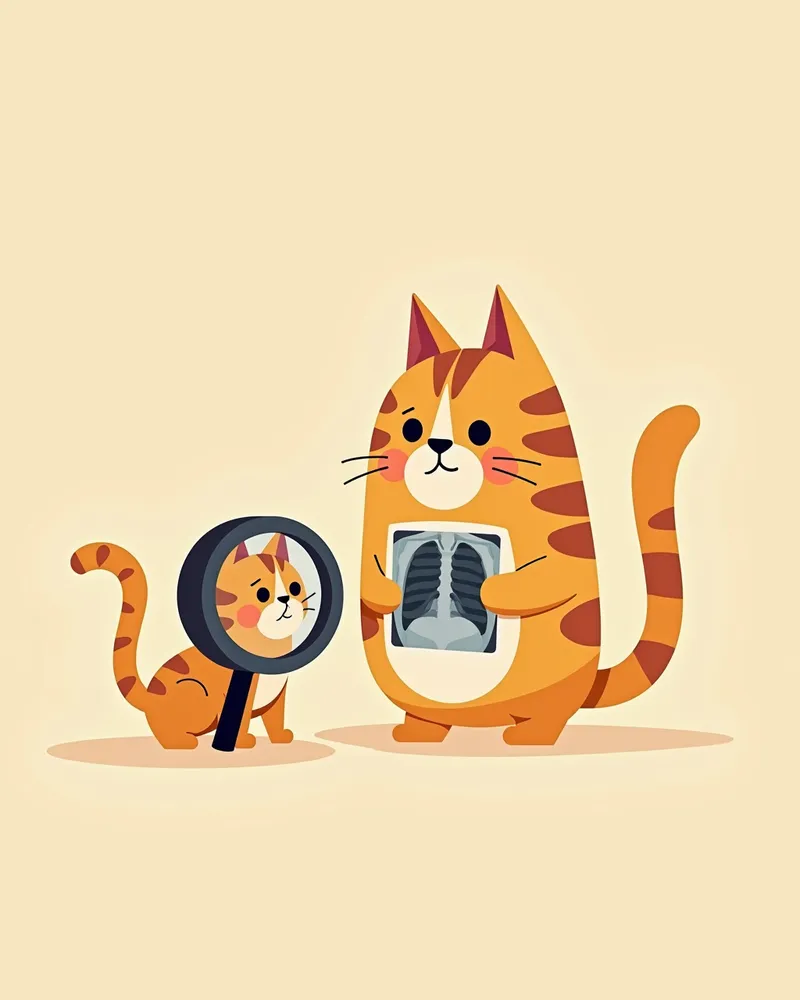
Even if X-ray results appear normal, further testing is essential if symptoms persist. - Early-stage pneumonia or bronchitis: Subtle lesions may not be visible on X-rays. - Functional abnormalities: Reduced lung function or increased bronchial reactivity cannot be detected through imaging alone. - Persistent coughing or difficulty breathing: These symptoms require a thorough evaluation by a veterinarian. - Additional tests needed: Procedures such as bronchoscopy, CT scans, and blood tests should be considered. Do not rely solely on X-ray results when symptoms are present; please consult your veterinarian.
A veterinarian carefully analyzing a cat's chest X-ray.

Comparison of Major Abnormality Types in Feline Thoracic X-ray Results

ItemKey FeaturesSuspected DiseaseAdditional Tests Needed
Lung OpacityThe lung area appears darkPneumonia, pulmonary edema, bronchitisBlood test, bronchial bacterial culture, CT scan
PneumothoraxAir accumulation in the thoracic cavity, lung compressionTrauma, alveolar rupture, chronic airway disease, lung tumorCT scan, thoracocentesis
CardiomegalyThe heart shadow becomes enlargedHeart disease, hypertensive heart disease, heart failureEchocardiography, blood pressure measurement, natriuretic peptide (BNP) test
Pulmonary NoduleSmall solid shadowTumor, infection, fibrosisCT scan, biopsy (tissue examination)

Abnormal patterns require a veterinarian's comprehensive judgment. Do not diagnose on your own.

Signs Requiring Immediate Veterinary Visit

If your cat suddenly starts breathing rapidly, pants with its mouth open, or develops blue-tinged lips or tongue, seek veterinary care immediately. These signs may indicate a medical emergency such as a pneumothorax or heart failure. Even if X-rays appear normal, these symptoms can be life-threatening, so prompt intervention is essential.

What Pet Owners Should Prepare Before an X-ray

Before an X-ray, it’s important to keep your cat calm and minimize stress. - Minimizing stress: Cats have limited tolerance for stress and restraint, so creating a calm environment before and after the procedure is essential. Fasting isn’t always required for a simple chest X-ray, so please confirm preparation steps with your clinic in advance. - Cat movement: Any movement can blur the images, potentially requiring a retake. - Difficulty breathing: If your cat is struggling to breathe, we may take just one image in a comfortable position rather than forcing a standard pose. - Procedure time: The process is usually quick and painless. - Owner’s role: Your main job is to help keep your cat as still and relaxed as possible.
A veterinarian gently positioning a cat

Key Points for Pet Owners to Understand When Interpreting X-ray Results

Chest X-ray results for cats should be interpreted by a veterinarian. - Do not interpret on your own: Misunderstandings can occur if you try to assess the images yourself. - Ask specific questions: Pose clear questions such as, “What does this shadow mean?” or “Is this condition serious?” - Record the explanation: It helps to take notes or record the veterinarian’s explanation. - Complexity of diagnosis: A comprehensive assessment of multiple factors is required, and it is difficult to reach a conclusion based on a single image alone. - Importance of communication: If you do not understand, please ask for further clarification.
The pet owner discusses the chest X-ray results with the veterinarian and takes notes.

Is Treatment Possible Even If Abnormalities Are Detected on X-rays?
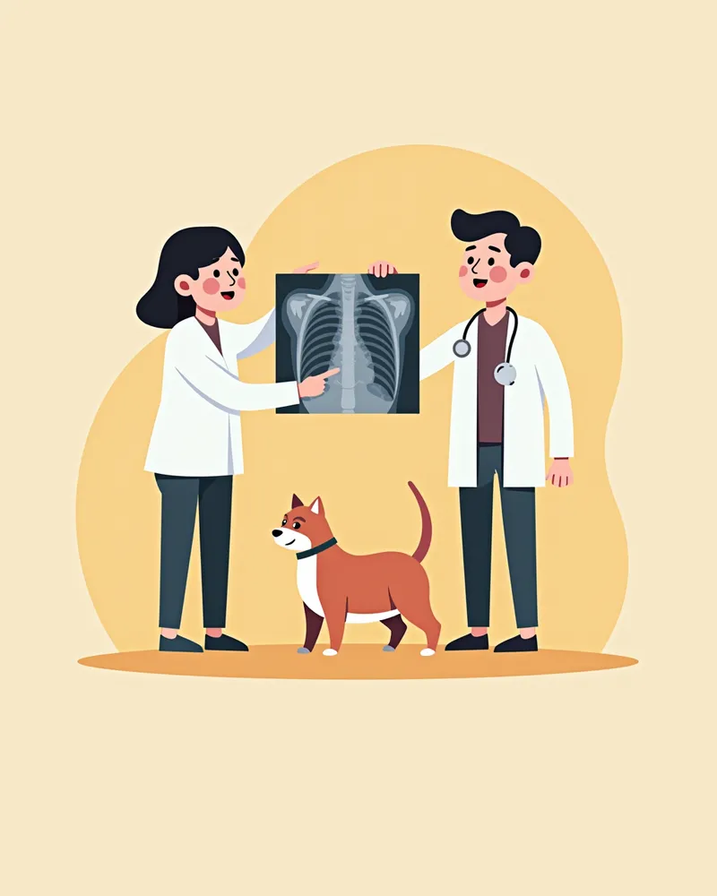
Even if abnormalities are detected on an X-ray, the potential for treatment varies depending on the type and stage of the disease. - Pneumonia: Can be effectively managed with antibiotics, and the likelihood of recovery is high when diagnosed early. - Pneumothorax: Respiratory function can be restored through treatments that remove air, such as aspiration or drainage. - Heart disease: Medication can help control symptoms and extend life expectancy, although a complete cure is often difficult to achieve. - Tumors: If detected early, tumors may be manageable through surgery or chemotherapy, but the prognosis depends on the nature of the lesion and whether it has metastasized. - Key to successful treatment: Early diagnosis, consistent care, and close collaboration with your veterinarian are essential. Treatment plans must be tailored individually to each patient’s condition.
A veterinarian explaining treatment options for a cat.

Reviewed by a veterinarian

Dr. Tony — Punnawat Phongkittirak

Dr. Tony — Punnawat Phongkittirak

Veterinarian

A veterinarian who majored in veterinary medicine at Khon Kaen University, Thailand, and completed the IVSA program at North Carolina State University in the United States. Drawing on clinical experience at animal hospitals, he works in the pet healthcare field and is dedicated to building a digital care environment that connects pet parents with veterinarians.

Frequently Asked Questions

Does a chest X-ray hurt cats?
No, X-rays are painless. Your cat just needs to lie still. However, stress or excessive movement can reduce image quality.
Is radiation exposure after an X-ray dangerous?
No, a feline chest X-ray is a relatively safe procedure performed with a low radiation dose. The risk from a single examination is very low, so please discuss any concerns you may have with your veterinarian.
Can I skip the X-ray for a coughing cat?
If the coughing persists, an X-ray is necessary. Without identifying the cause, it becomes difficult to provide appropriate treatment, and you may miss early signs of lung disease.
Even though the X-ray results are normal, the coughing continues.
Even if the initial results are normal, persistent symptoms may warrant further diagnostic tests such as a bronchoscopy, CT scan, or thoracic ultrasound. Some conditions, particularly those affecting the airways, can appear normal on X-rays in their early stages.
I’d like to see the X-ray results.
The veterinarian will explain the results, and you can request a PDF copy if needed. It’s important to discuss the detailed interpretation with the vet.

Share

Related Guides

References

[1] Canine and Feline Respiratory Medicine, 3rd Edition. 2020. Elsevier.

[2] The Cat, Clinical Medicine and Management, 2nd Edition. 2018. Wiley-Blackwell.

[3] Feline Respiratory Disease: Diagnosis and Management. Journal of Feline Medicine and Surgery, 2021. SAGE Publications.

This information is based on veterinary literature and does not replace diagnosis or treatment. Please consult a veterinarian for specific health concerns.

Metapet Co., Ltd. | CEO: Park Sung-yong | Business Reg. No. 417-88-02562 | Seoul, South Korea | Customer Center

Reading Your Cat's Chest X-ray: 5 Essential Questions for Pet Owners | Meongsiljang