Like
Share
멍실장
강아지 흉수(흉막삼출) 증상과 원인, 진단·치료와 관리 포인트 총정리

Canine Pleural Effusion: Symptoms, Causes, Diagnosis, Treatment, and Management Essentials

RespiratoryDiseasesMeongsiljang Veterinary Advisory Board

Canine pleural effusion is a condition in which excess fluid accumulates in the chest cavity, causing difficulty breathing. The underlying causes can vary, including heart disease, lung disease, infections, and cancer. Early detection and accurate diagnosis are key to effective treatment.

What Is Canine Pleural Effusion?

Puppies Showing Signs of Respiratory Distress: Visualizing Symptoms of Pleural Effusion
Canine pleural effusion is a condition in which excess fluid accumulates in the chest cavity, preventing the lungs from expanding normally. This can cause difficulty breathing, coughing, and fatigue, and in severe cases, it can be life-threatening. - Heart disease: Poor blood circulation due to heart failure can cause fluid to leak into the chest cavity. - Infectious causes: Conditions such as pneumonia, tuberculosis, or fungal infections can inflame the pleura, leading to fluid buildup. - Tumors: Lung cancer or heart tumors can invade the pleura and cause fluid accumulation. - Hypoproteinemia: Low protein levels in the blood make it easier for fluid to escape from blood vessels into surrounding tissues. Accurately identifying the underlying cause is key to effective treatment.

Main Causes of Pleural Effusion

Canine pleural effusion can result from a variety of underlying conditions. - Cardiac disease: Heart conditions, such as right-sided heart failure, are among the most common causes. When blood circulation is impaired, fluid tends to accumulate in the pleural cavity, leading to pleural effusion. - Infectious causes: Infections like pneumonia or bacterial pleuropneumonia can cause inflammation of the pleura, resulting in fluid production. Fungal infections and tuberculosis are less common causes. - Neoplastic causes: Thoracic tumors may infiltrate the pleura or cause malignant exudation, leading to fluid accumulation. - Trauma or vascular injury: Accidents or trauma can allow blood to enter the pleural cavity (hemothorax), which can manifest as pleural effusion. - Hypoproteinemia: Low blood protein levels reduce the oncotic pressure in blood vessels, making it easier for fluid to leak out into surrounding tissues. Accurately identifying the underlying cause is the first step toward effective treatment.
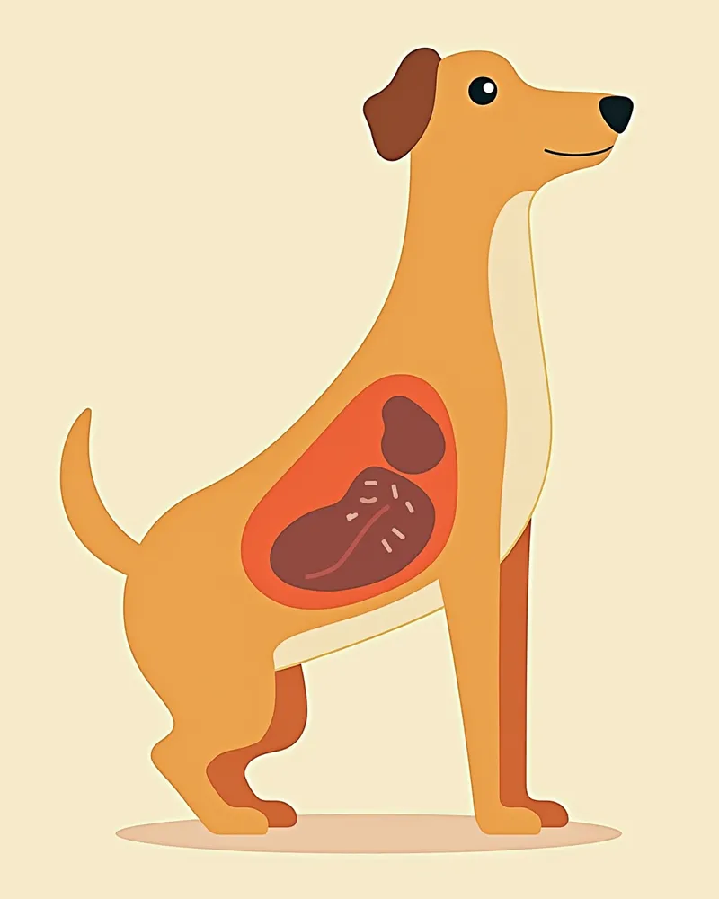
Anatomical diagram of the site where pleural effusion occurs in dogs, showing fluid accumulation within the thoracic cavity

Key Symptoms and Signs

The most common sign of pleural effusion in dogs is difficulty breathing. You may notice rapid, labored breathing or panting with the mouth open. The respiratory rate and effort increase, often accompanied by coughing and exercise intolerance, where the dog tires easily. Your dog may appear restless or reluctant to lie down. In severe cases, cyanosis—bluish discoloration of the lips or tongue—can occur. Because there is a risk of collapse, immediate veterinary care is essential.
Difficulty breathing: Repeated episodes of rapid, labored breathing or panting with the mouth open.
Coughing: Persistent coughing caused by irritation of the pleura.
Exercise intolerance and fatigue: The lungs cannot expand properly, leading to quick exhaustion and difficulty with activity.
Cyanosis of the lips or tongue: A sign of hypoxia due to insufficient oxygen.
Restlessness and reluctance to lie down: Discomfort from the fluid accumulation makes it hard for the dog to settle down, causing anxiety.
Blue lips and an open mouth in a puppy, symptoms of hypoxia

When to Seek Immediate Veterinary Care

If your dog’s panting or open-mouth breathing persists, seek veterinary care immediately. This is an emergency if the lips or tongue turn blue or if the dog appears at risk of collapsing. Because pleural effusion can obstruct breathing, prompt diagnosis and treatment are critical to saving your pet’s life.

Accurate Diagnostic Methods

An accurate diagnosis of pleural effusion in dogs requires a combination of several tests. - Chest X-ray: When fluid accumulates in the thoracic cavity, it compresses the lungs, making them appear smaller. - Thoracentesis: Fluid is collected from the thoracic cavity and analyzed to determine the underlying cause. - Blood and urine tests: These evaluate heart function, check for infections, and assess protein levels. - Echocardiogram: This ultrasound of the heart examines its structure and function to determine if heart failure is present. - CT scan: This provides detailed imaging to identify tumors or complex structural abnormalities. An accurate diagnosis is essential for effective treatment.
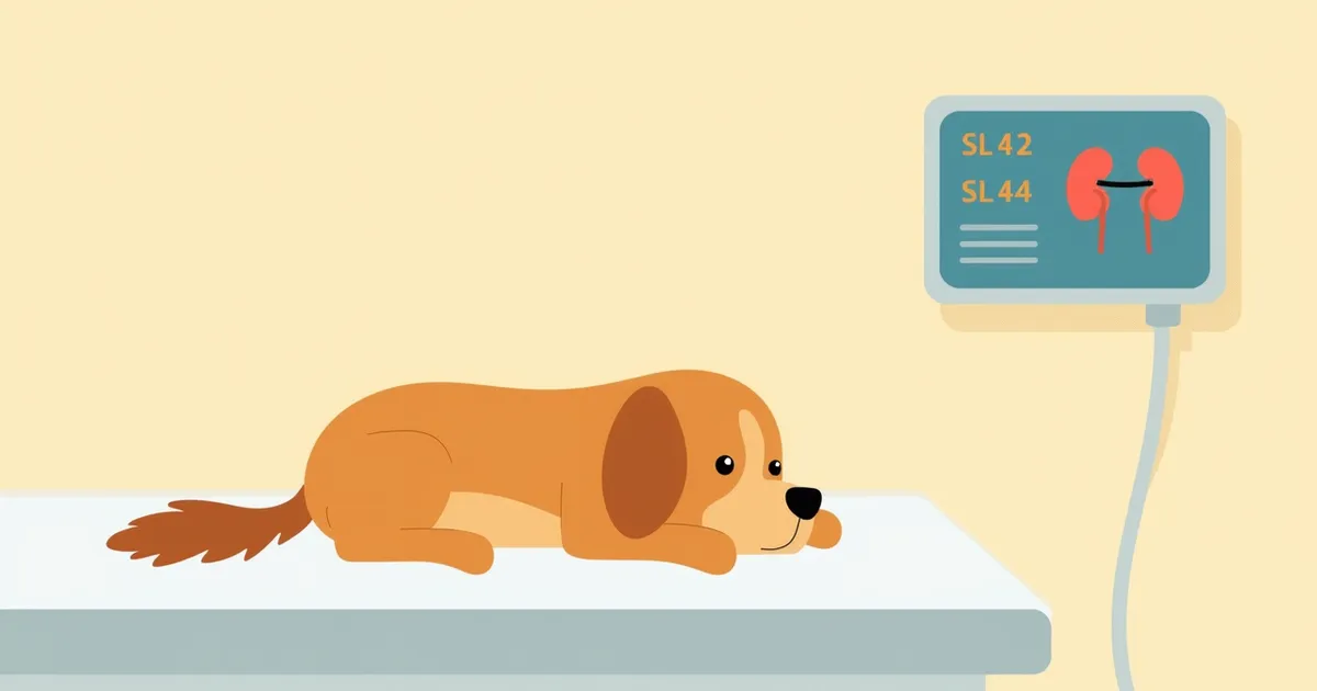
During a puppy’s chest X-ray, visualizing the diagnostic process

Treatment Options and Step-by-Step Management

Treatment depends on the underlying cause. First, thoracentesis is performed to remove pleural effusion, which provides immediate relief for breathing. If the cause is heart disease, cardiac medications are prescribed; if it’s an infection, antibiotics are used. If a tumor is suspected, a biopsy or surgery may be considered. Addressing the root cause is key to long-term management.
Thoracentesis: Fluid is removed from the chest cavity using a syringe to improve breathing.
Cardiac medications: For pleural effusion caused by heart failure, medications that support heart function are necessary.
Antibiotic therapy: If bacterial infection is the cause, antibiotics are prescribed.
Cancer treatment: If a tumor is the cause, radiation or chemotherapy may be considered.
A veterinarian performing thoracentesis on a puppy, including the diagnostic and treatment process

Home Care Essentials

Consistent care remains essential even after treatment. It’s crucial to administer medications exactly as prescribed. If supplemental oxygen is needed, options include oxygen capsules, an oxygen chamber, or a mask. Help your pet by managing their activity levels and providing a low-stress environment. If heart disease is present, a low-sodium diet may be necessary. Regular check-ups are important to monitor for recurrence. Clinically, it’s helpful to evaluate the characteristics of the pleural effusion (such as transudate, modified transudate, or chylothorax) alongside ultrasound echogenicity and the location of lesions.
A calm dog wearing an oxygen mask, visualizing home care

Preventing Recurrence and Breed-Specific Risks

Pleural effusion can recur depending on the underlying disease. Chylothorax is relatively common in Afghan Hounds, Shetland Sheepdogs, and Shiba Inus. Additionally, breeds with congenital heart defects may be at risk for pleural effusion due to right-sided heart failure. English Bulldogs and Boxers are prone to pulmonary valve stenosis, as are Mastiffs, Samoyeds, Miniature Schnauzers, and West Highland White Terriers. Labrador Retrievers tend to have tricuspid valve dysplasia. Regular cardiac check-ups and health management are essential. If you notice any unusual symptoms while your pet is on medication, consult your veterinarian immediately.

Reviewed by a veterinarian

Dr. Tony — Punnawat Phongkittirak

Dr. Tony — Punnawat Phongkittirak

Veterinarian

A veterinarian who majored in veterinary medicine at Khon Kaen University, Thailand, and completed the IVSA program at North Carolina State University in the United States. Drawing on clinical experience at animal hospitals, he works in the pet healthcare field and is dedicated to building a digital care environment that connects pet parents with veterinarians.

Frequently Asked Questions

Can pleural effusion be treated?
With accurate diagnosis and appropriate treatment, most cases can improve. The key is to address the underlying cause, such as heart disease, infection, or tumors.
Is thoracentesis painful?
Since the veterinarian will use anesthesia and pain medication, there will be little to no discomfort. The procedure is quick and safe.
Can it recur?
If the underlying cause isn’t addressed, there’s a risk of recurrence. Regular check-ups and consistent medication are essential.
Can oxygen be administered at home?
You can use oxygen capsules or an oxygen chamber. Please consult with your veterinarian to ensure safe use.
Do you need to adjust your diet?
Chest fluid associated with heart disease may require a low-sodium diet. Choose a food recommended by your veterinarian.

Share

Related Guides

References

[1] Evans, H.E. (1993). The respiratory system. In Miller’s Anatomy of the Dog, 3rd edition, pp. 463–493.

[2] Plumb's Drug Handbook for Dogs and Cats, 2023 Edition. Veterinary Pharmacology Press.

[3] Feldman, B.F., Nelson, R.W., & Reusch, C.E. (2013). Schalm's Veterinary Hematology, 6th Edition. Wiley-Blackwell.

This information is based on veterinary literature and does not replace diagnosis or treatment. Please consult a veterinarian for specific health concerns.

Metapet Co., Ltd. | CEO: Park Sung-yong | Business Reg. No. 417-88-02562 | Seoul, South Korea | Customer Center

Canine Pleural Effusion: Symptoms, Causes & Treatment | Meongsiljang