Like
Share
멍실장
강아지 조직구종 초기 증상과 진단·치료, 예후와 관리 가이드

Red Lump on Your Dog’s Skin — Distinguishing Benign Histiocytoma from Malignant Tumors and When to Visit the Vet

Oncology/CancerDiseasesMeongsiljang Veterinary Advisory Board

Canine histiocytoma is a benign tumor that originates from skin immune cells. While most cases regress naturally, it is essential to distinguish it from malignant tumors. This article covers everything from early symptoms to diagnosis, treatment, prognosis, and home care.

What Is Canine Histiocytoma?

Close-up of a young puppy with a histiocytoma in its ear
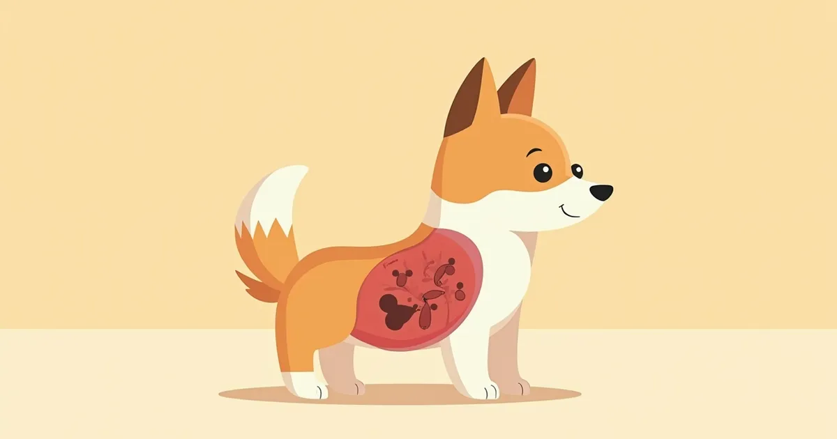
Canine histiocytoma is a benign skin tumor that develops when Langerhans cells, a type of skin immune cell, proliferate. The most important thing to know is that most of these tumors regress spontaneously without treatment within weeks to months after diagnosis, typically within 1 to 3 months. However, regression may be delayed if there is inflammation or secondary infection. Because it can be difficult to distinguish histiocytomas from malignant tumors based on appearance alone, it is safest to have any new lump evaluated through cytology or a biopsy rather than trying to diagnose it yourself. This condition is most common in young dogs, with about half of cases occurring in dogs under two years of age, and typically appears as pink, dome-shaped lumps on the head (especially the ear flaps), face, front legs, or trunk.

Causes and Mechanisms of Development

Histiocytomas are benign tumors that develop when Langerhans dendritic cells, which are distributed in the epidermis and dermis of the skin, increase in number. These cells are part of the immune system and respond to external stimuli, but the exact reason for their sudden proliferation remains unclear. However, since they occur most frequently in young dogs (with about half of cases appearing in dogs under two years old) and are more commonly reported in certain breeds, breed predisposition may play a role. Veterinary textbooks list Boxers and Dachshunds as breeds particularly prone to histiocytomas. Even though these tumors may grow rapidly and show numerous cell divisions under the microscope, they are essentially benign and often resolve on their own over time.

Checklist of Main Symptoms of Histiocytoma

Dome-shaped lump: Characterized by a hemispherical, raised appearance above the skin surface.
Pink to red color: Often appears reddish due to abundant blood vessels.
Rapid growth: Can grow quickly within a few weeks, reaching 1–2 cm in size.
Hairless surface: The tumor area may lose hair or develop ulcers.
Common locations: Frequently occurs on the ears, forehead, eyelids, and front legs.
No itching or pain: Most dogs do not show signs of discomfort.
The dome-shaped surface of a histiocytoma that has developed on a dog's ear

Go to the Vet Immediately If You Notice These Changes

If a lump grows rapidly within two to three weeks, ulcerates and bleeds, appears in multiple locations simultaneously, or fails to shrink at all after three months, there is a possibility it could be malignant. In such cases, it is important to promptly perform a cytology or histopathological biopsy.

How Is It Diagnosed?

The most basic diagnostic method is fine-needle aspiration cytology. This involves using a thin needle to collect cells from the lump and examining them under a microscope. It can be performed within minutes without anesthesia and offers high accuracy. If the results are inconclusive or malignancy is suspected, a tissue biopsy (pathological examination after partial or complete excision) is performed. Because it is difficult to distinguish malignant tumors based on photographs or visual inspection alone, it is most important to have the lump examined rather than trying to self-diagnose.

Treatment Options — From Watchful Waiting to Surgery

For histiocytomas, the first-line approach after diagnosis is often watchful waiting, as most lesions regress spontaneously within 8 to 12 weeks. However, surgical excision should be considered in the following cases: - The lesion continues to grow beyond 1 cm in size - The dog persistently licks the area, posing a risk of secondary infection - The lesion fails to shrink after three months - The location affects function, such as on the eyelids The prognosis after surgery is excellent, and the recurrence rate is low.
A veterinarian performing a fine-needle aspiration test on a dog's skin lump.

Home Care Points

If you’re monitoring the lump at home, here’s how to care for it: Keep your dog from licking or scratching the area by fitting them with an Elizabethan collar or protective wrap to prevent secondary bacterial infections. Take photos from the same angle every two weeks so you can compare the lump’s size during your next vet visit. If the surface becomes ulcerated, swelling worsens, or the color changes suddenly, schedule a follow-up appointment right away. If the lump begins to regress naturally, it will flatten and gradually disappear over time.

Owners of Predisposed Breeds Should Pay Special Attention

If you’re raising a Boxer or Dachshund, keep in mind that histiocytoma is reported more frequently in this breed than in others, so it’s important to regularly check for any new lumps. Don’t dismiss them with thoughts like, “It’s just a common lump, so it’s probably fine.” If you notice a new lump, the safest approach is to have a cytology test done rather than trying to assess it on your own. For reference, Bernese Mountain Dogs, Rottweilers, and Golden and Labrador Retrievers are predisposed to more serious histiocytic disorders (histiocytosis and histiocytic sarcoma), so if a lump appears in these breeds, it’s best to undergo careful differential diagnostics.

Reviewed by a veterinarian

Dr. Tony — Punnawat Phongkittirak

Dr. Tony — Punnawat Phongkittirak

Veterinarian

A veterinarian who majored in veterinary medicine at Khon Kaen University, Thailand, and completed the IVSA program at North Carolina State University in the United States. Drawing on clinical experience at animal hospitals, he works in the pet healthcare field and is dedicated to building a digital care environment that connects pet parents with veterinarians.

Frequently Asked Questions

Is histiocytoma a type of cancer?
Not at all. A histiocytoma is a benign skin tumor. In most cases, it resolves on its own within one to three months. However, because it can resemble a malignant tumor in appearance, it is essential to confirm the diagnosis through cytology or a biopsy.
Can it heal without surgery?
Yes, it’s possible. In most cases, they resolve on their own within a few weeks to a few months (commonly 1–3 months) after diagnosis, without treatment. However, if the lump continues to grow or doesn’t disappear after 3 months, we recommend surgical removal to prevent it from becoming malignant.
Can it recur?
The recurrence rate of histiocytomas themselves is low. However, new histiocytomas can develop in the same area or in different areas. In particular, predisposed breeds require regular skin checks.
Does it only happen in young puppies?
This condition most commonly affects young puppies, with about half of cases occurring in dogs under two years of age, though it can develop at any age. In older dogs, it is especially important to differentiate this condition from other types of tumors.
My dog keeps licking a lump. What should I do?
Licking can lead to secondary bacterial infections, which may slow down the healing process. It's a good idea to have your pet wear a neck collar, and if the surface becomes raw or the swelling worsens, it's best to have the wound treated at a veterinary clinic.

Share

Related Guides

References

[1] Withrow SJ, Vail DM, Page RL. Withrow and MacEwen's Small Animal Clinical Oncology, 5th Edition. Chapter 19: Tumors of the Skin and Subcutaneous Tissues. Elsevier Saunders, 2013.

[2] Gross TL, Ihrke PJ, Walder EJ, Affolter VK. Skin Diseases of the Dog and Cat: Clinical and Histopathologic Diagnosis, 2nd Edition. Chapter: Histiocytic Tumors. Blackwell Publishing, 2005.

[3] Moore PF. A review of histiocytic diseases of dogs and cats. Vet Pathol. 2014;51(1):167-184.

[4] Dobson JM, Lascelles BDX. BSAVA Manual of Canine and Feline Oncology, 3rd Edition. BSAVA, 2011.

This information is based on veterinary literature and does not replace diagnosis or treatment. Please consult a veterinarian for specific health concerns.

Metapet Co., Ltd. | CEO: Park Sung-yong | Business Reg. No. 417-88-02562 | Seoul, South Korea | Customer Center

Canine Histiocytoma: Benign vs. Malignant Skin Lumps | Meongsiljang