Like
Share
멍실장
고양이 흉부 X선 판독 증상과 원인, 진단·치료와 관리 포인트 총정리

Comprehensive Guide to Feline Thoracic Radiography: Symptoms, Causes, Diagnosis, Treatment, and Management

RespiratoryDiseasesMeongsiljang Veterinary Advisory Board

Chest X-rays are a key tool for the early detection of respiratory diseases in cats. Accurate interpretation allows us to identify the cause and severity of the condition, enabling us to initiate appropriate treatment.

What Is Feline Thoracic Radiography?

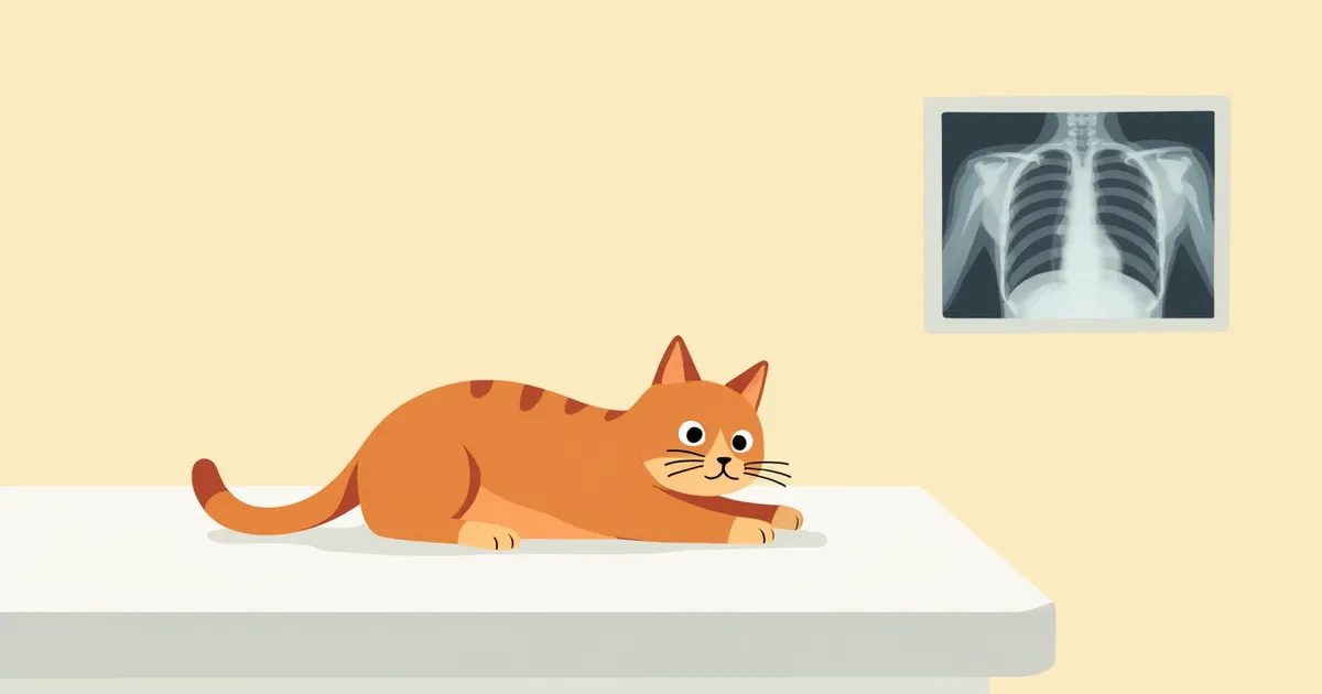
A cat undergoing a chest X-ray.
Feline thoracic radiography is a key diagnostic tool that allows for non-invasive assessment of the lungs, heart, and thoracic structures. It helps identify the underlying cause of symptoms such as dyspnea, coughing, and lethargy in cats. Veterinarians use a systematic approach to analyze changes in lung size, shape, and density to determine the presence of abnormalities. - Pneumonia: Inflammation of lung tissue caused by bacterial, viral, or fungal infections may appear on radiographs as increased, hazy opacity or signs of lobar abscessation. - Congestive heart failure: This condition can be accompanied by cardiomegaly and pulmonary edema, resulting in increased pulmonary opacity or obscuration of the cardiac silhouette. - Lower airway disease: Inflammatory conditions such as feline asthma and bronchitis may cause increased expiratory effort, abdominal breathing, and impaired airflow. Accurate interpretation is essential for early diagnosis and the development of an appropriate treatment plan.

Major Causes and Pathogenesis

Abnormal findings on a feline thoracic radiograph may be caused by pneumonia, pleural effusion, heart failure, pneumothorax, or bronchial disease. Pneumonia occurs when bacterial, viral, or fungal infections cause inflammation in the lung tissue, appearing on X-rays as hazy areas of increased density or signs of lobar abscesses. Heart failure can manifest as an enlarged heart and pulmonary edema, resulting in increased lung density and an indistinct cardiac silhouette. When pleural effusion accumulates, the edges of the lung lobes may appear rounded, the lobes may seem separated from the chest wall, or the cardiac silhouette may be obscured. In lower airway diseases, respiratory effort may increase during exhalation, and airflow can be obstructed. These structural changes in the lungs and thoracic cavity can present as characteristic findings on X-rays.
Feline thoracic anatomy diagram, showing lung and heart structures

Key Symptoms and Signs

When abnormalities appear on a cat’s chest X-ray, they are usually accompanied by symptoms such as difficulty breathing, coughing, decreased activity, weight loss, and a bluish discoloration of the lips or extremities. These signs indicate impaired lung function or insufficient oxygen supply, making immediate diagnosis essential. The veterinarian will comprehensively evaluate changes in the size, shape, and density of the lungs via X-ray to determine if there are any abnormalities. Accurate interpretation directly impacts early treatment and improved prognosis.
Difficulty breathing: The cat breathes rapidly or repeatedly pants with its mouth open.
Coughing: Persistent coughing occurs, often worsening at night or after activity.
Decreased activity: The cat no longer runs around as usual and tends to rest more frequently.
Cyanosis: The lips, extremities, and ear tips take on a bluish tint.
Weight loss: Appetite decreases, leading to rapid weight loss.
A cat showing signs of respiratory distress

Situations Requiring Immediate Veterinary Care

If your cat shows severe difficulty breathing, breathes with its mouth open, or exhibits cyanosis (a bluish tint to the lips or paw pads), seek veterinary care immediately. These signs may indicate life-threatening conditions such as pulmonary edema or heart failure. A veterinarian can promptly initiate emergency interventions, including X-ray imaging and oxygen therapy. Because delays significantly increase the risk of death, do not wait if you observe these symptoms.

Diagnostic Methods and Procedures
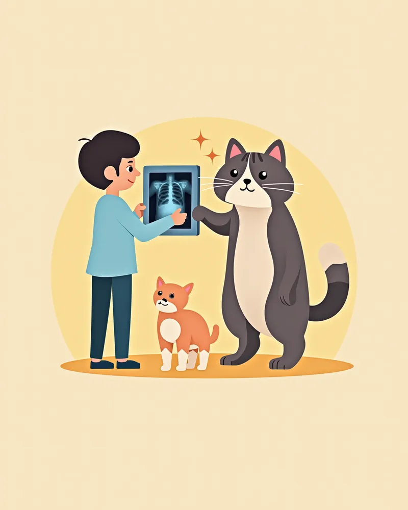
Feline thoracic radiographs are typically taken from two angles: ventrodorsal (or dorsoventral) and lateral. This allows for a comprehensive assessment of the size, shape, position, and density changes of the lungs and heart. If the radiographic findings are inconclusive, further diagnostic tests such as CT imaging or pathological examination may be necessary. The veterinarian analyzes factors such as pulmonary opacity, cardiac silhouette size, and the presence of fluid within the thoracic cavity to help determine the underlying cause of the disease. For an accurate diagnosis, these imaging studies are often combined with blood tests, bronchoalveolar lavage, or biopsy.
A veterinarian reviewing a cat's chest X-ray

Treatment Methods and Step-by-Step Management

Treatment depends on the underlying cause. Pneumonia is treated with antibiotics or antifungal medications, while congestive heart failure is managed with cardiac drugs and diuretics (e.g., furosemide). Lower airway diseases are controlled with bronchodilators or steroids, and tumors may require surgery, radiation therapy, or chemotherapy. Your veterinarian will develop a treatment plan based on X-ray results and monitor your pet’s response through regular follow-up X-rays. If significant pleural effusion is present, thoracentesis is performed to drain the fluid, and oxygen therapy is provided to ease respiratory distress. At home, maintaining a comfortable environment and ensuring consistent medication administration are essential.
A cat wearing an oxygen mask

Home Care and Daily Care

When a cat’s chest X-ray shows abnormalities, home care is crucial. Provide a quiet space to minimize stress and maintain an environment with adequate oxygen supply. Strictly adhere to prescribed medication schedules and keep all veterinary appointments. If your cat’s appetite decreases, offer easily digestible food and ensure fresh water is available frequently. Avoid sudden temperature changes and exposure to smoke. Following your veterinarian’s guidance for daily care is essential for improving the prognosis.
A cat resting in a calm and stable environment.

Breed-Specific Precautions and Relapse Prevention

Each cat may be more susceptible to specific respiratory conditions such as pleural effusion, lower airway disease, or pneumothorax. In these cases, regular check-ups and early detection are essential. Furthermore, if acute or chronic respiratory diseases recur, the risk of relapse increases, so it is important to establish a long-term management plan with your veterinarian. Following your vet’s guidance to minimize stressors and eliminate environmental irritants to maintain a clean living space can help prevent recurrence.

Reviewed by a veterinarian

Dr. Tony — Punnawat Phongkittirak

Dr. Tony — Punnawat Phongkittirak

Veterinarian

A veterinarian who majored in veterinary medicine at Khon Kaen University, Thailand, and completed the IVSA program at North Carolina State University in the United States. Drawing on clinical experience at animal hospitals, he works in the pet healthcare field and is dedicated to building a digital care environment that connects pet parents with veterinarians.

Frequently Asked Questions

When should a chest X-ray be taken for a cat?
If respiratory symptoms such as difficulty breathing, coughing, or decreased activity persist, your veterinarian may recommend an X-ray. Early detection is crucial for effective treatment.
If the X-ray results are abnormal, is it necessary to visit the hospital?
Yes, these abnormal findings suggest the possibility of serious conditions such as pneumonia, pleural effusion, heart failure, or airway disease, so prompt diagnosis and treatment are necessary.
My cat is scared of X-rays. What should I do?
The veterinarian helps position your cat in a stable posture for the imaging, ensuring the procedure is carried out while your cat is comfortable. In some cases, a sedative may be used.
Is X-ray imaging dangerous for cats?
Diagnostic chest X-rays involve relatively low radiation doses, making them generally safe. However, in special circumstances such as pregnancy, your veterinarian will carefully weigh the risks and benefits to determine whether an X-ray is necessary and the best approach to take.
My dog keeps coughing even though the X-ray results are normal. Why is that?
Coughing can also occur due to airway inflammation or allergies that don't show up on X-rays, as well as parasitic infections (such as lungworms). Additional tests, such as fecal examinations, are necessary.

Share

Related Guides

References

[1] Canine and Feline Respiratory Medicine, 3rd Edition. 2020. Elsevier.

[2] The Cat, Clinical Medicine and Management, 2nd Edition. 2018. Wiley-Blackwell.

[3] Feline Respiratory Disease: Diagnosis and Management. Journal of Feline Medicine and Surgery, 2021. SAGE Publications.

This information is based on veterinary literature and does not replace diagnosis or treatment. Please consult a veterinarian for specific health concerns.

Metapet Co., Ltd. | CEO: Park Sung-yong | Business Reg. No. 417-88-02562 | Seoul, South Korea | Customer Center

Feline Chest X-Ray (Thoracic Radiography): A Complete Guide | Meongsiljang