

Top Recommendations and Selection Criteria for Canine Thoracic X-ray Interpretation Management
Reading chest X-rays in dogs is key to the early detection of respiratory diseases. You can protect your pet’s health through management methods that combine expertise and accuracy.
Canine Thoracic X-ray Interpretation Is the Foundation of Accurate Respiratory Diagnosis

6 Key Criteria to Check When Choosing Canine Thoracic X-ray Interpretation
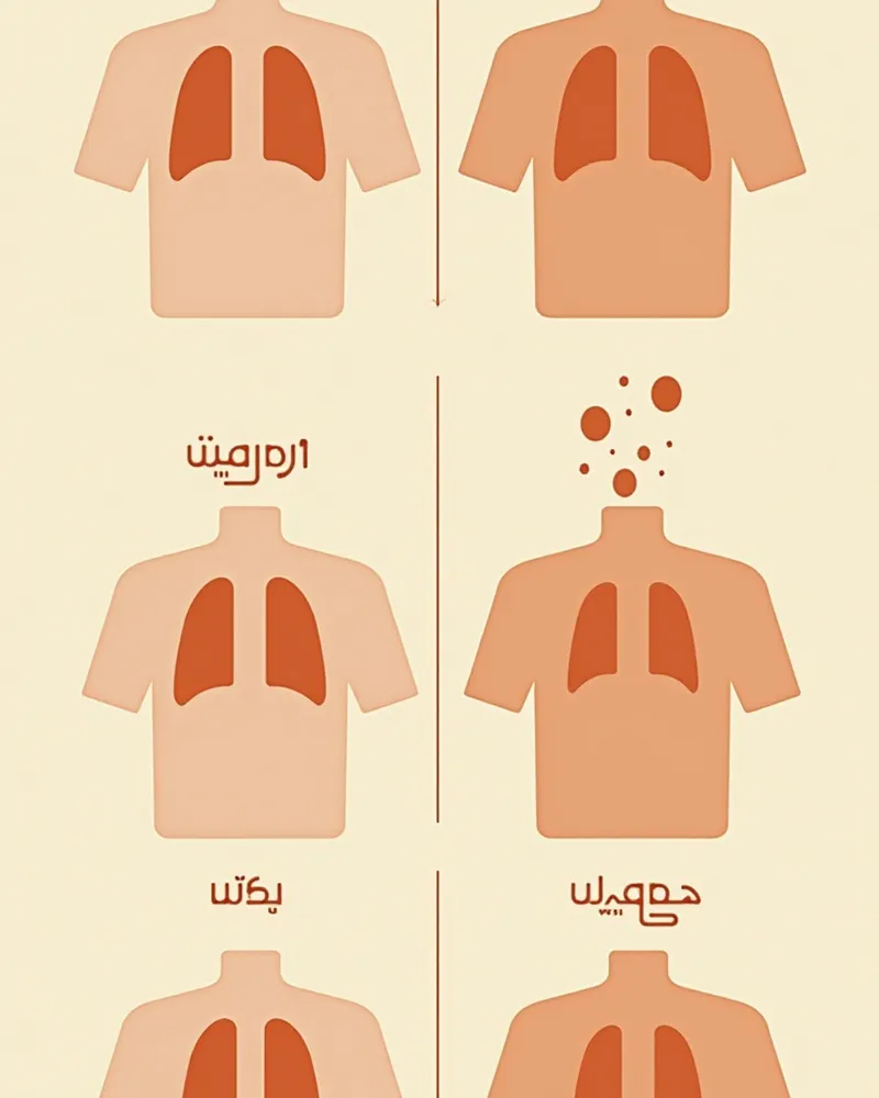
Check 1: Is It Interpreted by a Veterinarian with Extensive Radiology Experience?

Check 2: Is Comparison and Analysis with Past Images Possible?

Check 3: Are Retake Policies and Consultation (Remote Interpretation) Available?

Checklist for Selecting a Clinic for Thoracic X-ray Interpretation
| Item | Recommended criteria |
|---|---|
| Interpretation capability | A clinic that organizes the lungs, heart, airways, and pleura into a detailed report |
| Progress comparison | A clinic capable of comparative analysis with past imaging |
| Quality/collaboration | A clinic with a re-imaging policy and access to specialist interpretation collaboration |
| Result interpretation | A clinic where the veterinarian personally explains the test results and guides the management plan |
| Cost guidance | A clinic that informs you in advance of the estimated cost and the possibility of additional tests |
This is not a table recommending a specific clinic, but rather criteria worth checking when choosing a clinic to perform the testing. Prices and specific programs vary by clinic, so inquire directly before visiting.
Caution: Low X-ray Image Quality Increases the Risk of Diagnostic Errors
If the resolution or angle of an X-ray image is inadequate, important abnormalities such as lung nodules, bronchiectasis, or cardiomegaly may be missed. It is essential to obtain high-resolution images taken at appropriate angles. If you have any concerns about image quality, please request a retake.
5 Tips for Using Canine Thoracic X-ray Interpretation

Reviewed by a veterinarian

Dr. Tony — Punnawat Phongkittirak
VeterinarianA veterinarian who majored in veterinary medicine at Khon Kaen University, Thailand, and completed the IVSA program at North Carolina State University in the United States. Drawing on clinical experience at animal hospitals, he works in the pet healthcare field and is dedicated to building a digital care environment that connects pet parents with veterinarians.
Reviewed 2026-06-29
Frequently Asked Questions
When should a dog have a chest X-ray?
Are you still worried even if the X-ray results are normal?
Is the online platform's interpretation accurate?
Is X-ray imaging harmful to dogs?
Why is it important to compare with past X-rays?
Share
Related Guides

Top Recommended Feline Bronchodilators and Selection Criteria: A Complete Guide

Best Recommendations and Selection Criteria for Monitoring Your Cat’s Respiratory Rate (RR) During Sleep for Heart Care

Best Recommendations and Selection Criteria for Monitoring Your Dog’s Respiratory Rate (RR) During Sleep for Heart Care

Top Recommended Giardia Management Products for Dogs and How to Choose Them

Top Recommended Flea Control Products for Dogs and Selection Criteria

Top Recommended Products and Selection Criteria for Managing Flea Allergy Dermatitis in Dogs

The Best Fecal Parasite Testing Methods for Dogs: Management Recommendations and Selection Criteria

Top Recommended Products for Canine Ehrlichiosis Management and Key Selection Criteria

Top Recommended Products for Dog Ear Mite Management and Selection Criteria

Top 3 Recommended Treatments and Selection Criteria for Differentiating and Managing Dog Ringworm and Parasitic Infections
References
[1] Canine and Feline Respiratory Medicine, 3rd Edition. 2023. Elsevier.
[2] Clinical Medicine of the Dog and Cat, 4th Ed. 2021. Elsevier.
[3] Textbook of Respiratory Disease in Dogs and Cats. 2022. Wiley-Blackwell.