Like
Share
멍실장
강아지 귀 혈종 (이혈종) 응급 대처 완벽 가이드

Complete Emergency Guide to Canine Aural Hematomas

Skin & CoatQ&AMeongsiljang Veterinary Advisory Board

We’ve compiled everything pet owners need to know about canine ear hematomas, from the definition and emergency assessment criteria to a comparison of surgical versus conservative treatment and tips for preventing recurrence.

What Is a Canine Aural Hematoma?
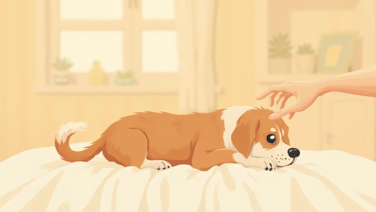
A guardian gently examining a puppy with swollen ears.
Aural hematomas in dogs occur when blood accumulates between the cartilage and skin of the ear flap, causing it to swell. The ear flap consists of two layers of skin separated by cartilage, and when a blood vessel crossing the cartilage ruptures, blood collects in the space between these layers. This condition typically appears suddenly as a smooth, round, plum-shaped swelling, most visible on the hairless, concave inner surface. It is essential to seek veterinary care as soon as possible before fibrosis (cartilage hardening) begins. If left untreated, the hematoma can cause permanent cartilage deformation, resulting in a wrinkled, hardened appearance known as "cauliflower ear," which also increases the risk of chronic ear inflammation. If the swelling feels soft and warm (or taut and elastic) and your dog repeatedly shakes its head or scratches at the ear, prompt veterinary evaluation is recommended for safety.

Causes and Pathogenesis

Aural hematomas most often result from vigorous head shaking or scratching. When a dog experiences persistent itching or discomfort in the ear, such as with otitis externa, it repeatedly shakes its head. The resulting impact ruptures small blood vessels running through the pinna cartilage, causing blood to accumulate. In fact, studies report that approximately half of the ears affected by aural hematomas also have concurrent otitis externa. Other potential causes include trauma, such as bite wounds, or coagulopathies. Although the exact etiology remains unclear, evidence suggests that degenerative changes weakening the ear cartilage may precede hematoma formation. Therefore, while the hematoma itself requires treatment, failure to address the underlying condition makes recurrence likely.

Symptom Checklist — Watch for These Signs

Swelling: The hairless, concave inner surface of the ear flap becomes smooth and round like a plum, swelling up softly like a balloon.
Warmth and Pain: The ear feels warm, firm, and elastic to the touch, and the dog may show signs of discomfort or pain when the ear is touched.
Head Shaking: The dog repeatedly shakes its head vigorously due to ear discomfort.
Repeated Scratching: The dog persistently scratches the same ear with its hind paw.
Secondary Discharge: If accompanied by otitis externa, there may be dark brown or black debris resembling coffee grounds, brown or yellow discharge, or an unpleasant odor coming from the ear.
If two or more of these symptoms persist for more than a day, please visit a veterinary clinic as soon as possible.
A puppy showing signs of ear discomfort by tilting its head to one side

⚠️ Criteria for Immediate Veterinary Visit

Do not delay veterinary care if you notice any of the following: • The swelling becomes noticeably larger within a day • Blood is visible in or draining heavily from the ear • Your dog loses its appetite and appears lethargic • Systemic symptoms such as fever or weakness are present • A strong foul odor or purulent discharge comes from inside the ear Never attempt home remedies like ice packs or draining the fluid with a needle. Improper needle aspiration can cause the hematoma to refill with blood, and may worsen cartilage damage and infection, leading to severe ear collapse or scarring.

How Is the Diagnosis Made?

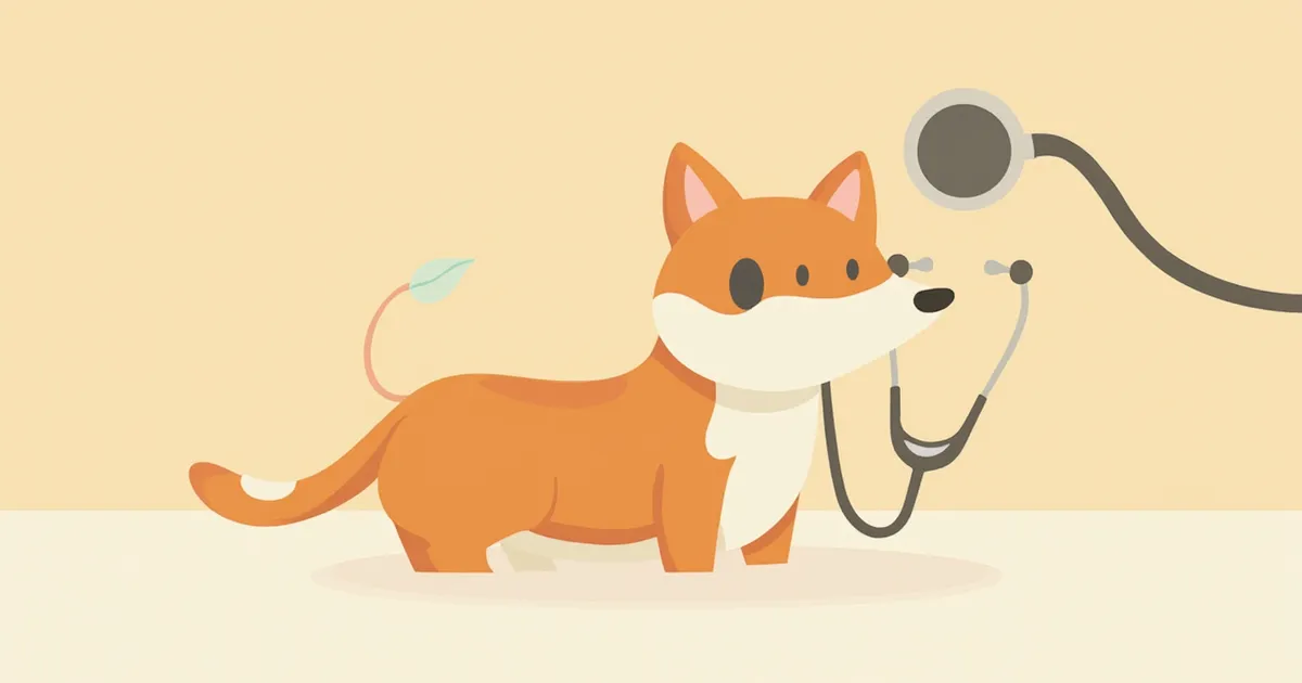
The veterinarian first uses visual inspection and palpation to determine whether the swelling is a blood-filled sac, then examines the ear canal with an otoscope to identify underlying conditions such as otitis externa. If necessary, a small sample of the fluid is aspirated with a syringe to confirm the presence of serosanguinous fluid, diagnosing a hematoma, while cytology and bacterial tests are performed to detect secondary infections or identify the cause. If otitis externa is present, dark brown or black, coffee-ground-like discharge may also be observed. If the condition is recurrent or a coagulation disorder is suspected, blood tests are also conducted. The diagnosis can typically be confirmed relatively quickly in the clinic.
A veterinarian examining the inside of a dog’s ear using an otoscope

Conservative Treatment vs. Surgery — Comparing Treatment Options

ItemNeedle AspirationDrain PlacementIncisional Surgery
Procedure MethodDraining blood with a syringeInserting a small tube for drainageIncising the hematoma and suturing
AnesthesiaLocal anesthesiaLocal or sedationGeneral anesthesia
Recurrence RateHighModerateLow
Suitable ForEarly · small hematomasMedium-sized hematomasLarge or recurrent hematomas
Recovery PeriodImmediate7–14 days14–21 days

The final treatment is decided by the veterinarian based on hematoma size, recurrence, age, and underlying disease. Veterinary surgery textbooks recommend incisional surgery as the first choice for medium or larger hematomas.

Post-Treatment Care — Key to Preventing Recurrence

After treatment, the most important step is addressing the underlying cause. Without resolving the root issue, auricular hematomas are likely to recur. If otitis externa is present, consistently use the prescribed ear cleanser and topical medication as directed by your veterinarian to reduce inflammation. Keep the Elizabethan collar (e-collar) in place until suture removal—typically around three weeks—to prevent your dog from scratching or shaking its ears. Regularly check any dressings or bandages, and postpone bathing until after suture removal to keep the suture site dry.
A dog wearing a recovery collar and resting comfortably

💡 Recurrence Prevention Checkpoints

Aural hematomas often recur in the same ear. • Visually inspect the ears once a month for redness, discharge, or odor. • For pets with a history of otitis externa, use a specialized ear cleanser every 2–3 weeks. • Thoroughly dry the inside of the ears after swimming or bathing. • If your pet has an allergic predisposition, work with your veterinarian to develop a dietary and environmental management plan. • For breeds with floppy ears (such as Cocker Spaniels and Basset Hounds), periodically lift and dry the ears to improve ventilation.

Reviewed by a veterinarian

Dr. Tony — Punnawat Phongkittirak

Dr. Tony — Punnawat Phongkittirak

Veterinarian

A veterinarian who majored in veterinary medicine at Khon Kaen University, Thailand, and completed the IVSA program at North Carolina State University in the United States. Drawing on clinical experience at animal hospitals, he works in the pet healthcare field and is dedicated to building a digital care environment that connects pet parents with veterinarians.

Frequently Asked Questions

Will a hematoma go away on its own if left untreated?
While the fluid may eventually be reabsorbed, the healing process can lead to the formation of granulation tissue and fibrosis, causing the cartilage to collapse into a permanent "cauliflower ear." This condition significantly increases the risk of recurrence and infection, so it's essential to seek veterinary care promptly rather than leaving it untreated.
Can I remove it at home with a needle?
Absolutely not. Improper needle aspiration can cause the hematoma to refill and may lead to severe ear deformity, scarring, cartilage damage, or bacterial infection. Drainage must be performed in a sterile environment at a veterinary clinic.
How long does my pet need to stay hospitalized after surgery?
For incision surgeries, same-day discharge is common, but depending on the size of the hematoma and the pet’s recovery from anesthesia, a hospital stay of one to two days may be necessary. Please follow your veterinarian’s guidance.
How much will it cost?
Costs can vary significantly depending on the clinic, location, hematoma size, and type of anesthesia, so it’s difficult to provide a one-size-fits-all estimate. I recommend getting a preliminary quote during your consultation.
Can my pet have surgery again if the condition recurs?
If the condition recurs, we first conduct a thorough diagnosis to identify the underlying cause (such as otitis externa) before deciding whether additional surgery is necessary. Without addressing the root cause, recurrence is likely.

Share

Related Guides

References

[1] Tilley, L.P., Smith, F.W.K., Blackwell's Five-Minute Veterinary Consult: Canine and Feline, 7th Edition, 2021 - Aural Hematoma Chapter

[2] Miller, W.H., Griffin, C.E., Campbell, K.L., Muller and Kirk's Small Animal Dermatology, 7th Edition, 2013 - Diseases of Eyelids, Claws, Anal Sacs, and Ear

[3] Fossum, T.W., Small Animal Surgery, 5th Edition, 2018 - Surgery of the Ear

[4] Ettinger, S.J., Feldman, E.C., Textbook of Veterinary Internal Medicine, 8th Edition, 2017 - Diseases of the Ear

This information is based on veterinary literature and does not replace diagnosis or treatment. Please consult a veterinarian for specific health concerns.

Metapet Co., Ltd. | CEO: Park Sung-yong | Business Reg. No. 417-88-02562 | Seoul, South Korea | Customer Center

Dog Aural Hematoma Emergency Care: Causes, Treatment, and Management | Meongsiljang