
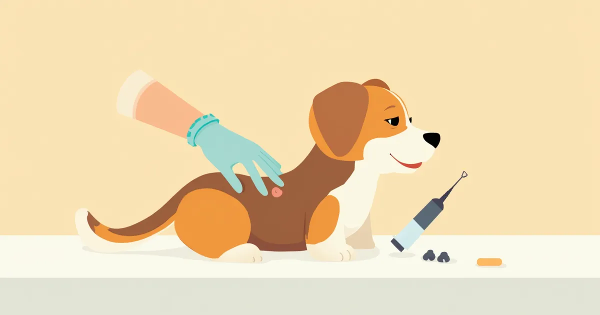
How much does a dog’s tumor biopsy cost? — How does accuracy vary by test type?
The cost of a dog’s biopsy ranges from $40 to $600 depending on the type, with significant differences in accuracy and anesthesia burden. We’ll walk you through how to choose step-by-step—from FNA to excisional biopsy—based on the tumor’s location, size, and suspected malignancy.
What is a canine biopsy, and why is choosing the right type important?
When is a biopsy necessary?
Comparison of biopsy types: cost, accuracy, and anesthesia
| Item | Fine-Needle Aspiration (FNA) | Core Needle Biopsy | Incisional Biopsy | Excisional Biopsy |
|---|---|---|---|---|
| Cost Range | $40 to $100 | $100 to $200 | $200 to $400 | $300 to $600 |
| Anesthesia Burden | Not needed | Sedation | General anesthesia | General anesthesia |
| Diagnostic Accuracy | Large variation by site/tumor (high for superficial, low for liver/spleen) | Histopathological confirmation when tissue is obtained | Histopathological confirmation | Histopathological confirmation |
| Grading Possible | False | True | True | True |
| Same-Day Performance | True | True | False | False |
| Recommended Situation | First-line test for lymph nodes/superficial masses | Deep masses, liver, spleen | Partial sampling of large masses | Simultaneous diagnosis + treatment of small masses |
Based on domestic animal hospital averages as of May 2026; pathology referral fees ($40 to $100) and immunostaining ($70 to $100) are separate. Accuracy varies by tumor type, location, and degree of inflammation, and definitive diagnosis and grading are confirmed by histopathology.
Cost structure by test and additional fees
Risk factors to check before a biopsy
A biopsy is relatively safe, but it does carry risks of anesthesia complications, bleeding, and infection. Biopsies of internal organs such as the spleen or liver pose a higher risk of bleeding and may require hospitalization for monitoring. For senior dogs or those with heart or kidney disease, pre-anesthetic blood tests and an echocardiogram are essential for safety. If mast cell tumor is suspected, antihistamines are administered beforehand to prevent histamine release during the procedure, which could trigger shock. Fasting from food for 12 hours and water for 8 hours prior to the procedure is standard protocol.
Biopsy procedure — How many days until results?
How to read the pathology report
Next steps after benign or malignant diagnosis
Even if the diagnosis is benign, we may still recommend surgical removal depending on the tumor’s location and size. If it’s malignant, we’ll perform imaging studies—such as chest X-rays, abdominal ultrasound, or CT scans—to check for metastasis, then choose the most appropriate treatment among chemotherapy, radiation therapy, or additional surgery. For rapidly progressing tumors like lymphoma, mast cell tumors, or osteosarcoma, starting treatment within one to two weeks after receiving the results significantly improves the prognosis. Be sure to keep a copy of your diagnostic report, and if the diagnosis is unclear or you’re uncertain about the treatment plan, don’t hesitate to seek a second opinion from another veterinary clinic.
Reviewed by a veterinarian

Dr. Tony — Punnawat Phongkittirak
VeterinarianA veterinarian who majored in veterinary medicine at Khon Kaen University, Thailand, and completed the IVSA program at North Carolina State University in the United States. Drawing on clinical experience at animal hospitals, he works in the pet healthcare field and is dedicated to building a digital care environment that connects pet parents with veterinarians.
Frequently Asked Questions
Is it okay to decide on surgery based solely on the FNA results?
Is it true that tumors grow faster after a biopsy?
What should I do if the pathology results are inconclusive?
Is it possible to perform a biopsy without anesthesia?
Can we start chemotherapy before the biopsy results are available?
Share
Related Guides

7 Factors Determining Prognosis and Life Expectancy in Dogs with Tumors, According to Veterinarians

7 Key Points of Canine Tumor Immunotherapy Selected by Veterinarians — From Principles to Selection Criteria

7 Criteria Veterinarians Recommend for Choosing Canine Tumor Biopsy and Fine-Needle Aspiration

How to Perform Pet CPR: Golden Time and Steps for Cardiac Arrest

How to Choose a Cat Litter Box? — A Guide to Size, Litter, and Location

Why Do Dogs Get Car Sickness? — Causes, Prevention, and Relief Explained

How to File a Pet Insurance Claim: A Guide to the Key Steps for Receiving Your Payout Without Denial

Why Are Your Pet Photos Blurry? — 10 Essential Tips for Great Pet Photos

If Your Cat Is Missing — Search Strategies and Golden Time for Indoor Escape and Outdoor Outing Cases

Is My Dog Okay Alone? How to Monitor Separation Anxiety with a Pet Camera
References
[1] Withrow & MacEwen's Small Animal Clinical Oncology, 6th Edition — Chapter on Diagnostic Cytology and Biopsy
[2] BSAVA Manual of Canine and Feline Oncology, 3rd Edition — Tissue Sampling Techniques
[3] Small Animal Cytologic Diagnosis Canine and Feline Disease, 2nd Edition — FNA vs Histopathology Concordance Studies
[4] BSAVA Manual of Canine and Feline Dermatology, 4th Edition — Cutaneous Mass Sampling