

Canine Nasolacrimal Duct Obstruction FAQ — Key Points Pet Owners Must Know
Canine nasolacrimal duct obstruction occurs when tears cannot drain properly, causing the area around the eyes to become wet and stained. Here’s a summary of the key questions and solutions pet owners should know.
Nasolacrimal Duct Obstruction in Dogs Is a Condition Where Tears Cannot Drain Properly

Identify the Main Causes of Nasolacrimal Duct Obstruction
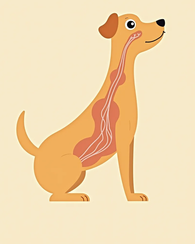
Check the 5 Main Symptoms of Nasolacrimal Duct Obstruction

Immediate Veterinary Visit Is Necessary If Tear Stains Appear
If tear stains appear and persist for more than a week, the risk of infection increases. In particular, if the skin around the eyes becomes flaky or red, you should seek veterinary care immediately. Ignoring the issue can lead to eye inflammation or complete blockage of the nasolacrimal duct. Early diagnosis and treatment are crucial, so please visit a clinic promptly if you notice any symptoms.
Comparison of Mild, Moderate, and Severe Symptoms of Nasolacrimal Duct Obstruction
| Item | Mild | Moderate | Severe |
|---|---|---|---|
| Tear Stains | Slight wet marks | Persistent tear stains | Severe staining, fur discoloration |
| Skin Reaction | None | Mild redness | Inflammation, peeling, ulceration |
| Behavioral Changes | None | Rubbing the eyes | Squinting, anxiety |
| Treatment Necessity | Observation | Medical treatment | Surgery required |
If symptoms worsen, surgical treatment may be necessary. Early intervention is important.
4 Home Care Measures You Can Take
5 Daily Habits for Prevention

Certain Breeds Are at Higher Risk for Nasolacrimal Duct Obstruction
Small breeds like Poodles and brachycephalic breeds with short muzzles, such as Pugs, are at higher risk for tear duct obstruction and tear staining. Small dogs often have excessive hair in the inner corners of their eyes, which can wick tears onto the skin, while brachycephalic breeds may have inward-rolling eyelids or curved nasolacrimal ducts that impede proper tear drainage. Regular grooming and veterinary check-ups are essential for these breeds, and any symptoms should be addressed promptly.
Reviewed by a veterinarian

Dr. Tony — Punnawat Phongkittirak
VeterinarianA veterinarian who majored in veterinary medicine at Khon Kaen University, Thailand, and completed the IVSA program at North Carolina State University in the United States. Drawing on clinical experience at animal hospitals, he works in the pet healthcare field and is dedicated to building a digital care environment that connects pet parents with veterinarians.
Reviewed 2026-06-03
5 Frequently Asked Questions
Does tear staining always mean I need to visit the vet?
Can a blocked tear duct be corrected with surgery?
How can I clean tear stains at home?
Is nasolacrimal duct obstruction genetically inherited?
Should I change my pet’s food if tear stains appear?
Share
Related Guides

Top 5 Dog Tear Supplements Recommended for 2026

What is Lutein? Effects on Dog Eye Health

The Best Canine Uveitis Management Products and Selection Criteria: A Comprehensive Guide

Complete Guide to IV Fluid Types (Normal Saline, Ringer’s Solution, 5% Dextrose)

Cyclosporine (Atopica) Usage Guide
Sedation and Anesthesia Medication Guide: Types of Pre-anesthetics and Safe Usage

What pathogens can be identified with a gastrointestinal PCR test?
Meaning and Interpretation of Urine Specific Gravity (USG) Testing

How to Read Cortisol Test Results

Comparison of Anemia Types (Regenerative, Non-Regenerative, IMHA)
References
[1] Gelatt KN et al. (2013). Veterinary Ophthalmology, 2nd ed. Wiley-Blackwell, Oxford, UK.
[2] Esson DW and Calvarese S (2022). Clinical Atlas of Canine and Feline Ophthalmic Disease, 2nd ed. John Wiley & Sons, Inc.
[3] Ny Y et al. (2006). Medial canthoplasty for epiphora in dogs: a retrospective study of 23 cases. J Am Anim Hosp Assoc, 42, 435–439.