Like
Share
멍실장
강아지·고양이 생검 가이드 | 세포진 검사와 조직생검 차이 총정리

Dog and Cat Biopsy Guide: A Complete Overview of Cytology vs. Histopathology

ImmunityQ&AMeongsiljang Veterinary Advisory Board

We’ve made it easy for pet owners to understand the differences between cytology and histopathology, the testing process, how to interpret results, and management strategies.

What is a Biopsy? Understanding Cytology and Histopathology at a Glance
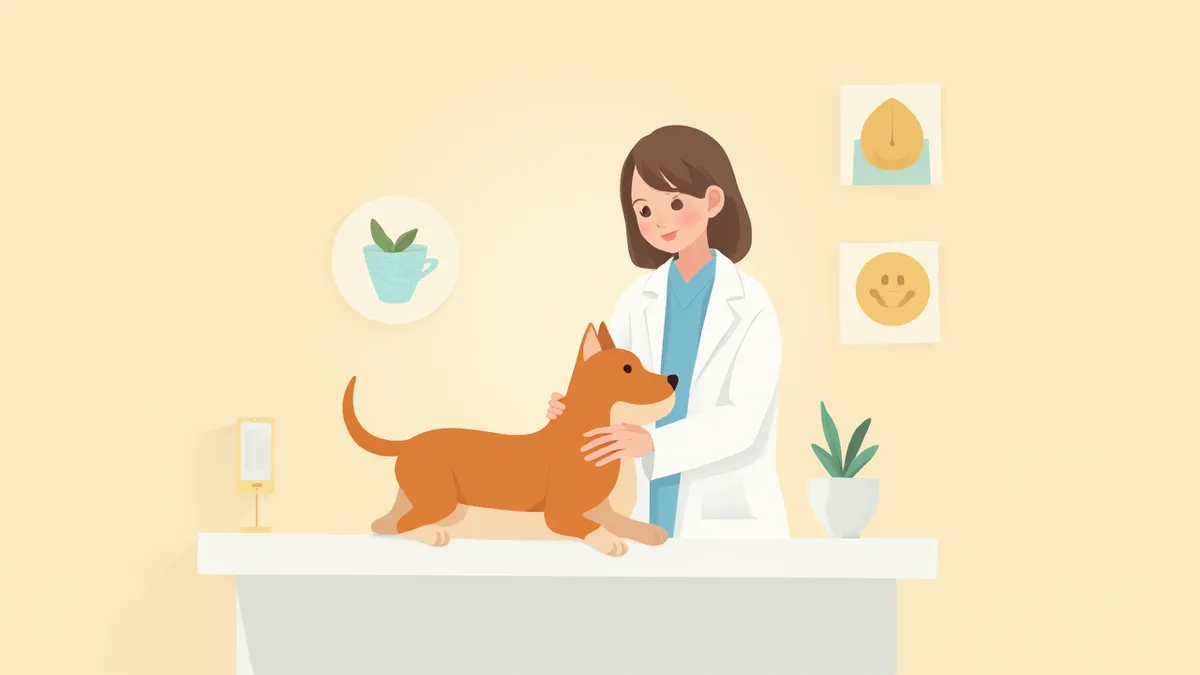
A veterinarian gently examining a dog in a bright, welcoming animal clinic.
A biopsy is a precise diagnostic test in which a small sample of cells or tissue is taken from a lump, organ, or lymph node in your pet and then examined under a microscope to assess cellular morphology and diagnose disease. There are two main types: fine-needle aspiration, which extracts only cells using a needle, and tissue biopsy, which removes a small piece of tissue. The most important point is that when a lump is detected, getting it tested promptly—rather than adopting a “wait-and-see” approach—is key to not missing the optimal treatment window. Understanding which test is appropriate for which situation will help you better comprehend your veterinarian’s recommendations and actively participate in treatment decisions.

What is Cytology? Quick Diagnosis with a Single Needle

A fine-needle aspiration (FNA) biopsy involves inserting a thin needle into a lump to extract cells for examination. In veterinary medicine, this procedure is known as fine-needle aspiration. It can usually be performed without anesthesia, takes only a short amount of time, and causes relatively little discomfort to your pet.
Quick results: You can receive initial diagnostic results more quickly than with a tissue biopsy.
Minimal burden: Since it requires no anesthesia or incisions, it causes less stress for your pet.
Screening tool: It serves as an initial step to quickly determine whether a mass is benign or malignant.
However, because this test examines only individual cells, a tissue biopsy may be recommended if a more detailed assessment of the tissue architecture is needed.

What is Histopathology? Precise Diagnosis by Removing Tissue
A biopsy involves taking a small sample of a lump or organ, either by cutting out a piece or using a special needle to extract tissue fragments, which are then examined under a microscope. This test provides much more information than a cytology test, allowing for an accurate assessment of the tumor type, malignancy, and extent of invasion.
Accurate Diagnosis: It can confirm the tumor type and malignancy grade.
Determining Treatment Direction: It serves as a basis for deciding the extent of surgery and whether chemotherapy is needed.
Anesthesia Required: Most procedures are performed under sedation or general anesthesia.
Because the process requires tissue processing and specialized pathological interpretation, it takes longer to receive results compared to a cytology test. It is also common to send samples to external pathology laboratories for analysis.
A view of a veterinary pathology laboratory with microscopes and tissue sample slides laid out.

Cytology vs. Histopathology: A Side-by-Side Comparison

ItemCytologyTissue biopsy
Collection methodAspirate cells with a fine needleCollect tissue by incision or biopsy needle
AnesthesiaUsually not requiredSedation or general anesthesia required
Test durationRelatively shortLonger, including sedation & anesthesia
Time for resultsRelatively shortLonger, due to tissue processing & pathology reading
AccuracyScreening level (distinguishing benign vs. malignant)Definitive diagnosis (including type & grade)
PainPerformed without anesthesia, momentary stimulusPerformed after anesthesia
CostRelatively lowRelatively high

The testing method is decided by the veterinarian based on the mass's location, size, and condition

Do Not Delay a Biopsy in These Situations

If a lump under the skin doesn’t go away after a while or grows quickly, don’t delay getting a biopsy. Especially if the surface of the lump is ulcerated or bleeding, or if nearby lymph nodes are swollen, please visit a vet right away. Waiting to see if it gets better can cause you to miss the best time for diagnosis and treatment.

How is the Test Performed?
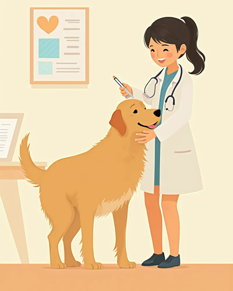
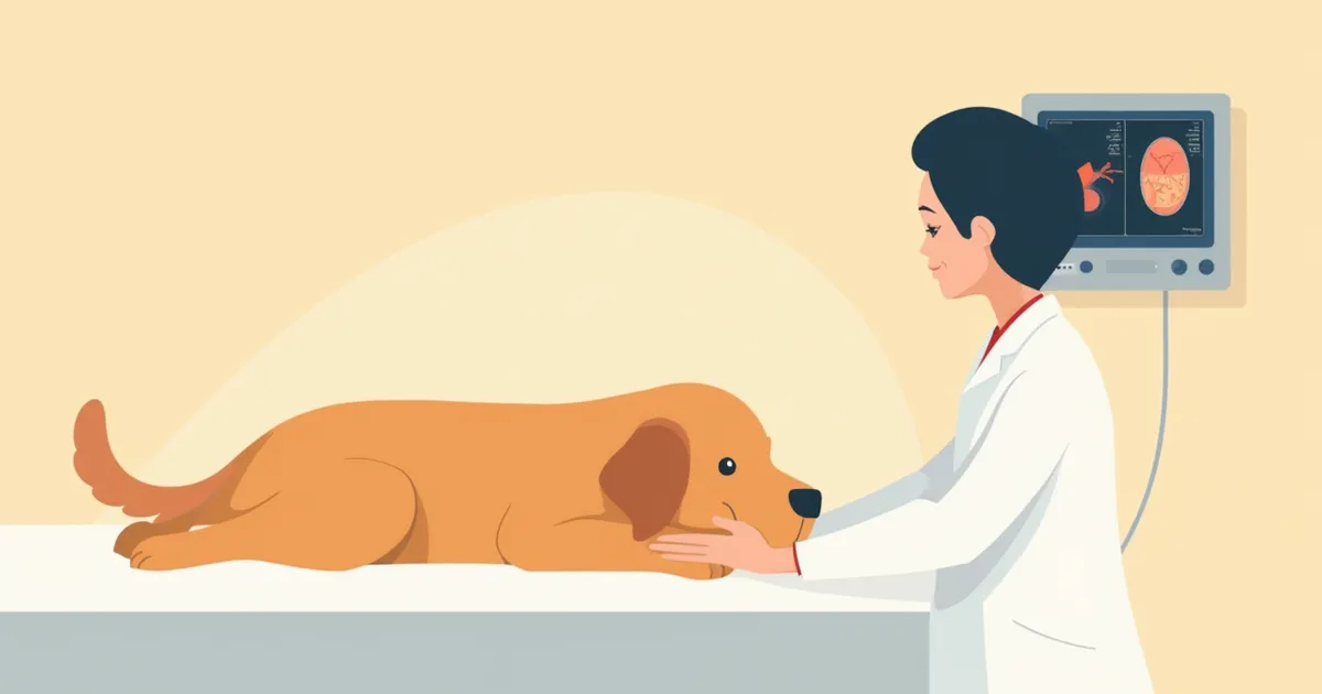
Fine-needle aspiration is often performed while the owner gently restrains their pet. The veterinarian inserts a thin needle into the lump to collect cells, which are then spread onto a glass slide and examined under a microscope. For internal organs or other areas not visible to the naked eye, ultrasound guidance may be used to direct the needle accurately. Biopsies are performed after sedation or general anesthesia. A small incision is made in the skin, or a specialized biopsy needle is used to obtain a tissue sample, which is then sutured closed. The collected tissue is placed in a formalin solution and sent to the pathology laboratory for analysis.
A veterinarian performing a fine-needle aspiration biopsy on a Golden Retriever that has undergone corrective treatment.

How to Interpret the Test Results

Cytology results typically fall into one of three categories: "highly likely benign," "suspected malignant," or "inconclusive." An inconclusive result means that an insufficient number of cells were collected, so a repeat test may be necessary. The histopathology report will include details such as the tumor type, malignancy grade, and surgical margins. Based on these findings, your veterinarian will guide you toward the most appropriate course of action, whether that involves additional surgery, chemotherapy, or monitoring. Once the results are available, it’s a good idea for you to review them together with your vet to discuss the treatment plan. If anything is unclear, don’t hesitate to ask your veterinarian for further explanation.

Key Management Points Before and After the Test
Before the procedure: Since a tissue biopsy requires anesthesia, please follow your veterinarian’s instructions for proper fasting beforehand. Let your vet know about any medications your pet is currently taking. Some drugs can interfere with blood clotting and may need to be stopped prior to the test, so be sure to consult with your veterinarian in advance.
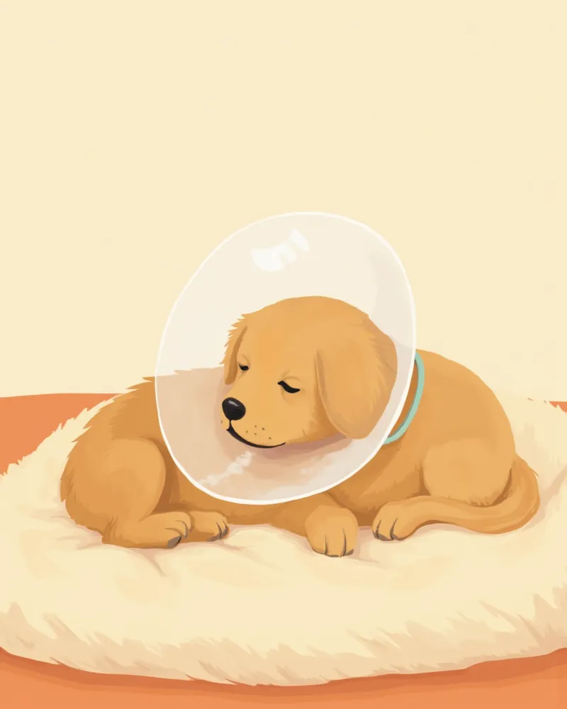
After the procedure: A fine-needle aspiration (FNA) typically requires no special aftercare, but following a tissue biopsy, your pet should wear an Elizabethan collar to prevent licking or scratching the incision site. Contact the clinic immediately if you notice swelling or discharge at the suture site. Your veterinarian will advise you on when to remove the sutures based on how well the wound is healing.
A puppy wearing a neck collar rests comfortably at home.

Special Precautions for Cat Owners

Cats can be highly sensitive to handling and examination, so veterinarians often recommend sedation to ensure safe and accurate sample collection. In cases requiring prompt diagnosis and treatment decisions, such as with tumors, it’s best not to delay testing. If a fine-needle aspirate isn’t definitive, proceeding to a biopsy can provide more conclusive diagnostic information. However, since anesthesia and biopsies carry both risks and benefits, your veterinarian will determine the most appropriate method and timing based on your cat’s individual condition. If you notice a lump on your cat, please consult your veterinarian about whether a biopsy is needed rather than opting to wait and see.

Reviewed by a veterinarian

Dr. Tony — Punnawat Phongkittirak

Dr. Tony — Punnawat Phongkittirak

Veterinarian

A veterinarian who majored in veterinary medicine at Khon Kaen University, Thailand, and completed the IVSA program at North Carolina State University in the United States. Drawing on clinical experience at animal hospitals, he works in the pet healthcare field and is dedicated to building a digital care environment that connects pet parents with veterinarians.

Frequently Asked Questions

The cytology results came back as “inconclusive.” Does that mean there’s a problem?
A "cannot be interpreted" result occurs when there are too few cells collected or when excessive blood contamination makes analysis difficult. This doesn't indicate disease—it's simply a sample quality issue. Please consult your veterinarian about arranging a retest.
I’ve heard that a biopsy can cause a tumor to spread. Is that true?
There may be concerns about the potential for cells to spread along the needle path during a biopsy. Your veterinarian will carefully weigh the risks and benefits for your pet’s specific situation before deciding whether to proceed. In most cases, an accurate diagnosis through biopsy is crucial for determining the appropriate treatment plan, and delaying the test too long could cause you to miss the optimal window for treatment. Please discuss any questions or concerns with your veterinarian so you can make an informed decision together.
How much does a biopsy typically cost?
Costs vary depending on the type of test, whether anesthesia is required, and the clinic, so it’s difficult to give a one-size-fits-all answer. Fine-needle aspiration is generally more affordable, while a biopsy incurs additional costs for anesthesia and pathological analysis. Please consult your veterinarian in advance for an accurate estimate.
Do I need to wait for the biopsy results before starting treatment?
Even while waiting for the results, necessary treatments such as pain management and infection prevention can be carried out. Your veterinarian will guide you through appropriate care during the waiting period, depending on the situation.
Are there cases where a cytology test alone is sufficient?
Yes, we do. In some cases, a fine-needle aspiration biopsy can provide a clear diagnosis. However, if malignancy is suspected or the results are inconclusive, it’s safer to proceed with an additional tissue biopsy. While cytology is a useful screening tool, the final diagnosis and treatment plan are determined by your veterinarian after a comprehensive review of all findings.

Share

Related Guides

References

[1] Garrett L, Berent L, Barger AM. Small Animal Cytologic Diagnosis: Canine and Feline Disease, 2nd Edition. Wiley-Blackwell.

[2] BSAVA Manual of Canine and Feline Dermatology, 4th Edition. BSAVA.

[3] Karadsheh Z, Al-Haddad M. Techniques for improving diagnostic yield of EUS-FNA of solid pancreatic lesions. Gastrointestinal Endoscopy, 2014.

This information is based on veterinary literature and does not replace diagnosis or treatment. Please consult a veterinarian for specific health concerns.

Metapet Co., Ltd. | CEO: Park Sung-yong | Business Reg. No. 417-88-02562 | Seoul, South Korea | Customer Center

Dog & Cat Biopsy: Cytology vs. Histopathology Guide | Meongsiljang