Like
Share
멍실장
강아지 흉부 X선 판독 증상과 원인, 진단·치료와 관리 포인트 총정리

Dog Chest X-ray Interpretation: Symptoms, Causes, Diagnosis, Treatment, and Care Points Summary

RespiratoryDiseasesMeongsiljang Veterinary Advisory Board

Chest X-ray interpretation is a key tool in diagnosing respiratory diseases in dogs. Accurate interpretation and an appropriate treatment plan are essential.

What is Dog Chest X-ray Interpretation?

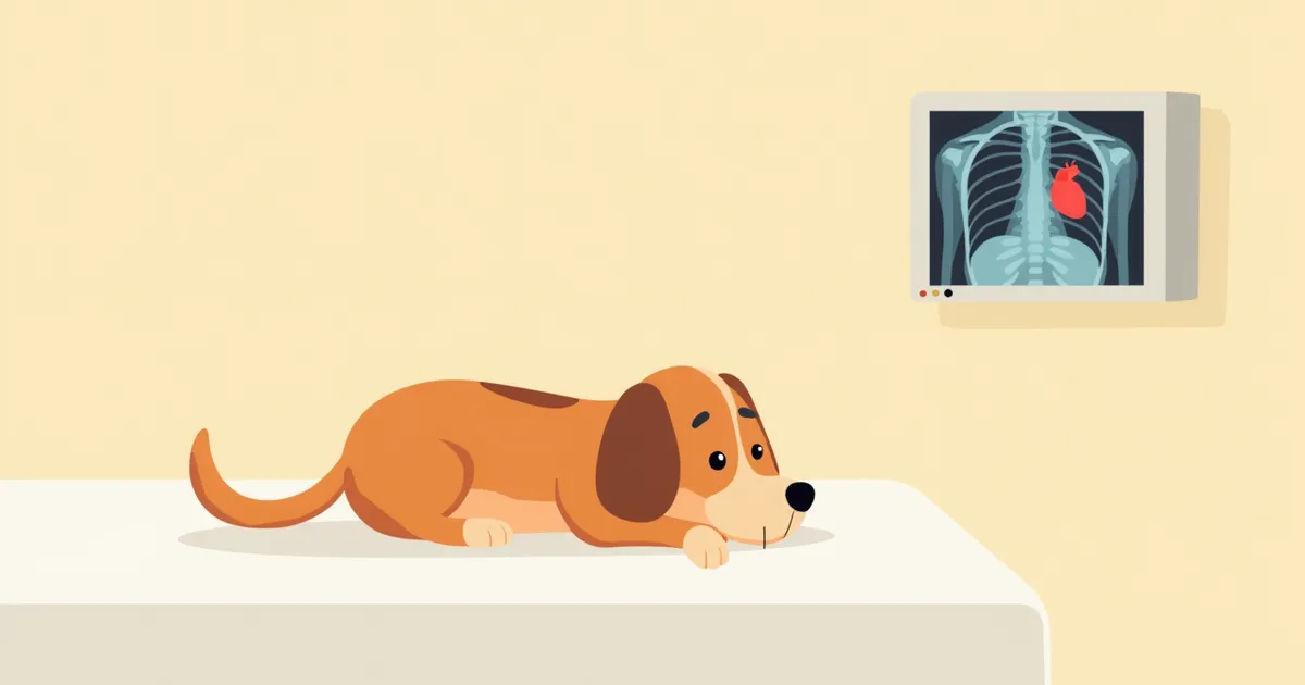
A veterinarian reviewing a dog’s chest X-ray
Chest X-ray interpretation is a key imaging diagnostic tool that allows for non-invasive visualization of internal thoracic structures, including the lungs, heart, and airways. This test is essential for identifying the underlying cause of symptoms such as difficulty breathing, coughing, and lethargy. - Pneumonia: Inflammation of lung tissue appears on X-rays as hazy opacities or increased density. - Heart failure: Cardiomegaly, or overall enlargement of the heart, may be visible, often accompanied by pulmonary edema or pleural effusion. - Pleural effusion: Fluid accumulation in the thoracic cavity compresses the lungs, making them appear smaller, and obscures the cardiac silhouette. - Pneumothorax: Air accumulation in the thoracic cavity causes the lungs to collapse away from the chest wall. Accurate interpretation depends on proper imaging techniques, knowledge of thoracic anatomy, and skilled reading expertise, so professional assessment is always required.

Main Causes and Lesion Types

Abnormalities seen on a dog’s chest X-ray can stem from a variety of causes. - Pneumonia: Inflammation of the lung tissue due to bacterial or viral infection appears as hazy shadows or increased density on X-rays. In cases of bacterial pneumonia, blood tests may show an elevated white blood cell count. - Bronchial disease and bronchiectasis: Chronic inflammation of the airways can cause the bronchial pattern to appear prominent on X-rays, with the bronchi failing to taper normally and instead appearing abnormally dilated. - Heart failure: When heart function declines, the heart may appear enlarged overall, and pulmonary edema or pleural effusion may develop. Increased opacity around the bronchi can sometimes be an early sign. - Pleural effusion: Fluid accumulation in the chest cavity compresses the lungs, making them appear smaller, and blurs the heart’s outline. - Pneumothorax: Air enters the chest cavity, causing the lung to collapse and pull away from the chest wall. - Tumors: These appear as high-density masses with borders that may be ill-defined or irregular. Each lesion exhibits a unique imaging pattern, so veterinarians analyze these features comprehensively to determine the underlying cause.
Chest X-ray images showing hazy lung shadows and an enlarged heart

Main Symptoms and Signs

Abnormalities on a puppy’s chest X-ray typically manifest as symptoms such as difficulty breathing, coughing, lethargy, and an increased respiratory rate. In particular, situations involving labored breathing, foaming at the nose, or bluish discoloration of the lips constitute medical emergencies. Structural abnormalities visible on the X-ray provide critical information for identifying the underlying causes of these symptoms.
Difficulty breathing: This refers to labored breathing or panting with the mouth open. If pleural effusion is present, it may occur alongside restrictive breathing difficulties and a rapid heart rate.
Persistent coughing: A cough that worsens at night or after exercise.
Increased lethargy: Behaviors such as appearing sleepier than usual or avoiding play activities.
Bluish lips: This indicates a lack of sufficient oxygen supply.
Increased respiratory rate: When the breathing rate is faster than normal, even while at rest.
A puppy showing signs of difficulty breathing

Signs Requiring Immediate Veterinary Visit

If your dog is panting with its mouth open, or if its lips or tongue turn bluish and its breathing becomes rapid, you should seek veterinary care immediately. These symptoms may indicate a medical emergency such as severe pleural effusion, pneumothorax, or heart failure. The longer you wait, the fewer treatment options may be available, so prompt action is crucial.

Diagnostic Procedures and Additional Tests
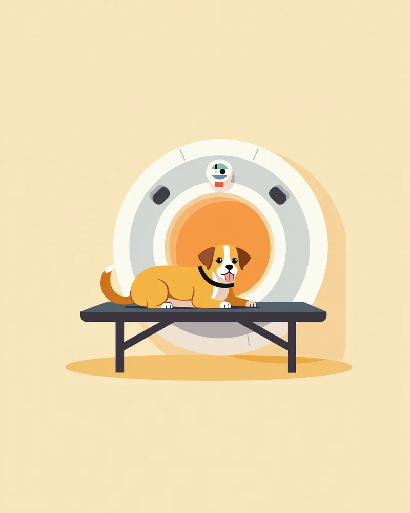
A chest X-ray is the starting point for diagnosis, but additional tests are essential to identify the exact cause. - Mucosal cell sampling: This procedure is performed to detect pathogens in coughs or respiratory secretions. - Thoracentesis (chest fluid aspiration): Fluid from the chest cavity is collected to analyze for inflammation, infection, or tumors. If infection is suspected, blood and bacterial tests may be conducted concurrently. - CT scan: This provides a more precise evaluation of detailed structures that are unclear on X-rays. - Biopsy: Suspected lesions or tumors are directly sampled to determine whether they are malignant. The veterinarian will develop a testing plan by integrating the patient’s symptoms, X-ray findings, and blood test results. These additional tests play a crucial role in determining the treatment direction.
A veterinarian is performing a chest CT scan on a puppy.

Treatment Methods and Step-by-Step Approach

Treatment is approached systematically based on the underlying cause of the lesion. - Pneumonia: Antibiotics are used to eliminate the infection, while coughing and difficulty breathing are alleviated. - Heart failure: Medications that improve cardiac function help regulate fluid balance and blood circulation. - Pleural effusion or pneumothorax: A chest tube is inserted to drain fluid or air, allowing the lungs to re-expand. - Tumors: A comprehensive treatment strategy involving surgery, radiation therapy, and chemotherapy is often required. Treatment should combine short-term symptom relief with long-term management. The veterinarian will continuously adjust the treatment plan according to changes in the patient’s condition.
A veterinarian administering medication to a puppy

Home Care and Daily Management Points

Dogs undergoing treatment require care in a stable, controlled environment. - Well-ventilated space: Ensure fresh indoor air circulation and avoid irritants such as dust and cigarette smoke. - Maintain optimal humidity: Keep humidity levels between 40% and 60%, as environments that are too dry or too humid can make breathing more difficult. - Regulate exercise: Avoid excessive activity if the dog is experiencing labored breathing or fatigue; limit outings to short walks only. - Adhere to medication instructions: Administer medications at the prescribed times and dosages, and do not discontinue them without veterinary guidance. - Attend regular follow-up examinations: Keep all scheduled appointments to monitor treatment efficacy and detect any changes in symptoms early. Consistent daily care significantly influences the speed of recovery.
A dog resting comfortably in a quiet environment

Breed-Specific Precautions and Prevention of Recurrence

Certain breeds are more prone to respiratory issues due to their chest structure. For example, Pekingese, Bulldogs, and Toy Poodles have a higher risk of airway obstruction. Regular check-ups and environmental management are essential for these breeds. Additionally, it’s important to maintain healthy lifestyle habits to prevent symptoms from recurring after treatment, and to stay in consistent communication with your veterinarian.

Reviewed by a veterinarian

Dr. Tony — Punnawat Phongkittirak

Dr. Tony — Punnawat Phongkittirak

Veterinarian

A veterinarian who majored in veterinary medicine at Khon Kaen University, Thailand, and completed the IVSA program at North Carolina State University in the United States. Drawing on clinical experience at animal hospitals, he works in the pet healthcare field and is dedicated to building a digital care environment that connects pet parents with veterinarians.

Frequently Asked Questions

What is a chest X-ray?
A chest X-ray is a diagnostic imaging test that allows us to visualize the structures of the lungs, heart, and airways. It helps detect respiratory diseases and heart problems at an early stage.
If the X-ray results show abnormalities, is it necessary to visit the hospital?
Yes, if any abnormalities are detected on an X-ray, it is essential to consult with a veterinarian. Identifying the cause and initiating appropriate treatment is crucial.
Should I get a chest X-ray if my dog is coughing?
If your pet has persistent coughing, a chest X-ray is recommended. There could be various causes, such as pneumonia, airway issues, or tumors.
Does an X-ray hurt?
It won’t hurt. Your dog just needs to stay still, and in most cases, no sedation is needed. If your dog is anxious, the veterinarian will help keep them calm.
When should we schedule a follow-up X-ray after treatment?
Your veterinarian will schedule a follow-up exam at the most appropriate time to assess treatment effectiveness, depending on your pet’s specific condition. The timing may vary based on how your pet responds to treatment.

Share

Related Guides

References

[1] Evans HE, Miller's Anatomy of the Dog, 1993, WB Saunders.

[2] Clinical Medicine of the Dog and Cat, 4th Ed, 2020, Elsevier.

[3] Canine and Feline Respiratory Medicine, 3rd Edition, 2021, Wiley-Blackwell.

This information is based on veterinary literature and does not replace diagnosis or treatment. Please consult a veterinarian for specific health concerns.

Metapet Co., Ltd. | CEO: Park Sung-yong | Business Reg. No. 417-88-02562 | Seoul, South Korea | Customer Center

Dog Chest X-ray Interpretation: Symptoms, Causes & Treatment | Meongsiljang