

The Complete Guide to the Schirmer Tear Test for Dogs — Key to Early Detection of Dry Eye
The Schirmer tear test for dogs measures tear production and helps identify the cause of dry eyes or excessive tearing. Regular testing is essential for maintaining eye health.
The Schirmer Tear Test for Dogs Is a Diagnostic Method That Measures Tear Production
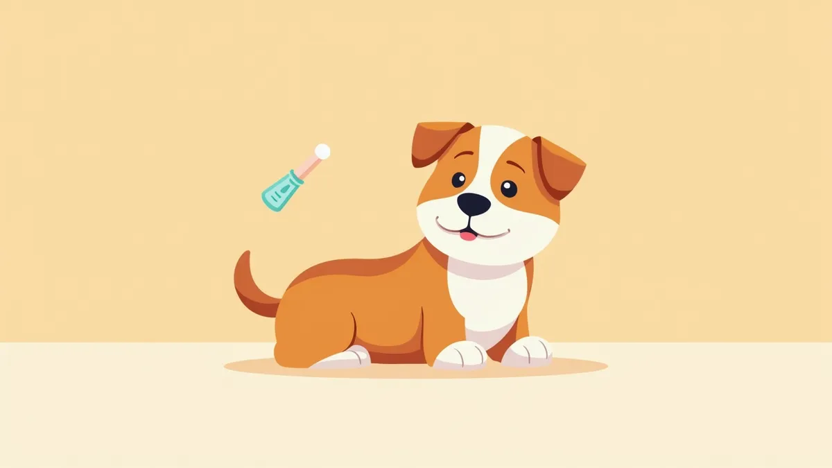
The Main Causes of Abnormal Tear Production Are Structural Eye Abnormalities and Diseases

Main Symptoms Include Excessive Tearing, Tear Stains, Eye Redness, and Fur Pigmentation

If Excessive Tearing Is Accompanied by Eye Redness, Pain, or Vision Loss, Immediate Veterinary Visit Is Required
If your pet’s eyes are persistently watery, red, or if they’re rubbing or squinting more than usual, or if their vision seems impaired, seek veterinary care immediately. These symptoms could indicate corneal damage, keratitis, or inflammation of the front part of the eye. Left untreated, these conditions can lead to vision loss, so prompt diagnosis and treatment are essential. In some cases, long fur may obstruct the eyes, or tear drainage may be misdirected, causing irritation. A veterinarian needs to identify the exact cause and begin appropriate treatment.
Diagnosis Is Performed Through the Schirmer Tear Test, Structural Eye Examination, and Imaging If Necessary

Treatment Is Divided Into Medication, Surgery, and Management Depending on the Cause

At-Home Care Consists of Cleaning Around the Eyes, Grooming Fur, and Regular Examinations
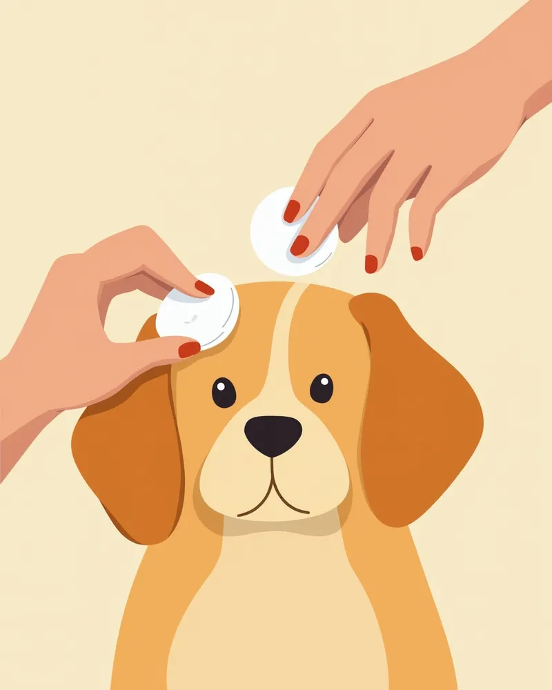
Breeds With Lots of Hair Around the Eyes, Such as Poodles, Chartreux, and Terriers, Require Attention to Tear Issues
In dogs with light or white coats, tear staining appears more prominently as brown discoloration when tears soak into the fur. While this may seem like a purely cosmetic concern, it can actually signal an underlying issue with the tear film. For these dogs, it is essential to regularly groom the hair around the eyes and keep the area clean. If excessive tearing persists, a Schirmer tear test is important to assess tear production. In cases of severe staining, it is crucial to identify and manage contributing irritants such as trichiasis. Early diagnosis and consistent care are highly effective preventive measures.
Reviewed by a veterinarian

Dr. Tony — Punnawat Phongkittirak
VeterinarianA veterinarian who majored in veterinary medicine at Khon Kaen University, Thailand, and completed the IVSA program at North Carolina State University in the United States. Drawing on clinical experience at animal hospitals, he works in the pet healthcare field and is dedicated to building a digital care environment that connects pet parents with veterinarians.
Reviewed 2026-06-03
Frequently Asked Questions
Does the Schirmer tear test hurt?
When should you take your pet to the vet for excessive tearing?
Can tear stains be treated?
What are some ways to manage tear staining at home?
What should I do if the Schirmer test result is low?
Share
Related Guides

Top 5 Dog Tear Supplements Recommended for 2026

What is Lutein? Effects on Dog Eye Health

The Best Canine Uveitis Management Products and Selection Criteria: A Comprehensive Guide

If Your Dog Has Frequent Eye Discharge or Tearing — Safe Eye Cleaning Methods and Precautions

Foreign Bodies in a Dog’s Eye — First Aid and When to See a Vet

If Your Dog’s Eyeball Suddenly Protrudes — Emergency First Aid for Globe Prolapse and the Golden Time

If Your Dog’s Vision Suddenly Blurs — Causes of Glaucoma and Treatment Timing

If Your Dog’s Pupil Has Turned Dark — Causes of Corneal Pigmentation and Warning Signs of Vision Loss

If Your Dog’s Eye Is Discharging Fluid — Warning Signs of Corneal Perforation and the Emergency Golden Hour

Canine Cataracts: Symptoms, Causes, Diagnosis, Treatment, and Management Essentials
References
[1] Gelatt KN et al. (2013). Veterinary Ophthalmology, 2nd ed. Wiley-Blackwell, Oxford, UK.
[2] Esson DW and Calvarese S (2022). Clinical Atlas of Canine and Feline Ophthalmic Disease, 2nd ed. John Wiley & Sons, Inc.
[3] Ny Y et al. (2006). Medial canthoplasty for epiphora in dogs: a retrospective study of 23 cases. Journal of the American Animal Hospital Association, 42(5), 435–439.