Like
Share
멍실장
강아지 망막 박리 증상과 원인, 진단·치료와 관리 포인트 총정리

If Your Dog Suddenly Loses Vision — Warning Signs of Retinal Detachment and Emergency Response

Eye HealthDiseasesMeongsiljang Veterinary Advisory Board

Canine retinal detachment is a serious eye condition that can lead to vision loss or even blindness. Early detection and appropriate treatment are crucial.

Canine Retinal Detachment Is a Serious Eye Condition That Can Lead to Vision Loss

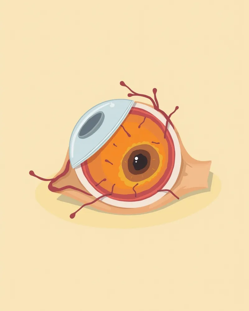
Cross-sectional diagram of a dog’s eye with retinal detachment
Canine retinal detachment occurs when the neurosensory retina separates from the underlying retinal pigment epithelium (RPE), disrupting the transmission of visual information and potentially leading to vision loss or blindness. - Causes of Retinal Detachment: Trauma, genetic or congenital abnormalities, and vascular or systemic diseases are the primary causes. In particular, traumatic detachment can occur if a dog falls from a height or is hit by a car. - Genetic and Congenital Risk Factors: Certain breeds, such as Collies (associated with Collie Eye Anomaly, or CEA), Labrador Retrievers, German Shepherds, and Australian Shepherds, have a congenital predisposition that increases their risk of developing this condition. - Vascular and Systemic Disease Associations: Hypertension, in particular, is a common cause that affects retinal blood vessels and increases the risk of detachment. While partial detachment may not result in blindness, complete detachment carries a poor prognosis for vision. Therefore, early detection and appropriate treatment are essential for preserving sight.

The Main Causes of Retinal Detachment Include Trauma, Genetics, and Vascular Diseases
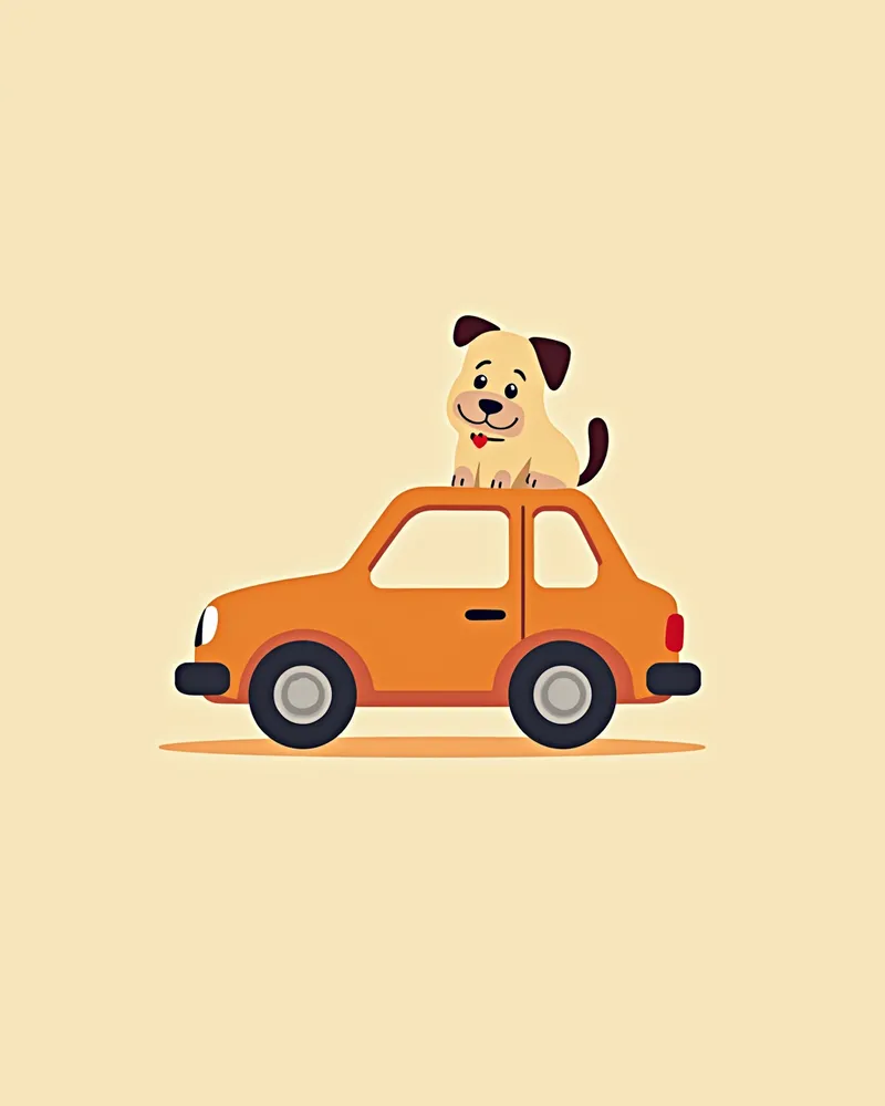
The main causes of retinal detachment are diverse, including trauma, genetic or congenital abnormalities, and vascular or systemic diseases. - Traumatic causes: Falls from heights or being hit by a car can cause the retina to detach due to the impact. - Genetic and congenital factors: Collies are prone to "Collie Eye Anomaly (CEA)," while Labrador Retrievers, German Shepherds, and Australian Shepherds have a higher risk of retinal detachment due to congenital conditions such as retinal dysplasia. - Vascular and systemic diseases: Hypertension can damage retinal blood vessels, leading to detachment, and may also be associated with kidney disease or infectious chorioretinitis. - Other factors: Drug side effects (e.g., phenylpropanolamine overdose), coagulation disorders, and severe uveitis can also contribute. Early diagnosis and identifying the underlying cause are the first steps in treatment.
A scene of a puppy undergoing an eye examination after being hit by a car.

The Main Symptoms of Retinal Detachment Include Vision Loss and Wandering in the Dark

The main symptoms of retinal detachment are sudden vision loss and changes in the pupils. - Sudden vision loss: When the retina detaches completely, your pet may suddenly struggle to see and wander around in the dark. - Abnormal pupils: Both pupils may become significantly dilated and unresponsive to light. - Behavioral changes: Your pet may have difficulty recognizing their surroundings, leading to bumping into walls or walking backward. - Eye abnormalities: Retinal blood vessels may be visible behind the lens, and bleeding may occur. While partial detachment may not lead to blindness, complete detachment has a poor prognosis for vision, making early detection crucial.
A puppy wandering around in the dark

Emergency Signs Requiring Immediate Veterinary Visit

If your pet suddenly loses vision or has both pupils widely dilated and unresponsive to light, consult a veterinarian immediately. This is especially important if there has been trauma or if your pet has systemic conditions like hypertension, as these increase the risk of acute retinal detachment. The sooner appropriate intervention is provided—ideally within 1 to 3 weeks—the higher the chance of preserving vision, making early diagnosis and treatment critical.

Retinal Detachment Is Accurately Diagnosed Through Ophthalmic Examination and Ultrasound
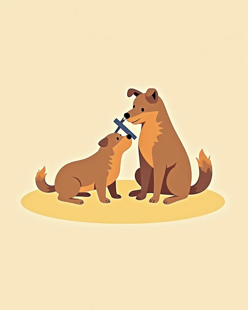
Diagnosing retinal detachment requires a specialized ophthalmic examination. - Fundoscopic examination (ophthalmoscopy): This basic test involves directly observing the retina inside the eye to confirm whether detachment has occurred and to assess its appearance. - B-mode ultrasonography: When direct visualization with an ophthalmoscope is difficult, this imaging technique is useful for accurately determining the location and extent of the retinal detachment. - Concurrent systemic evaluation: Blood work, biochemical analysis, infectious antibody testing, and blood pressure measurements are performed to identify underlying causes such as hypertension or infections. - Accurate diagnosis: Establishing a precise diagnosis is essential for determining the appropriate treatment plan. Early detection significantly improves the likelihood of successful treatment, making consultation with a veterinary specialist crucial.
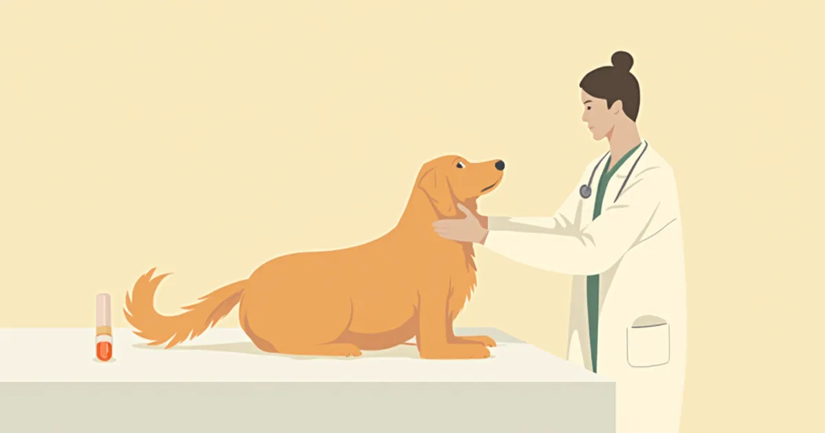
A veterinarian examines a dog’s eyes using an ophthalmic retinal scanner.

Treatment for Retinal Detachment Proceeds in Stages: Surgery, Medication, and Conservative Management
Treatment for retinal detachment is carried out in stages, depending on the cause, type, and severity of the condition. - Surgical treatment: Rhegmatogenous detachment caused by a retinal tear is difficult to resolve with medication alone, so procedures such as laser retinopexy, vitrectomy, or silicone oil tamponade are used to reattach the retina. - Medical treatment: Bullous (primary) detachment, which is presumed to be immune-mediated, typically responds well to anti-inflammatory therapy, such as systemic corticosteroids. - Treatment of underlying conditions: If a systemic disease such as hypertension is the underlying cause, that condition must be managed concurrently, with regular monitoring of the patient’s progress. - Early intervention: The sooner treatment begins—ideally within 1 to 3 weeks of symptom onset—the more favorable the prognosis for vision recovery.
A puppy undergoing retinal reattachment surgery.

Home Care Focuses on Environmental Stability and Stress Reduction

After retinal detachment, it’s important to maintain a stable environment at home. - Environmental Stability: Minimize unnecessary movement and keep the arrangement of items consistent. - Provide a Quiet Space: Prepare a quiet room to reduce stress. - Maintain Daily Routines: Keep feeding and walking times consistent. - Medication Management: Administer medications exactly as directed by your veterinarian. - Observation and Record-Keeping: Carefully document any changes in symptoms and schedule regular vet visits.
A puppy resting comfortably in a quiet environment

Certain Breeds Are at Higher Risk for Retinal Detachment. Caution Is Advised.

Certain breeds, such as Collies, Labrador Retrievers, German Shepherds, and Australian Shepherds, have a genetic predisposition that puts them at higher risk for retinal detachment. In Collies, this condition can be caused by Collie Eye Anomaly (CEA). It is advisable to have your dog tested for breed-specific genetic disorders before adoption.

Reviewed by a veterinarian

Dr. Tony — Punnawat Phongkittirak

Dr. Tony — Punnawat Phongkittirak

Veterinarian

A veterinarian who majored in veterinary medicine at Khon Kaen University, Thailand, and completed the IVSA program at North Carolina State University in the United States. Drawing on clinical experience at animal hospitals, he works in the pet healthcare field and is dedicated to building a digital care environment that connects pet parents with veterinarians.

Frequently Asked Questions

Can vision be restored after treatment for retinal detachment?
The outcome depends on the timing of treatment and the extent of the detachment. In cases of partial detachment, vision is typically less affected, and if intervention such as retinal reattachment surgery is performed relatively soon (within 1–3 weeks), there is a good chance of preserving vision. However, complete detachment often carries a poor prognosis for vision.
Can retinal detachment be completely cured?
Treatment depends on the underlying cause. In cases of retinal detachment due to a tear, surgery is attempted to reattach the retina. Serous detachment typically responds to corticosteroid therapy, though long-term treatment may be necessary. Genetic or congenital causes require ongoing management.
Is it possible to prevent retinal detachment?
Prevention is possible through injury prevention and genetic disease screening. In particular, care tailored to the breed’s characteristics and regular ophthalmic examinations are important.
Is surgery always necessary for retinal detachment?
The decision to perform surgery depends on the extent of the retinal detachment and the degree of vision loss. Rhegmatogenous detachment is difficult to treat with medication alone and typically requires surgery, whereas bullous detachment may be managed with medical treatment. Please consult with your veterinarian to determine the best course of action.
Can my dog go outside again after retinal detachment?
Recovery after treatment depends on the veterinarian's assessment. To minimize stress when going outside, it's best to start gradually in a safe environment.

Share

Related Guides

References

[1] Esson DW, Calvarese S. Clinical Atlas of Canine and Feline Ophthalmic Disease, 2nd Ed. Wiley; 2022.

[2] Gelatt KN. Veterinary Ophthalmology. 5th ed. Wiley; 2018.

[3] Plumb's Veterinary Drug Handbook, 9th ed. Wiley; 2021.

This information is based on veterinary literature and does not replace diagnosis or treatment. Please consult a veterinarian for specific health concerns.

Metapet Co., Ltd. | CEO: Park Sung-yong | Business Reg. No. 417-88-02562 | Seoul, South Korea | Customer Center

Sudden Vision Loss in Dogs: Retinal Detachment Warning Signs | Meongsiljang