
Like
Share
멍실장
강아지 폐동맥협착증 증상과 원인, 진단·치료와 관리 포인트 총정리

If Your Dog Keeps Fainting — Symptoms, Causes, and Treatment Timing for Pulmonary Stenosis

HeartDiseasesMeongsiljang Veterinary Advisory Board

Pulmonary stenosis in dogs is a congenital valve malformation, and the symptoms and treatment options vary depending on the severity of the narrowing. Early detection and balloon dilation can significantly improve the prognosis.

What Is Pulmonary Stenosis in Dogs?

Illustration of the site of pulmonic valve stenosis in a dog's heart
Pulmonary stenosis in dogs is a congenital heart defect in which the pulmonary valve, responsible for pumping blood from the right ventricle to the lungs, is abnormally narrowed, forcing the right ventricle to work excessively. The core issue lies in the abnormal formation of the valve structure at birth, which obstructs blood flow and causes pressure to build up in the right ventricle. While mild cases may remain asymptomatic, severe cases can lead to fainting or heart failure. Once a heart murmur is detected, regular follow-up with echocardiograms is essential.

Causes of Pulmonary Stenosis — Why Does It Occur?

Pulmonary stenosis is almost always congenital. It occurs when the pulmonary valve or surrounding structures develop abnormally during fetal heart development, and in certain breeds like Boxers, a hereditary predisposition has been observed, often running in families. The condition is classified based on where the narrowing occurs along the right ventricular outflow tract—the pathway from the right ventricle to the pulmonary artery.
Valvular type: This is the most common form, where the pulmonary valve itself is thickened or abnormally formed, resulting in a narrowed opening.
Subvalvular type: This occurs when the right ventricular outflow tract below the valve (the infundibulum) becomes narrowed and obstructed.
Supravalvular type: This is the rarest form, characterized by narrowing of the pulmonary artery itself above the valve.

Key Symptom Checklist — Check for These Signs

Symptoms of pulmonic stenosis vary widely depending on the severity of the narrowing. If you notice any of the following signs, please have your pet undergo a cardiac examination.
Exercise intolerance: Your pet tires more easily than usual or suddenly refuses to go for walks.
Syncope (fainting): Your pet collapses suddenly after excitement or exercise.
Ascites (abdominal swelling): Fluid accumulates in the abdomen due to increased pressure in the right ventricle.
Dyspnea and cyanosis: Your pet breathes rapidly or laboredly, and their gums or tongue turn blue.
Poor growth: Puppies are significantly smaller and more lethargic than their peers.
A puppy showing signs of exercise intolerance and lying down weakly

When to Go to the Vet Immediately

If your dog suddenly faints during a walk or play, shows cyanosis (bluish gums or tongue), experiences rapid abdominal swelling due to fluid accumulation, or pants heavily even while resting, it is a medical emergency. Contact a 24-hour veterinary clinic immediately, and be careful not to agitate your dog during transport.

Diagnostic Methods — How Is It Confirmed?

Auscultation: The first clue is when the veterinarian detects a heart murmur. Pulmonary stenosis produces an ejection systolic murmur caused by obstruction of the right ventricular outflow tract, which is audible at the left base of the chest.
Thoracic Radiography: This confirms right ventricular hypertrophy (right heart enlargement) and dilation of the post-stenotic pulmonary artery.
Echocardiography: This allows direct visualization of the valve structure and the degree of stenosis, while Doppler measures the pressure gradient between the right ventricle and the pulmonary artery. A larger pressure gradient indicates more severe stenosis, which is classified as mild, moderate, or severe.
Electrocardiography: This checks for arrhythmias that may accompany right ventricular hypertrophy.

Treatment Options — Varies by Severity of Stenosis
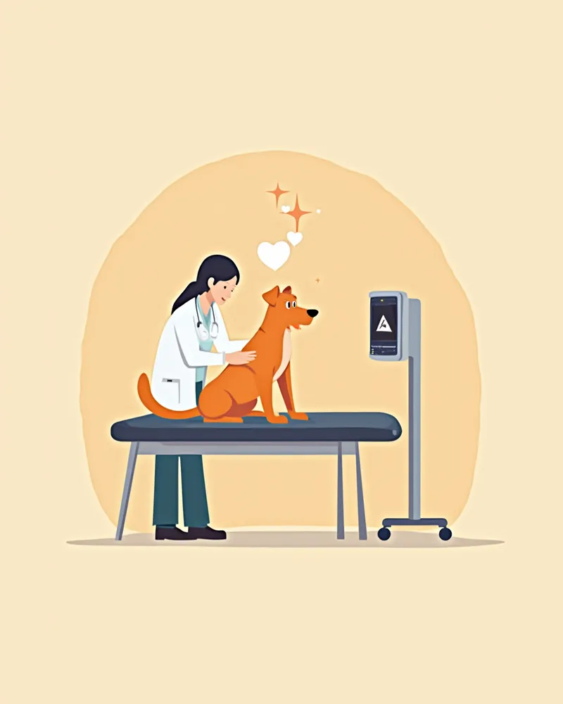
Mild stenosis: In many cases, no specific treatment is required, and regular monitoring with echocardiography is sufficient. Avoid strenuous exercise and schedule check-ups every six months to once a year.
Moderate to severe stenosis: Balloon valvuloplasty (balloon dilation) is the primary treatment. This procedure involves inserting a catheter through the femoral vein to widen the narrowed valve, allowing it to be performed without opening the chest. When carried out by an experienced veterinary cardiologist, it is generally successful.
Subvalvular and atypical stenosis: Balloon dilation alone may not be sufficient, and surgical intervention (open-heart surgery) may be necessary. Please consult thoroughly with a veterinary cardiologist.
A veterinarian performing an echocardiogram on a dog

Home Care Points — What Owners Should Monitor

Consistent care remains essential even after treatment.
Exercise restriction: If the condition is severe or before treatment begins, limit running and vigorous play. Light walks should be adjusted according to your veterinarian's guidance.
Regular check-ups: Have an echocardiogram performed once or twice a year to monitor for disease progression and changes in heart function.
Medication adherence: Do not arbitrarily reduce or discontinue medications prescribed by your veterinarian.
Minimize stress: Excitement and tension place strain on the heart. Please maintain a quiet and comfortable environment for your pet.
A dog resting comfortably at home

Breeds That Require Special Attention

Pulmonary artery stenosis occurs significantly more frequently in certain breeds. It is particularly common in English Bulldogs, French Bulldogs, and Boxers, with Boxers even showing a familial tendency to develop it alongside subaortic stenosis (SAS). Since breed-specific predispositions vary widely, we recommend that owners of these high-risk breeds have their pets undergo annual cardiac auscultation and examinations from an early age, even if no symptoms are present. Early detection can make a substantial difference in prognosis.

Reviewed by a veterinarian

Dr. Tony — Punnawat Phongkittirak

Dr. Tony — Punnawat Phongkittirak

Veterinarian

A veterinarian who majored in veterinary medicine at Khon Kaen University, Thailand, and completed the IVSA program at North Carolina State University in the United States. Drawing on clinical experience at animal hospitals, he works in the pet healthcare field and is dedicated to building a digital care environment that connects pet parents with veterinarians.

Frequently Asked Questions

Can pulmonary artery stenosis be cured?
"Management" is a more accurate term than "cure." If the narrowing is significantly improved through balloon dilation, your pet can lead a nearly normal life without symptoms. However, since the valve may narrow again, regular follow-up examinations are essential.
How risky is balloon angioplasty?
General anesthesia is required, but the procedure is far less invasive than open-chest surgery. When performed by a board-certified veterinary cardiologist, success rates are high. Before the procedure, a thorough assessment of heart function is conducted to carefully evaluate and mitigate any risks.
Do I need to completely restrict exercise for mild stenosis?
For mild cases, light exercise such as short walks is generally permitted. However, it’s safest to consult your veterinarian before engaging in running, swimming, or vigorous play, as recommendations may vary depending on your dog’s individual cardiac function.
What is the life expectancy of a dog with pulmonary artery stenosis?
In mild cases, life expectancy is nearly the same as normal. For moderate to severe cases, if the pressure drops significantly after balloon dilation, many dogs can live without a shortened lifespan. However, untreated severe cases can lead to heart failure and a shorter life expectancy.
My vet said my puppy has a heart murmur. Could it be pulmonary artery stenosis?
There are many possible causes of a heart murmur in dogs, including but not limited to pulmonic stenosis. An echocardiogram is essential for an accurate diagnosis. The correct approach is to combine the murmur’s grade and location with the echocardiographic findings to reach a definitive diagnosis.

Share

Related Guides

References

[1] Johnson MS, Martin M, Edwards D, French A, Henley W. Pulmonic stenosis in dogs: balloon dilation improves clinical outcome. J Vet Intern Med. 2004;18(5):656-662.

[2] Bussadori C, DeMadron E, Santilli RA, Borgarelli M. Balloon valvuloplasty in 30 dogs with pulmonic stenosis: effect of valve morphology and annular size on initial and 1-year outcome. J Vet Intern Med. 2001;15(6):553-558.

[3] Ware WA. Cardiovascular Disease in Small Animal Medicine. 2nd ed. London: Manson Publishing; 2011.

This information is based on veterinary literature and does not replace diagnosis or treatment. Please consult a veterinarian for specific health concerns.

Metapet Co., Ltd. | CEO: Park Sung-yong | Business Reg. No. 417-88-02562 | Seoul, South Korea | Customer Center

Dog Pulmonary Stenosis: Symptoms, Causes & Treatment Timing | Meongsiljang