
Like
Share
멍실장
강아지 안내 종양 증상과 원인, 진단·치료와 관리 포인트 총정리

If Your Dog’s Eye Color Has Changed — Symptoms of Intraocular Tumors and When to Visit the Vet

Eye HealthDiseasesMeongsiljang Veterinary Advisory Board

Canine intraocular tumors are growths that develop inside the eye, making early detection crucial. Key symptoms include vision loss, bulging of the eye, and changes in the eye’s appearance.

Canine Intraocular Tumors Develop Inside the Eye

An anatomical illustration showing the internal structures of a dog's eye
Intraocular tumors in dogs develop inside the eye, specifically within the intraocular structures such as the uvea and ciliary body. Early stages often show no obvious signs, making early detection difficult, but they may be observed as slowly growing pink to yellowish-brown masses behind the pupil or around the iris. - Masses inside the eye or iris changes: A pink to yellowish-brown mass may be seen growing slowly behind the pupil or in the iris, or the shape of the iris may change. - Vision loss: Your dog may avoid light or bump into objects. - Secondary complications: Complications such as uveitis, retinal detachment, anterior chamber hemorrhage (bleeding inside the eye), and glaucoma may occur. - Redness and pain: The blood vessels in the white of the eye may become red, and eye pain may be present. Early detection helps with treatment and preserving the eye.

The Main Causes of Intraocular Tumors Are Genetic Predisposition and Age

The exact cause of canine intraocular tumors remains unclear, and breed predispositions have not been fully established. However, they are more commonly reported in middle-aged to senior dogs. - Breed predisposition: Spindle cell tumors associated with blue eyes are relatively frequently reported in Siberian Huskies, Australian Shepherds, Border Collies, Labrador Retrievers, and German Shepherds. - Age: These tumors generally occur more often in middle-aged to senior dogs. - Secondary changes: The tumor itself often causes intraocular inflammation, such as uveitis (note that the inflammation does not cause the tumor). - Tumor characteristics: The behavior varies widely, ranging from benign adenomas to adenocarcinomas with metastatic potential. These factors may act in combination.
A photo showing changes in the eyes of an aging dog

Be Sure to Check for Key Symptoms and Signs

Symptoms of canine intraocular tumors manifest as changes within the eye and secondary complications. - Vision loss: Your dog may walk unsteadily in dim light or frequently bump into obstacles. - Intraocular mass: A pink to yellowish-brown tumor may gradually grow behind the pupil or on the iris, or the shape of the iris may change. - Hyphema (anterior chamber hemorrhage): Blood may accumulate in the front of the eye, causing it to appear red. - Redness and pain: The blood vessels in the white of the eye may become red, and your dog may frequently squint or blink due to pain. - Signs of glaucoma: Increased intraocular pressure can lead to corneal edema or changes in pupil size. If you notice any of these signs, consult your veterinarian immediately.
A photo of a puppy showing proptosis (bulging eyes)

Emergency Signs Requiring Immediate Veterinary Visit

If your pet’s eyeball suddenly bulges, if they show severe eye pain, if their vision deteriorates rapidly, or if blood accumulates in the front of the eye making it appear red (anterior chamber hemorrhage), immediate veterinary care is required. These symptoms may indicate elevated intraocular pressure due to glaucoma or secondary complications from a tumor.

Multiple Tests Are Needed for an Accurate Diagnosis
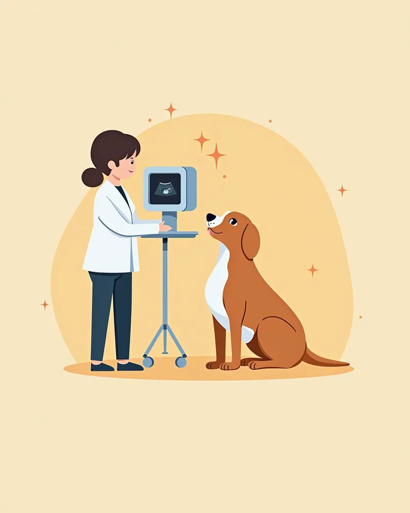
To accurately diagnose a tumor, several tests are typically performed together. - Ocular ultrasound: Precisely determines the tumor’s location, size, and its relationship to internal eye structures. - Intraocular pressure measurement: Checks for elevated eye pressure to assess for related conditions such as glaucoma. - Histopathology (biopsy): Examines excised or resected tissue to definitively determine whether the tumor is benign or malignant and to evaluate its biological characteristics. A definitive diagnosis is established through histopathology. - MRI or CT imaging: Helps assess the structures surrounding the eye and check for metastasis. - Systemic evaluation: Includes blood tests (CBC and biochemistry), thoracic radiographs, and fine-needle aspiration of regional lymph nodes to confirm whether the cancer has spread.
A veterinarian performing an ultrasound examination on a puppy's eye.

Treatment Methods Vary Depending on the Tumor’s Nature and Location
Treatment for tumors depends on the tumor’s nature, location, size, and your dog’s overall health. - Surgical excision: If the tumor is localized and removable, it can be surgically excised. - Diode laser photocoagulation: When appropriate, a laser can be used to coagulate and remove the tumor tissue. - Enucleation/evisceration: If complications are severe or preservation of the eye is not feasible, the eye may be removed (enucleation) or its contents extracted (evisceration). - Chemotherapy/metronomic therapy: For malignant tumors or those with a risk of metastasis, chemotherapy or metronomic therapy may be administered alongside surgery. - Regular monitoring: Periodic check-ups are necessary even after treatment.
A medical illustration showing the surgical removal of a tumor from a dog's eye

Home Care and Precautions

After treatment, protect your pet’s eyes and maintain a stable environment. - Eye protection mask: Use a mask to prevent frequent eye rubbing. - Maintain cleanliness: Keep the surgical site and the area around the eyes clean to prevent infection. - Medication administration: Administer prescribed medications exactly as directed by your veterinarian, adhering to the specified schedule. - Regular check-ups: Schedule follow-up examinations every 1 to 3 months after treatment. - Environmental adjustments: Keep lighting bright and provide a safe space free of obstacles. These measures support recovery and help prevent recurrence.
A puppy wearing an eye protection mask

Certain Breeds Have a Higher Risk of Developing Intraocular Tumors

Spindle cell tumors (spindleoid tumors) associated with blue eyes are relatively commonly reported in Siberian Huskies, Australian Shepherds, Border Collies, Labrador Retrievers, and German Shepherds. Middle-aged to senior dogs require particular attention. Regular ophthalmic examinations can help with early detection.

Reviewed by a veterinarian

Dr. Tony — Punnawat Phongkittirak

Dr. Tony — Punnawat Phongkittirak

Veterinarian

A veterinarian who majored in veterinary medicine at Khon Kaen University, Thailand, and completed the IVSA program at North Carolina State University in the United States. Drawing on clinical experience at animal hospitals, he works in the pet healthcare field and is dedicated to building a digital care environment that connects pet parents with veterinarians.

Frequently Asked Questions

Can canine intraocular tumors be treated?
The prognosis depends on the nature of the tumor and whether it is detected early. Benign adenomas rarely metastasize, but they often lead to loss of the eye due to secondary complications, so surgical removal or enucleation may be necessary. Malignant tumors (such as adenocarcinomas) carry a risk of metastasis, making early diagnosis and treatment crucial.
Can transmissible venereal tumors be passed to other dogs?
No. Nasal tumors are not contagious. They are influenced by genetic predisposition, age, and environmental factors.
Will vision return after surgery?
Whether vision recovers after surgery depends on the tumor’s location and the extent of damage. In some cases, vision is preserved, but if the damage is severe, full recovery may not be possible.
When do intraocular tumors typically appear?
This condition is primarily seen in middle-aged to senior dogs. Since the exact age of onset varies, regular eye examinations are important for dogs in middle age and beyond.
Can tumors be prevented?
While prevention can be challenging, regular eye exams can help catch issues early. A healthy lifestyle and proper nutrition also play a supportive role.

Share

Related Guides

References

[1] Jubb, K. et al. (2017) Pathology of Domestic Animals, 6th ed. Elsevier.

[2] Plumb's Veterinary Drug Handbook, 9th ed. (2022). Wiley-Blackwell.

[3] Veterinary Ophthalmology, 5th ed. (2020). Wiley-Blackwell.

This information is based on veterinary literature and does not replace diagnosis or treatment. Please consult a veterinarian for specific health concerns.

Metapet Co., Ltd. | CEO: Park Sung-yong | Business Reg. No. 417-88-02562 | Seoul, South Korea | Customer Center

Dog's Eye Color Changed? Intraocular Tumor Signs & Timing | Meongsiljang