Like
Share
멍실장
강아지 녹내장 약물·수술 치료 증상과 원인, 진단·치료와 관리 포인트 총정리

If Your Dog’s Vision Suddenly Blurs — Causes of Glaucoma and Treatment Timing

Eye HealthDiseasesMeongsiljang Veterinary Advisory Board

Glaucoma in dogs is a condition where increased intraocular pressure damages the optic nerve, making early diagnosis and appropriate treatment essential. With medication and surgery, the progression of the disease can be halted and vision preserved.

Glaucoma in Dogs Is a Condition Where Elevated Intraocular Pressure Damages the Optic Nerve
A veterinarian measuring intraocular pressure in a dog's eye.
Glaucoma in dogs is a condition in which abnormally elevated intraocular pressure compresses and damages the optic nerve. Primary glaucoma is linked to genetic predisposition and often presents as a sudden, rapid spike in intraocular pressure due to impaired drainage of aqueous humor. If left untreated, it can lead to vision loss or blindness. - Elevated intraocular pressure: When aqueous humor, the fluid that fills the eye, fails to drain properly, intraocular pressure rises, causing damage to the optic nerve. - Optic nerve damage: Sustained high intraocular pressure disrupts the transmission of visual signals, leading to progressive vision loss. - Importance of early diagnosis: Regular ophthalmic examinations that measure intraocular pressure enable early detection, helping to reduce pain and preserve remaining vision. Prompt veterinary attention is essential if signs indicating an urgent need for medical care appear.

The Main Cause of Glaucoma Is Impaired Drainage of Aqueous Humor
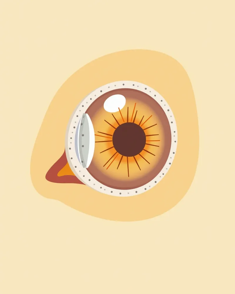
The main cause of glaucoma in dogs is the improper drainage of aqueous humor, the fluid that fills the eye. - Genetic factors: Certain breeds are genetically predisposed to elevated intraocular pressure, making them more susceptible to glaucoma (primary glaucoma). - Trauma or inflammation: Eye injuries or inflammation can block the drainage pathways for aqueous humor, leading to secondary (acquired) glaucoma. - Other eye conditions: Issues such as lens luxation or intraocular bleeding (hyphema) can also obstruct aqueous humor flow and cause glaucoma. - Mechanism of increased intraocular pressure: When impaired drainage of aqueous humor raises intraocular pressure, the optic nerve becomes compressed, resulting in vision loss. Early detection and identifying the underlying cause are key to effective treatment.
An anatomical diagram showing the drainage pathways of aqueous humor in a dog's eye and the sites where blockages can occur.

Main Symptoms Include Tearing, Redness, Blinking, and Vision Loss
The main symptoms of glaucoma in dogs include excessive tearing, redness of the eyes, and frequent blinking (blepharospasm) or rubbing due to pain. - Vision loss: If your dog seems disoriented or bumps into objects, it may indicate declining vision. - Eye swelling and corneal clouding: Elevated intraocular pressure can cause the cornea to swell and appear cloudy. In advanced cases, the eyeball itself may enlarge (buphthalmos). - Pupil changes: The pupil may dilate or constrict abnormally, and its response to light may become sluggish. - Restless behavior: Since glaucoma is painful, your dog may appear anxious or overly sensitive to its surroundings. - Prompt veterinary care is essential: These symptoms can also occur with other eye conditions, so accurate diagnosis through tests like intraocular pressure measurement is crucial. Seek veterinary ophthalmic care as soon as symptoms appear, as timely intervention is key to preserving vision.
A dog’s eye swollen due to glaucoma

Signs Requiring Immediate Veterinary Visit

If your dog suddenly starts rubbing its eyes, or if its eyes become swollen, red, or its vision deteriorates rapidly, you should take it to the vet immediately. In primary glaucoma, intraocular pressure can spike within just a few hours, and this painful increase in pressure can quickly lead to optic nerve damage. The sooner the intraocular pressure is lowered, the greater the chance of reducing pain and preserving remaining vision, so it is crucial to begin treatment as quickly as possible.

Diagnosis Is Made Through Tonometry and Fundus Examination
Canine glaucoma is diagnosed by accurately measuring intraocular pressure with a tonometer; manual palpation is not a reliable method. - Tonometry: This fundamental test checks whether intraocular pressure exceeds normal levels and is central to diagnosing glaucoma. In dogs, the goal is to keep intraocular pressure at or below 19–20 mmHg. - Gonioscopy: This procedure directly examines the structure of the eye’s drainage angle (iridocorneal angle), where aqueous humor exits, to determine the type of glaucoma. - Ocular Ultrasound: When corneal edema or other conditions obscure the view inside the eye, ultrasound helps visualize internal structures and deep drainage pathways, including the trabecular meshwork and ciliary body. - Clinical Symptom Assessment: Diagnosis involves a comprehensive evaluation of symptoms alongside the dog’s breed and medical history. Combining multiple diagnostic tests is essential for an accurate diagnosis.
A veterinarian using an intraocular pressure gauge on a puppy's eye.

Treatment Proceeds Stepwise with Medication and Surgery
Glaucoma treatment typically begins with medications to lower intraocular pressure. In emergency situations where pressure spikes rapidly, mannitol may be administered intravenously to quickly reduce it. - IOP-lowering agents: Eye drops such as carbonic anhydrase inhibitors (dorzolamide, brinzolamide) that decrease aqueous humor production, or medications that facilitate aqueous humor drainage, are used. - Anti-inflammatory agents: If inflammation is present, corticosteroids can help alleviate pain and swelling. - Combination therapy: Multiple medications are often prescribed together, tailored to the specific symptoms. - Surgical treatment: If medication alone cannot adequately control intraocular pressure, surgery to improve aqueous humor drainage pathways may be considered. - Enucleation: In cases where vision is nearly lost and pain is severe, removing the eye may be chosen to eliminate suffering. The treatment plan is determined by your veterinarian based on the individual case.
A puppy undergoing glaucoma surgery

Home Management Centers on Medication Administration and Environmental Control
Managing glaucoma in dogs at home centers on consistent medication administration and maintaining a stable environment. - Medication administration: Administer prescribed intraocular pressure-lowering drops or oral medications exactly on schedule, without missing any doses. - Environmental adjustments: Remove obstacles that could cause collisions and minimize sudden noises or shocks to ensure your dog’s safety. - Pain management: Since glaucoma is painful, follow your veterinarian’s instructions to manage discomfort effectively and maintain a calm, comfortable setting. - Daily monitoring: Regularly check for changes in eye appearance or behavior to detect issues early. - Regular veterinary visits: Even during treatment, schedule routine check-ups with your veterinarian to confirm that intraocular pressure remains well-controlled. Consistent care plays a major role in preserving vision.
A puppy resting comfortably in a calm, stable environment.

Certain Breeds Are More Susceptible to Hereditary Glaucoma

Breed-specific predispositions to glaucoma are well-documented: American Cocker Spaniels, Basset Hounds, Boston Terriers, and Shar-Peis are prone to primary closed-angle glaucoma, while Beagles and Norwegian Elkhounds are more susceptible to open-angle glaucoma. For these genetically predisposed breeds, regular ophthalmic examinations to monitor intraocular pressure are highly recommended. Early detection is the key to preserving your pet's vision.

Reviewed by a veterinarian

Dr. Tony — Punnawat Phongkittirak

Dr. Tony — Punnawat Phongkittirak

Veterinarian

A veterinarian who majored in veterinary medicine at Khon Kaen University, Thailand, and completed the IVSA program at North Carolina State University in the United States. Drawing on clinical experience at animal hospitals, he works in the pet healthcare field and is dedicated to building a digital care environment that connects pet parents with veterinarians.

Frequently Asked Questions

Can canine glaucoma be cured?
While a complete cure is difficult, the goal is to stabilize intraocular pressure and preserve remaining vision for as long as possible through medication and surgery. Early treatment is key.
Is medication alone sufficient?
Medications are effective in the early stages, but surgery is necessary if the condition has progressed. Medications are also used as an adjunct before and after surgery.
Can vision return even after surgery?
Once the optic nerve is damaged, it cannot be repaired, so restoring vision is generally difficult. However, we can manage intraocular pressure to prevent further deterioration.
Is glaucoma contagious to other dogs?
No, glaucoma is not contagious. It develops due to genetic factors, trauma, or other eye conditions, so contact with other dogs is safe.
How often should regular check-ups be performed?
For certain breeds or pets with a relevant medical history, regular eye exams are recommended, and any symptoms should prompt immediate veterinary evaluation.

Comparison Table of Medication and Surgical Treatments

ItemTreatment MethodEfficacyTiming of ApplicationPrecautions
IOP-Lowering Agent (Medication)Reduces intraocular pressure, relieves painEarly and mild stagesRequires daily regular dosingPossible ocular side effects
Surgery (Improving Aqueous Humor Drainage)Enables long-term IOP controlWhen difficult to control with medicationRequires recovery period after surgeryRisk of infection exists
Eye Removal Surgery (Enucleation)Complete elimination of painWhen vision is already lost and pain is severeAimed at relieving pain in a blind eyeChange in appearance, psychological adjustment needed

The treatment method is decided by the veterinarian based on the dog's condition and the degree of vision damage.

Share

Related Guides

References

[1] Fossum TW. (2007). Small Animal Surgery. 3rd ed. Elsevier.

[2] Hosgood G, Scholl DT. (1998). Evaluation of age as a risk factor for perianesthetic morbidity and mortality in the dog. J Vet Emerg Crit Care, 8(3):222-236.

[3] Reader RC, McCarthy RJ, Schultz KL, et al. (2020). Comparison of liposomal bupivacaine and 0.5% bupivacaine hydrochloride for control of postoperative pain in dogs undergoing tibial plateau leveling osteotomy. J Am Vet Med Assoc, 256:1011–1019.

This information is based on veterinary literature and does not replace diagnosis or treatment. Please consult a veterinarian for specific health concerns.

Metapet Co., Ltd. | CEO: Park Sung-yong | Business Reg. No. 417-88-02562 | Seoul, South Korea | Customer Center

Sudden Blurry Vision in Dogs: Glaucoma & Treatment Timing | Meongsiljang