Like
Share
멍실장
강아지 형광 염색 각막 검사 증상과 원인, 진단·치료와 관리 포인트 총정리

Complete Guide to Canine Fluorescein Corneal Staining: Why It’s Necessary and How to Interpret Results

Eye HealthDiseasesMeongsiljang Veterinary Advisory Board

Fluorescein staining of the cornea is a key diagnostic method for checking for corneal damage in dogs. If your dog has persistent tearing or repeatedly squints, they should be examined immediately.

What Is Fluorescein Corneal Staining in Dogs?
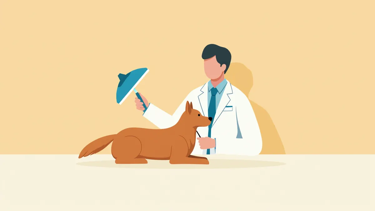
A scene of applying a fluorescent dye to a dog's eye
Fluorescein staining of the cornea is a key diagnostic method for accurately assessing corneal damage in dogs. After applying a fluorescent dye (fluorescein) to the eye and illuminating it with cobalt blue light, the dye adheres to areas where the corneal epithelium has been damaged, glowing bright green. The dye does not bind to healthy corneal epithelium, so no color appears in those areas. This test is minimally painful and provides relatively quick results. It is particularly recommended if your dog exhibits persistent tearing or frequent blinking. The results reveal the location and extent of corneal ulcers, serving as essential baseline information for developing a treatment plan. This test is effective for the early detection of ophthalmic conditions such as corneal ulcers.

Main Causes and Pathogenesis

Corneal injuries in dogs can occur for a variety of reasons. - Trauma: The cornea can be scratched when the dog bumps into sharp objects or gets into a fight with another animal. - Infection: If a bacterial infection accompanies a corneal wound, the ulcer can deepen and worsen rapidly. - Dry eye (Keratoconjunctivitis sicca): Insufficient tear production leaves the corneal surface dry and less protected, making it more susceptible to damage. - Breed predisposition: Brachycephalic breeds with short muzzles and protruding eyes are at higher risk of corneal injury due to increased exposure. - Self-trauma: Persistent rubbing or scratching of the eyes due to pain or itching can deepen the injury. Since these factors can interact, early detection is crucial.
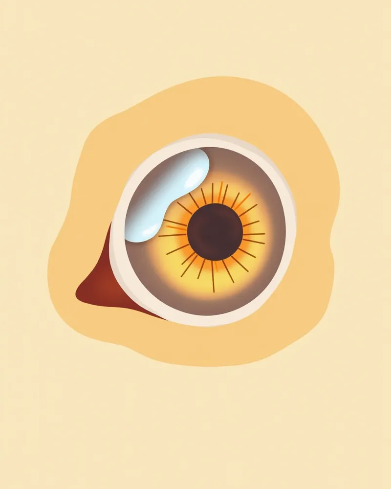
An anatomical illustration showing corneal damage in a dog’s eye and the corresponding fluorescein staining reaction.

Key Symptoms and Warning Signs

During a canine fluorescein corneal stain test, the following key symptoms may indicate an abnormality:
Frequent blinking or squinting: This behavior often results from eye pain.
Excessive tearing: Pain in the eye can lead to increased tear production.
Rubbing or scratching at the eye: This can further irritate the affected area and worsen discomfort.
Sensitivity to light: The eye becomes more sensitive, causing the dog to avoid bright environments.
Redness in the eye: This occurs due to inflammation and the resulting dilation of blood vessels.
If these behaviors persist, it is important to seek veterinary care immediately.
Your dog rubbing its eyes and showing signs of discomfort

When to Seek Immediate Veterinary Care

If your dog’s eyes become red, tear excessively, and remain squinted or closed, seek veterinary care as soon as possible. Corneal ulcers can rapidly deepen due to bacterial infection or persistent eye rubbing, and without timely treatment, they may lead to corneal melting or perforation.

Diagnostic Methods and Procedures

Fluorescein staining of the cornea is a simple and quick procedure. First, a drop of fluorescent dye is placed in your dog’s eye, and the eye is blinked a few times to spread the dye evenly. The excess dye is then thoroughly rinsed away with a sterile saline solution. Next, a cobalt blue light is shone on the eye to examine the cornea for any damage. Areas where the corneal epithelium has been lost will glow bright green, clearly indicating the presence of a corneal ulcer. The test is virtually painless and provides results quickly. If necessary, a corneal swab or bacterial culture may also be performed. It is important to properly rinse away the excess dye to avoid false-positive results, so it is best to have the test performed accurately at a veterinary clinic.
A scene showing a dog’s eyes being examined with blue light after applying a fluorescent dye.

Treatment Options and Step-by-Step Management

Treatment depends on the severity of the corneal injury. - Mild (superficial) cases: All corneal ulcers are initially treated with topical antibiotic eye drops. Medications like erythromycin are commonly used as a first-line treatment to prevent infection and support healing. - Deep or infected cases: Fluoroquinolone-based topical antibiotics are used. If reflex uveitis is present, atropine eye drops are administered concurrently, and systemic antibiotics may be added if necessary. - Severe cases (melting ulcers/descemetoceles): If the wound is deep or there is a risk of perforation, prompt surgical intervention, such as a conjunctival flap procedure, may be required. During treatment, it is essential to manage the environment to prevent the dog from scratching its eye and to strictly adhere to the medication regimen. Regular follow-up examinations are crucial to monitor healing progress, typically within 5–7 days for superficial ulcers and over 2–4 weeks for deep ulcers. Early treatment significantly aids recovery.
A puppy wearing glasses to protect its eyes.

Home Care and Prevention Tips

Here are some key tips to help keep your dog’s eyes healthy:
Clean around the eyes: Gently wipe the area around the eyes with a soft cloth to keep it clean.
Protect the eyes: During treatment, use an Elizabethan collar (neck cone) to prevent your dog from scratching or rubbing their eyes.
Follow medication instructions: Administer eye drops at the correct times and in the proper dosage as directed by your veterinarian.
Keep the environment safe: Remove sharp objects or toys that could irritate or injure the eyes.
Schedule follow-up exams: For corneal ulcers, follow your veterinarian’s advice for rechecks—superficial ulcers should be re-evaluated every 5–7 days, while deeper ulcers require weekly check-ups to ensure proper healing.
Gently wiping around your puppy’s eyes

Breed-Specific Precautions and Recurrence Prevention

Brachycephalic breeds with short muzzles and prominent eyes, such as Bulldogs, Pugs, Shih Tzus, and Pekingese, are at higher risk for corneal damage and ulcers because their corneas are more easily exposed. For these breeds, it’s important to regularly monitor and protect their eyes. After treatment, environmental adjustments and consistent care are essential to prevent recurrence. Follow your veterinarian’s guidance and attend follow-up visits to monitor the healing of corneal ulcers.

Reviewed by a veterinarian

Dr. Tony — Punnawat Phongkittirak

Dr. Tony — Punnawat Phongkittirak

Veterinarian

A veterinarian who majored in veterinary medicine at Khon Kaen University, Thailand, and completed the IVSA program at North Carolina State University in the United States. Drawing on clinical experience at animal hospitals, he works in the pet healthcare field and is dedicated to building a digital care environment that connects pet parents with veterinarians.

Frequently Asked Questions

Is fluorescent staining painful?
It’s hardly painful at all. Since we only place a single drop of dye in the eye, there’s minimal discomfort, and the procedure itself is quick.
Does the eye turn yellow after the test?
The damaged area of the cornea will glow bright green under blue light. Although the dark dye may temporarily stain the skin around the eye, it will wash away with sterile saline or water, so please avoid rubbing your pet's eye.
Is one test enough?
No. If symptoms recur or during treatment, periodic re-examinations are necessary to confirm recovery. Superficial ulcers are typically rechecked after 5–7 days; please follow your veterinarian’s instructions.
Can playing with other dogs cause eye injuries?
Yes, rough play with other animals can scratch or injure the cornea from claws or impact. Please ensure your pet plays gently.
Is it safe for my pet to go home after a fluorescent staining test?
Yes, you can head home right after the exam. Just be sure to administer the prescribed eye drops at the exact times and doses indicated.

Share

Related Guides

References

[1] Gelatt KN. Essentials of veterinary ophthalmology. Ames, IA: John Wiley & Sons, Inc.; 2014.

[2] Clinical Atlas of Canine and Feline Ophthalmic Disease, 2nd Ed. Wiley-Blackwell; 2018.

[3] Englar RE. Performing the small animal physical examination. Hoboken, NJ: Wiley; 2017.

This information is based on veterinary literature and does not replace diagnosis or treatment. Please consult a veterinarian for specific health concerns.

Metapet Co., Ltd. | CEO: Park Sung-yong | Business Reg. No. 417-88-02562 | Seoul, South Korea | Customer Center

Dog Fluorescein Corneal Staining: Why & How to Read It | Meongsiljang