Like
Share
멍실장
강아지 각막 천공 증상과 원인, 진단·치료와 관리 포인트 총정리

If Your Dog’s Eye Is Discharging Fluid — Warning Signs of Corneal Perforation and the Emergency Golden Hour

Eye HealthDiseasesMeongsiljang Veterinary Advisory Board

Corneal perforation in dogs is an emergency condition in which the eye’s outer protective layer ruptures, posing a high risk of vision loss and infection. Early detection and appropriate treatment are essential.

Corneal Perforation in Dogs Is an Acute Condition Where the Eye’s Protective Layer Ruptures

A medical image showing the condition of corneal perforation in a dog's eye
A corneal perforation in dogs is an acute condition in which the cornea, the outermost layer of the eye, develops a full-thickness hole. It causes severe pain, vision loss, and a rapid increase in the risk of infection. This is an emergency requiring immediate veterinary care. Common causes include trauma, rapidly progressing severe corneal ulcers, and rupture of the deepest corneal layer (the Descemet’s membrane) after it has bulged. - Trauma: The eye can be injured by tree branches, another dog’s claws, or toys. - Severe corneal ulcer: If a corneal ulcer deepens quickly due to bacterial infection or other factors, the cornea weakens and may perforate. - Rupture of a Descemetocoele: When an ulcer reaches the deepest layer of the cornea, the bulging area may rupture, leading to perforation. Early detection and prompt surgical intervention are essential to preserving vision.

The Main Causes Include Trauma, Infection, and Genetic Predisposition
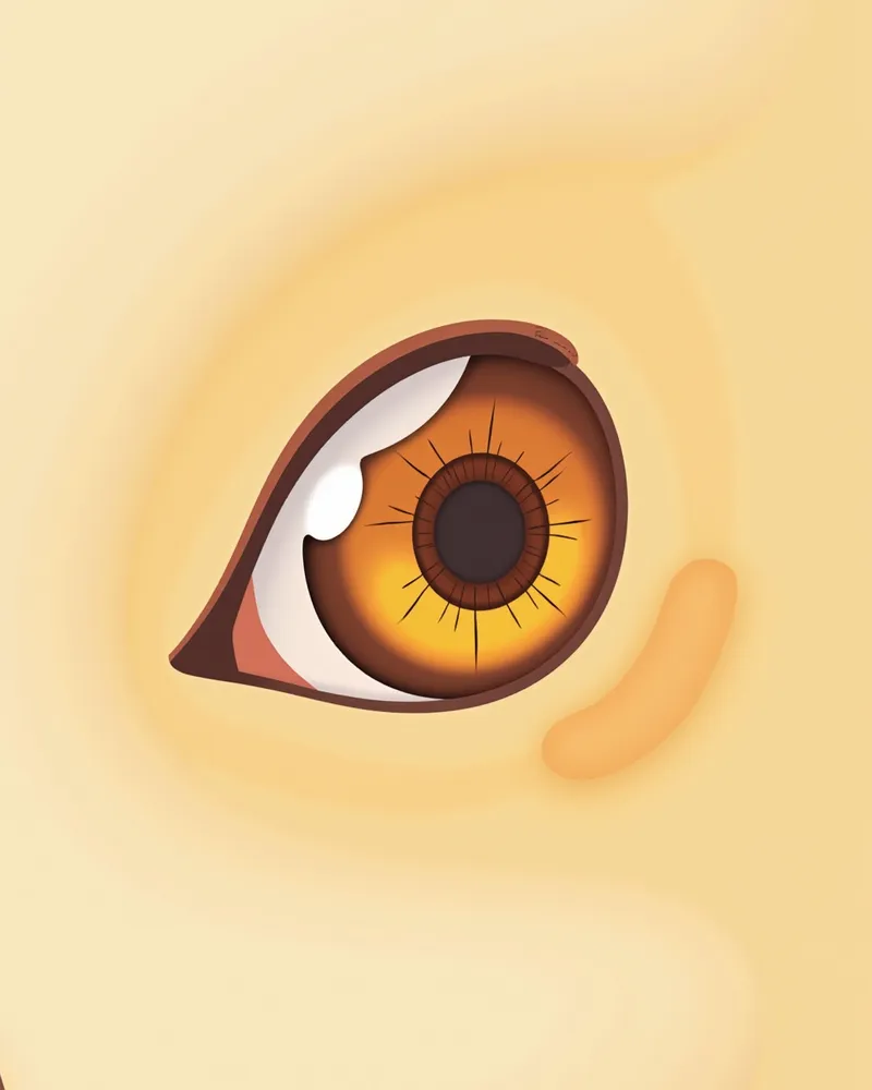
The most common cause of corneal perforation in dogs is trauma from external impacts or scratches. For example, the eye can be injured by tree branches, another dog’s claws, or toys. Additionally, if a corneal ulcer progresses rapidly and severely due to bacterial infection or other factors, the cornea can weaken and perforate. A descemetocele, which forms when an ulcer deepens, can also rupture and lead to perforation. - Trauma: Impacts or scratches from external objects are the most frequent cause. - Severe corneal ulcers: If an ulcer deepens quickly due to infection or inflammation, the cornea can weaken and rupture. - Rupture of a descemetocele: If damage extends to the deepest layer of the cornea, that area can burst, resulting in perforation. It is important to be aware of these causes and to detect eye injuries promptly.
Medical image of a dog’s eye showing inflammation following trauma

Main Symptoms Include Tear Production, Redness, Squinting, and Vision Loss

When a dog develops a corneal perforation, tear production increases dramatically, the eye becomes red and bloodshot, and the dog may avoid approaching you or keep its eyes tightly closed. Due to severe pain, the dog might scratch at its head or rub it against the ground. Vision loss can also occur, leading to difficulty seeing surroundings or misjudging directions. - Increased tearing: Tear production rises sharply when the cornea is damaged. - Redness: Blood vessels in the eye dilate, causing a red appearance. - Squinting or keeping eyes closed: Pain often causes the dog to frequently squint or keep its eyes shut. - Vision loss: Damage to the eye’s structure can impair vision. If you notice these symptoms, immediate veterinary care is essential.
A medical image showing a dog with its eyes closed, appearing to be in pain.

This Is an Emergency Situation Requiring Immediate Veterinary Care

A perforation in the eye significantly increases the risk of vision loss and intraocular infection. Because a perforated eye is structurally very fragile, improper handling can lead to further complications such as iris prolapse or retinal detachment. Do not attempt to administer medication, touch, or press on the eye yourself. Instead, prevent your dog from rubbing or scratching the eye and seek immediate veterinary care for prompt surgical intervention.

Diagnosis Is Confirmed Through Ophthalmic Examination and Microscopic Examination
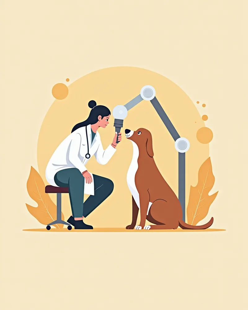
Veterinarians carefully examine your dog’s eyes using a specialized lamp, such as a slit lamp, to check for any perforations. Additionally, a fluorescein stain test helps visualize corneal damage. If the dye adheres to the affected area and appears green under cobalt blue light, it indicates a corneal defect. When necessary, further tests like cytology, Gram staining, bacterial culture, and sensitivity testing are conducted to identify the causative bacteria and determine the most effective antibiotic. - Ophthalmic Examination: A detailed inspection of the eye’s structure to assess whether there is a perforation. - Fluorescein Stain: The dye highlights damaged areas, making them easier to identify. - Cytology and Gram Staining: These tests evaluate the presence of infection and determine the type of bacteria involved. - Bacterial Culture and Sensitivity Testing: Used to pinpoint the exact bacteria causing the infection and select the most appropriate antibiotic. Accurate diagnosis ensures an effective treatment plan can be developed.
A medical image showing a veterinarian examining a dog's eye with a slit lamp

Treatment Proceeds in Stages Through Surgery, Medication, and Protective Devices

Treatment for corneal perforation varies depending on the severity, but because it is an emergency, the standard approach is to surgically repair the cornea as quickly as possible. Common surgical options include tectonic grafts to reinforce the cornea or conjunctival flap procedures. Alongside surgery, or in milder cases, we manage infection and inflammation with antibiotic eye drops, systemic antibiotics, and anti-inflammatory medications, while also stabilizing the cornea using agents like autologous serum eye drops. Since a perforated eye is extremely fragile, referral to a veterinary ophthalmologist is highly recommended. - Surgical treatment: Repair the perforated cornea using a corneal graft or conjunctival flap. - Medication: Prevent infection and inflammation with antibiotic eye drops, systemic antibiotics, and anti-inflammatory drugs. - Protective measures: Fit an Elizabethan collar after surgery to prevent the pet from scratching or rubbing its eye. - Medication administration: Administer medications at precise times to maximize therapeutic efficacy. Consistent care throughout the treatment period is essential.
A medical image showing a puppy wearing an eye protection mask

Home Care Centers on Administering Eye Drops and Environmental Control

During the treatment period, it is essential to administer eye drops at the exact prescribed times. To prevent your dog from scratching or rubbing its eyes, fit it with an Elizabethan collar and maintain a clean indoor environment with appropriate humidity levels. Avoid rough play in parks or under trees, and minimize contact with other dogs. Additionally, handle any foreign matter around the eyes with care, ensuring you do not press or rub the eye itself. - Eye drop administration: Missing the scheduled time can reduce treatment efficacy. - Environmental control: Maintain a clean environment and minimize irritants such as dust. - Activity restriction: Limit outdoor activities and ensure your dog gets adequate rest. - Contact management: Avoid contact with other dogs to reduce the risk of infection. - Foreign matter removal: Carefully remove dust or hair around the eyes without irritating them. Meticulous care significantly aids recovery.
A medical image showing a dog resting in a calm, stable environment

Certain Breeds Are at Higher Risk for Corneal Perforation

Corneal perforation is most commonly caused by trauma or rapidly progressing, severe corneal ulcers. Therefore, extra caution is needed for dogs in environments with a high risk of eye injury, as well as for breeds with prominent eyes that are more susceptible to trauma and ocular surface diseases. For any dog, if you notice frequent blinking or abnormalities such as excessive tearing or redness, do not delay—seek prompt veterinary care and schedule regular ophthalmic examinations.

Reviewed by a veterinarian

Dr. Tony — Punnawat Phongkittirak

Dr. Tony — Punnawat Phongkittirak

Veterinarian

A veterinarian who majored in veterinary medicine at Khon Kaen University, Thailand, and completed the IVSA program at North Carolina State University in the United States. Drawing on clinical experience at animal hospitals, he works in the pet healthcare field and is dedicated to building a digital care environment that connects pet parents with veterinarians.

Frequently Asked Questions

Why should you rush your dog to the vet if a perforation occurs in its eye?
If the eye’s protective membrane ruptures, bacteria can enter the eye and cause a rapidly spreading infection. This can lead to serious complications such as vision loss, iris prolapse, or retinal detachment, so immediate veterinary care is essential.
Can corneal perforation be completely cured?
Corneal perforation is a medical emergency, so surgery to repair the damage as quickly as possible is the standard approach. In mild cases, medication may be used alongside surgery, but because the cornea is highly sensitive to injury, the extent of recovery depends on the size of the perforation and how promptly treatment is administered. The sooner treatment begins, the better the prognosis, making it crucial to seek veterinary ophthalmic care as early as possible.
How should I care for my dog after surgery?
After surgery, it is important to administer eye drops as directed and fit your dog with an Elizabethan collar to prevent them from scratching or rubbing their eyes. Keep the indoor environment clean and quiet, and avoid contact with other dogs.
Can corneal perforation be prevented?
To prevent eye injuries, it’s important to create a safe play environment and schedule regular veterinary eye exams. This is especially crucial for breeds with prominent, protruding eyes.
I find it difficult to administer eye drops. What should I do?
When applying eye drops, gently lift your dog’s head and place the drops directly onto the eye. Avoid touching the eye with your fingers, and after administering the drops, gently close the eye to help distribute the medication.

Share

Related Guides

References

[1] Plumb's Veterinary Drug Handbook, 9th ed. (2022). Wiley-Blackwell.

[2] Kirk's Current Veterinary Therapy XIV: Small Animal Practice. Elsevier, 2022.

[3] Veterinary Ophthalmology, 6th ed. (2021). Wiley-Blackwell.

This information is based on veterinary literature and does not replace diagnosis or treatment. Please consult a veterinarian for specific health concerns.

Metapet Co., Ltd. | CEO: Park Sung-yong | Business Reg. No. 417-88-02562 | Seoul, South Korea | Customer Center

Canine Corneal Perforation: Warning Signs & Golden Hour | Meongsiljang