Like
Share
멍실장
고양이 3안검 돌출 증상과 원인, 진단·치료와 관리 포인트 총정리

If a White Film Covers Your Cat’s Eye — Health Signals and Causes of Third Eyelid Prolapse

Eye HealthDiseasesMeongsiljang Veterinary Advisory Board

Feline cherry eye occurs when the third eyelid on the inner corner of the eye protrudes outward. Because it can cause pain and infection, early detection is crucial. The underlying causes vary, including genetics, trauma, and inflammation, so an accurate diagnosis and appropriate treatment are essential.

What Is Third Eyelid Prolapse in Cats?

A cat's protruding third eyelid
Third eyelid prolapse in cats is a condition involving the third eyelid (nictitating membrane) located on the inner corner of the eye. The most common form is "cherry eye," where the tear gland (lacrimal gland) inside the third eyelid slips out of its normal position and appears as a smooth, pink-to-red mass. This condition can impair the eye’s protective functions and tear production, increasing the risk of conjunctivitis, ocular discharge, and secondary corneal ulcers, so early detection is crucial. It typically occurs in young cats and is more frequently reported in breeds with a genetic predisposition, such as Burmese cats. - Third eyelid (nictitating membrane) and lacrimal gland: Normally hidden from view, the third eyelid becomes visible as a smooth, red mass when the lacrimal gland prolapses. - Causes of prolapse: The condition often results from congenital weakness in the connective tissue that supports the lacrimal gland. It can also be triggered by pain, trauma, or inflammation causing the third eyelid to elevate. - Need for treatment: Beyond cosmetic concerns, prolapse can lead to reduced tear production, chronic inflammation, and corneal damage, making prompt veterinary care essential.

What Are the Main Causes?

The most common cause of third eyelid prolapse in cats, particularly cherry eye (prolapse of the third eyelid gland), is congenital weakness of the connective tissue that supports the gland, allowing it to slip out of place. This condition is most often seen in young cats, and certain breeds with a known genetic predisposition, such as Burmese cats, are at higher risk. - Genetic and connective tissue defects: Weakness in the supportive structures of the gland can allow it to prolapse naturally. - Trauma and pain: If the eye experiences impact, irritation, or pain, the cat may reflexively elevate (prolapse) the third eyelid. - Inflammatory responses: Inflammation around the eye due to conditions like conjunctivitis or keratitis can make the third eyelid appear more prominent. - Systemic and neurological factors: Third eyelid elevation can also occur in conditions such as Horner’s syndrome (accompanied by miosis and ptosis). Accurate differential diagnosis by a veterinarian is essential to determine the underlying cause.
Anatomy of the third eyelid in cats

Main Symptoms and Signs

The hallmark sign of a cat’s third eyelid prolapse (cherry eye) is a smooth, pink-to-red tissue mass visible on the inner corner of the eye. This is not pus, but rather the prolapsed glandular tissue of the third eyelid. It may be accompanied by conjunctivitis, increased tearing and ocular discharge, eye-rubbing behavior, and squinting or keeping the eyes partially closed. Because prolonged symptoms increase the risk of corneal ulcers and secondary infections, early intervention is essential.
Third eyelid (nictitating membrane) prolapse: A smooth, red tissue protruding from the inner corner of the eye.
Increased tearing and discharge: Persistent tearing or an increase in ocular secretions.
Conjunctival hyperemia and swelling: Redness and swelling of the conjunctiva surrounding the eye.
Eye-rubbing behavior: Repeated scratching or rubbing of the eyes due to discomfort.
Risk of corneal damage: Prolonged prolapse can lead to secondary corneal ulcers.
A cat experiencing discomfort due to third eyelid protrusion

When to Seek Immediate Veterinary Care

If your cat keeps its eyes closed, experiences persistent tearing, or has severely swollen eyes, seek veterinary care immediately. These symptoms may indicate corneal damage or a serious infection. Early diagnosis is crucial, as delayed treatment can lead to vision loss.

Accurate Diagnosis Methods
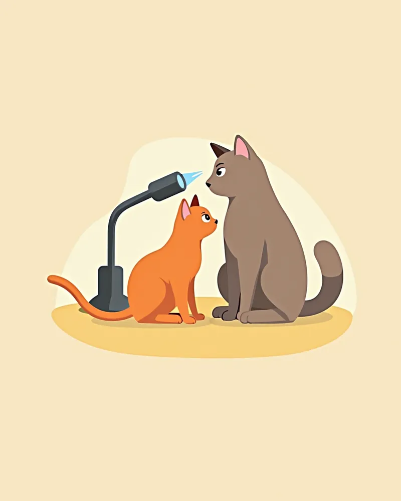
Accurate diagnosis of a cat’s everted third eyelid relies most heavily on a veterinarian’s clinical examination. Most feline eye conditions are diagnosed based on clinical signs, with additional tests performed as needed. - Visual and slit-lamp examination: The vet directly observes and assesses whether the protruding tissue is the nictitating gland, the degree of prolapse, and the condition of the conjunctiva and cornea. If pain causes the third eyelid to cover the eye, topical anesthesia may be necessary for examination. - Schirmer tear test: This measures tear production. However, since normal cats can also have low readings, results must be interpreted with caution. - Corneal fluorescein staining: A dye is applied to check for secondary corneal ulcers. - Additional testing and referral: If other causes such as Horner’s syndrome are suspected, or if the condition does not respond to treatment, referral to a veterinary ophthalmologist or further advanced diagnostics should be considered. Starting treatment without an accurate diagnosis increases the risk of recurrence or complications.
A veterinarian examining a cat's eyes

Treatment Methods and Step-by-Step Management

Treatment depends on the underlying cause and severity. If there is inflammation and pain, symptoms are managed with eye drops and similar medications. However, in cases of gland of the third eyelid prolapse (cherry eye), medication alone cannot return the gland to its proper position, so surgical repositioning is usually required. The standard treatment is a repositioning surgery, such as the pocket technique, which secures the prolapsed gland back in its original location. Because the gland produces a significant portion of the tear film, removal (excision) should be avoided whenever possible to prevent complications like dry eye (keratoconjunctivitis sicca).
Medical Management: Eye drops and other medications are used as supportive care to relieve inflammation and pain.
Surgical Repositioning: Repositioning surgery to return and secure the gland in its original place is the standard of care.
Gland Preservation: Excision is avoided whenever possible due to the risk of dry eye.
Post-Operative Care: Medications, such as antibiotics, may be prescribed by your veterinarian to prevent infection.
Protective E-Collar: An Elizabethan collar (E-collar) is worn after surgery to prevent the pet from scratching or rubbing its eyes.
A cat wearing protective glasses after surgery

Home Care Points

Proper management of third eyelid prolapse in cats is key to successful treatment and preventing recurrence. - Maintain cleanliness: Gently wipe the area around the eyes with a soft cloth one to two times daily to prevent infection. - Use eye drops as directed: Administer eye drops at the correct dosage and frequency as instructed by your veterinarian, and avoid overuse. Excessive use can lead to side effects. - Post-surgical care: After surgery, always use a protective Elizabethan collar to prevent the cat from scratching its eyes. - Nail care: Regularly trim your cat’s nails to prevent accidental scratches and injuries to the eyes. - Environmental adjustments: Provide a quiet, low-stress environment to help keep your cat calm and comfortable. - Regular check-ups: Since kittens and predisposed breeds may develop the condition in the other eye, schedule regular eye examinations with your veterinarian to continuously monitor their condition.
A cat resting in a calm, stable environment

Breed-Specific Precautions and Recurrence Prevention

Certain breeds, such as the Burmese, have been reported to have a genetic predisposition to third eyelid gland prolapse (cherry eye). These breeds require regular eye examinations, and because the condition can develop in the opposite eye if it occurs in one, close monitoring is essential. It is best to establish a management plan in consultation with your veterinarian.

Reviewed by a veterinarian

Dr. Tony — Punnawat Phongkittirak

Dr. Tony — Punnawat Phongkittirak

Veterinarian

A veterinarian who majored in veterinary medicine at Khon Kaen University, Thailand, and completed the IVSA program at North Carolina State University in the United States. Drawing on clinical experience at animal hospitals, he works in the pet healthcare field and is dedicated to building a digital care environment that connects pet parents with veterinarians.

Frequently Asked Questions

Can a cat’s third eyelid prolapse be treated?
Yes, it can be treated. However, because the third eyelid gland (cherry eye) won’t return to its normal position with medication alone, most cases require surgical repositioning and fixation of the gland. The specific approach depends on the underlying cause and severity, so please consult your veterinarian.
Can the eye become proptotic again after surgery?
While there is a risk of recurrence, it can usually be minimized with appropriate surgical correction and ongoing care. For breeds with a genetic predisposition, regular check-ups are essential, and owners should be aware that if the condition develops in one eye, it may also occur in the other.
Is using only eye drops sufficient?
Eye drops can help reduce inflammation and pain, but they won’t restore a prolapsed gland to its normal position. If the prolapse persists, surgical correction is necessary, so please follow your veterinarian’s recommendation.
Why does my cat rub its eyes?
Your cat may rub its eyes due to pain or irritation. Cats can also evert their third eyelid in response to discomfort, so if the symptoms persist, early diagnosis is essential.
Is it necessary to put a protective collar (Elizabethan collar) on after surgery?
Your pet may feel uncomfortable at first, but they will gradually get used to it. The protective neck collar (Elizabethan collar) is essential for preventing your pet from scratching their eyes after surgery, so it’s important to keep it on for the duration specified by your veterinarian.

Share

Related Guides

References

[1] Maggs DJ. Third eyelid. In: Maggs DJ, Miller PE, Ofri R, Slatter DH, editors. Slatter’s fundamentals of veterinary ophthalmology. 5th ed. St. Louis, MO: Elsevier; 2013. p. 151-6.

[2] Chahory S, Crasta M, Trio S, Clerc B. Three cases of prolapse of the nictitans gland in cats. Vet Ophthalmol. 2004;7(6):417–419.

[3] Dugan SJ, Severin GA, Hungerford LL, Whiteley HE, Roberts SM. Clinical and histologic evaluation of the prolapsed third eyelid gland in dogs. J Am Vet Med Assoc. 1992;201(12):1861–1867.

This information is based on veterinary literature and does not replace diagnosis or treatment. Please consult a veterinarian for specific health concerns.

Metapet Co., Ltd. | CEO: Park Sung-yong | Business Reg. No. 417-88-02562 | Seoul, South Korea | Customer Center

White Film Over Cat's Eye? Third Eyelid Prolapse Causes & Signs | Meongsiljang