Like
Share
멍실장
고양이 비만세포종 등급·치료 증상과 원인, 진단·치료와 관리 포인트 총정리

Is That Lump on Your Cat’s Skin a Mast Cell Tumor? — A Complete Guide to Treatment and Prognosis by Grade

Oncology/CancerDiseasesMeongsiljang Veterinary Advisory Board

Feline mast cell tumors are malignant growths that can develop in the skin and internal organs, with treatment options and prognosis varying depending on the tumor grade. Early diagnosis and proper management are essential.

Feline Mast Cell Tumors Are Malignant Growths That Develop in the Skin or Internal Organs

Early lesions of mast cell tumor on a cat's skin
Feline mast cell tumors are growths that develop when mast cells, a type of immune cell, proliferate abnormally. In cats, these tumors most commonly appear as single lesions on the skin, though they can also occur in internal organs such as the spleen or intestines. Fortunately, many feline cutaneous mast cell tumors are well-differentiated and benign, often curable with surgical removal alone. However, some can follow a malignant course, meaning treatment plans and prognosis vary depending on the tumor’s grade. In the early stages, these tumors typically present as small, firm lumps, making careful monitoring by pet owners essential. Veterinarians usually begin with a fine-needle aspirate cytology and may perform a biopsy to accurately determine the tumor’s grade and nature. Early detection is especially important if the lump grows rapidly, or if the skin becomes red or ulcerated. Early diagnosis and appropriate treatment are key to achieving a favorable outcome.

The Cause of Mast Cell Tumors Is Unclear, but Genetic and Environmental Factors Play a Role

The exact cause of feline mast cell tumors remains unclear. However, certain breeds have been reported to have a higher risk of developing the disease, suggesting a possible genetic predisposition. In fact, mutations in the c-KIT tyrosine kinase receptor, similar to those reported in canine mast cell tumors, have been identified in some cases of feline cutaneous mast cell tumors, prompting ongoing research into their association with genetic mutations. Nevertheless, how these mutations contribute to tumor development has not yet been sufficiently validated through large-scale studies. There is also insufficient evidence to definitively link other potential triggers, such as chronic inflammation, infections, or hormonal changes. Since no established preventive measures are currently available, early diagnosis and consistent management are more important than prevention.
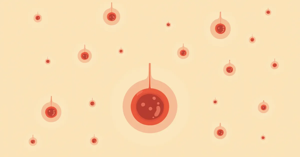
The location and role of mast cells in the feline immune system

Understand the Main Symptoms and Signs

The most common sign of feline mast cell tumor is a lump on the skin. These lumps often appear on the head, neck, and trunk, and may gradually grow, become red, or swell. When histamine is released from the tumor, the size of the lesion may change or ulcers may develop, and the cat may frequently lick or scratch the area. If the tumor occurs in internal organs such as the spleen or intestines, vomiting and loss of appetite are common symptoms, and intestinal bleeding can lead to anemia. More careful evaluation is needed if the tumor grows rapidly or if satellite nodules are present nearby. Early detection is a crucial first step toward appropriate treatment.
Scratching behavior in cats with skin lesions

Signs That Require Immediate Veterinary Attention

If a skin lump grows rapidly or develops bleeding, ulceration, or swelling, immediate veterinary attention is necessary. Additionally, if systemic symptoms such as loss of appetite, weight loss, vomiting, or diarrhea are present, the condition may be highly malignant. Early diagnosis and treatment are essential if tumors appear in multiple locations or if internal organ dysfunction is suspected.

Histopathology and Imaging Are Necessary for an Accurate Diagnosis

To diagnose mast cell tumors, we first perform a fine-needle aspiration cytology on the mass. Mast cells are relatively easy to identify due to their characteristic cytoplasmic granules, making preoperative cytology a useful initial test. However, since cytology alone cannot accurately determine the tumor grade, we analyze the excised biopsy tissue histologically to assess both the grade and the surgical margins. These histopathology results are crucial in determining the treatment plan. Additionally, imaging studies such as ultrasound may be performed to evaluate the patient’s overall condition and check for metastasis. It is especially important to thoroughly assess for metastasis when a high degree of malignancy is suspected. An accurate diagnosis is the first step toward appropriate treatment.
The process of a cat skin biopsy

Treatment Options Vary Depending on Tumor Grade and Location

Treatment for feline mast cell tumors depends on the tumor’s grade, location, and whether it has metastasized. For solitary cutaneous mast cell tumors, surgical excision is the primary treatment of choice. Most feline cutaneous mast cell tumors are well-differentiated, benign (Group 1) forms, so surgery alone is often curative. Cats with stage I–III disease rarely die from mast cell tumors, and many achieve long-term survival with surgery alone. Therefore, the need for chemotherapy is limited in most cases and is carefully considered only in malignant (Group 2) or surgically challenging situations. Your veterinarian will comprehensively evaluate your pet’s condition to create a tailored treatment plan, with regular check-ups and ongoing care being essential throughout the process.
Recovering after surgery for feline mast cell tumor

Home Care Is Key to Improving Treatment Success Rates

After surgery, wound care and infection prevention are the top priorities. Owners should check the incision site frequently and keep an Elizabethan collar on the cat to prevent licking. Medications must be given exactly as prescribed, and any side effects should be reported to the veterinarian immediately. Monitor food intake and nutritional status, and schedule regular check-ups to screen for recurrence. Emotional well-being is also important, so maintaining a quiet environment and a consistent daily routine will help. Consistent care at home is a key factor in improving treatment success rates.
Wearing a protective collar after cat surgery

Regular Checkups Are Essential for Cases at High Risk of Recurrence

Malignant (Group 2) mast cell tumors or those located in internal organs carry a relatively higher risk of recurrence and metastasis. However, in cats with cutaneous mast cell tumors, recurrence occurs only in some cases, and it has not been proven that the completeness of the surgical margins predicts the recurrence rate. Therefore, after surgery, it is important to follow your veterinarian’s recommended schedule for regular check-ups and, when necessary, to confirm whether metastasis has occurred through blood tests and imaging studies. Consistent monitoring is key to early detection.

Reviewed by a veterinarian

Dr. Tony — Punnawat Phongkittirak

Dr. Tony — Punnawat Phongkittirak

Veterinarian

A veterinarian who majored in veterinary medicine at Khon Kaen University, Thailand, and completed the IVSA program at North Carolina State University in the United States. Drawing on clinical experience at animal hospitals, he works in the pet healthcare field and is dedicated to building a digital care environment that connects pet parents with veterinarians.

Frequently Asked Questions

Can feline mast cell tumors be transmitted to humans?
No, feline mast cell tumors are not contagious to humans. They do not spread between animals, and the tumors in humans and cats are entirely separate conditions.
Are there certain breeds that are more prone to developing mast cell tumors?
While it tends to be more common in certain breeds, it can occur in any breed. The risk of developing this condition has been reported to be somewhat higher in several breeds, particularly Siamese, Burmese, Maine Coon, Ragdoll, Oriental Blue, Russian Blue, and Havana.
What is the prognosis after surgery?
Most feline cutaneous mast cell tumors are well-differentiated and benign, often resolving with surgical excision alone and allowing for a long, healthy life. However, malignant (Grade 2) tumors or those located in the gastrointestinal tract carry a risk of recurrence and metastasis, necessitating further evaluation and regular check-ups.
What medications are used for treatment?
In cats, surgery is the primary treatment, and most cases can be cured with surgery alone, so the need for chemotherapy is limited. In some cases, the potential benefit of tyrosine kinase inhibitors such as masitinib and toceranib is being considered in relation to cKIT mutations, but this has not yet been confirmed by large-scale studies. When using these medications, side effects must be closely monitored.
What should I do if my pet develops mast cell tumors?
If you feel a lump on your pet’s skin, consult a veterinarian right away. Accurate diagnosis through preoperative cytology and excisional biopsy histopathology significantly impacts the prognosis. Treatment primarily involves surgical excision, with additional therapies considered comprehensively based on the situation.

Comparison of Characteristics and Treatment Approaches by Mast Cell Tumor Grade

ItemCharacteristicsTreatment methodPrognosis
Group 1 (benign)Low mitotic count and well-differentiated morphology; often solitary cutaneous tumorsSurgical excision is the primary treatment (most are treated with surgery alone)Good (most cats in stages I–III achieve long-term survival with surgery alone)
Group 2 (malignant)High mitotic count and atypical cell morphology; may follow a malignant courseSurgical excision is the basis; additional treatment carefully considered depending on the situationCaution needed (evaluation and follow-up for possible recurrence/metastasis required)

Feline mast cell tumors are classified into Group 1 (benign) and Group 2 (malignant) based on mitotic count and cell morphology (Sabattini & Bettini, 2018), and the final grade is determined by the veterinarian according to the biopsy results.

Share

Related Guides

References

[1] Hawkins E, et al. (2018) Feline mast cell tumors: A review of clinical presentation, grading, and treatment. Journal of Feline Medicine and Surgery, 20(1), 5-15.

[2] Lloyd WH, et al. (2020) Evaluation of the WHO grading system for feline cutaneous mast cell tumors. Veterinary Pathology, 57(4), 567-575.

[3] American Veterinary Medical Association (2022) Guidelines for the Management of Feline Mast Cell Tumors. AVMA Position Statement.

This information is based on veterinary literature and does not replace diagnosis or treatment. Please consult a veterinarian for specific health concerns.

Metapet Co., Ltd. | CEO: Park Sung-yong | Business Reg. No. 417-88-02562 | Seoul, South Korea | Customer Center

Cat Skin Lump: Mast Cell Tumor? Treatment & Prognosis by Grade | Meongsiljang