
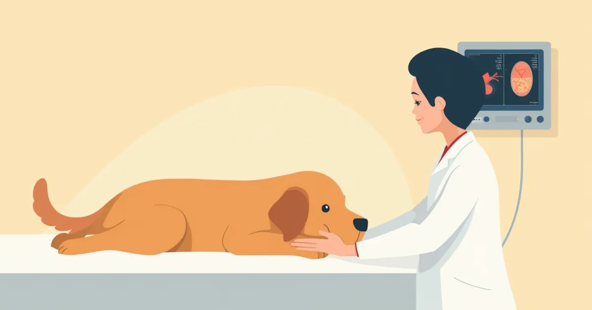
What is an ultrasound exam? — How are abdominal and cardiac conditions diagnosed?
This guide provides an easy-to-understand overview of the types of ultrasound exams for pets (abdominal and cardiac), the examination process, preparation steps, and the differences between ultrasound and X-ray imaging.
What is an ultrasound exam?

Abdominal ultrasound — What can be assessed?
Echocardiography — The key test for heart disease
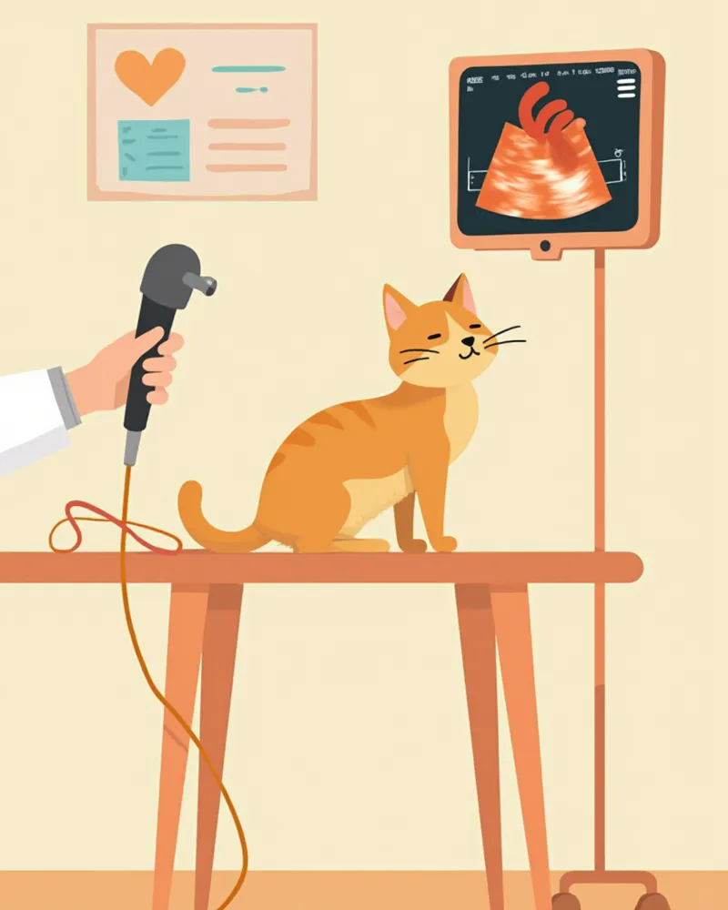
How is the exam performed?
Important points to check before the exam
Fasting before an abdominal ultrasound is essential for accurate results. If food remains in the stomach, it can obscure internal organs and hinder clear visualization. The exact fasting duration may vary depending on your pet’s condition and the purpose of the exam, so be sure to confirm the specific guidelines with your veterinarian when scheduling. Note that fasting instructions for cardiac ultrasounds may differ from those for abdominal ultrasounds, so it’s best to ask your vet in advance. Since the area being examined will need to be shaved, it’s advisable to schedule the ultrasound before, rather than after, a grooming session.
Ultrasound vs. X-ray comparison
| Item | Ultrasound | X-ray |
|---|---|---|
| Areas that can be assessed | Internal organ structures, blood flow | Bones, organ size & position, lung condition |
| Real-time imaging | Possible (observe movement & blood flow) | Not possible (still image) |
| Anesthesia needed | Usually not required | Usually not required |
| Radiation exposure | None | Yes (very small amount) |
| Test duration | 20–40 minutes | 5–10 minutes |
| Suitable situations | Internal organ abnormalities, cardiac function assessment | Fractures, foreign bodies, lung & chest assessment |
The two tests are complementary. Depending on the situation, they are often performed together.
When is an ultrasound exam needed?

Breed-specific recommendations for regular echocardiography
Certain cat breeds, such as Maine Coons, Ragdolls, Sphynx, Norwegian Forest Cats, Siberians, British Shorthairs, and Devon Rexes, are known to have a predisposition to hypertrophic cardiomyopathy. According to veterinary cardiology textbooks, echocardiography is the most sensitive and specific test for detecting structural heart disease during breed screening, while screening based solely on auscultation has limited diagnostic value. Therefore, regular cardiac echocardiographic screening is recommended for predisposed breeds or kittens intended for breeding, and the timing and frequency of these exams should be determined in consultation with your veterinarian. For dogs, if a breed is suspected of having structural abnormalities such as mitral valve insufficiency, discuss the need for regular cardiac echocardiograms starting in middle age with your veterinarian. If you are planning to breed, pre-breeding cardiac echocardiography is especially important to check for phenotypic abnormalities in advance.
Reviewed by a veterinarian

Dr. Tony — Punnawat Phongkittirak
VeterinarianA veterinarian who majored in veterinary medicine at Khon Kaen University, Thailand, and completed the IVSA program at North Carolina State University in the United States. Drawing on clinical experience at animal hospitals, he works in the pet healthcare field and is dedicated to building a digital care environment that connects pet parents with veterinarians.
Reviewed 2026-04-19
Frequently asked questions
How can I find out the cost of an ultrasound examination?
Does my pet feel pain during an ultrasound?
Is fasting absolutely necessary before an ultrasound?
Can an ultrasound alone provide an accurate diagnosis?
Can cats undergo ultrasound examinations?
Share
Related Guides

Primary vs. Secondary Veterinary Hospitals: A Comparison

5 Easy-to-Raise Cat Breeds for First-Time Owners — Grooming Difficulty Comparison

Top 7 Short-Legged Dog Breeds

Guide to Prescription Abbreviations — Meanings of SID, BID, and PRN at a Glance

Early Signs of Congestive Heart Failure in Dogs and Management

Doggy Skin Itch and Red Ears — A Complete Guide to Choosing Food for Food Allergies

Can Cats Get Heartworm Disease? — Why It’s More Dangerous Than in Dogs and How to Prevent It

Guide to Traveling with Pets on KTX and Trains

How to Check Lymph Nodes at Home

Recognizing End-of-Life Signs in Cats
References
[1] Virginia Luis Fuentes et al., Textbook of Cardiovascular Medicine in Dogs and Cats, Ch. 11 – Echocardiography
[2] Silverstein & Hopper, Small Animal Critical Care Medicine, 3rd Ed, Ch. 43 – Echocardiography Indications
[3] Schaer & Gaschen, Clinical Medicine of the Dog and Cat, 4th Ed, Ch. 23 – Point-of-Care Ultrasound
[4] Burkitt Creedon & Davis, Feline Emergency and Critical Care Medicine, 2nd Ed, Ch. 14 – Cardiac Ultrasound in Dyspneic Cats