Like
Share
멍실장
강아지·고양이 피부 종괴 세포진 검사, 꼭 해야 할까요?

Is a Fine-Needle Aspiration of a Skin Mass Necessary for Dogs and Cats?

Skin & CoatQ&AMeongsiljang Veterinary Advisory Board

We’ve compiled everything pet owners need to know about fine-needle aspiration (FNA)—the first test performed when a lump is felt on the skin—including its principles, procedure, and how to interpret the results.

What Is a Fine-Needle Aspiration of a Skin Mass?
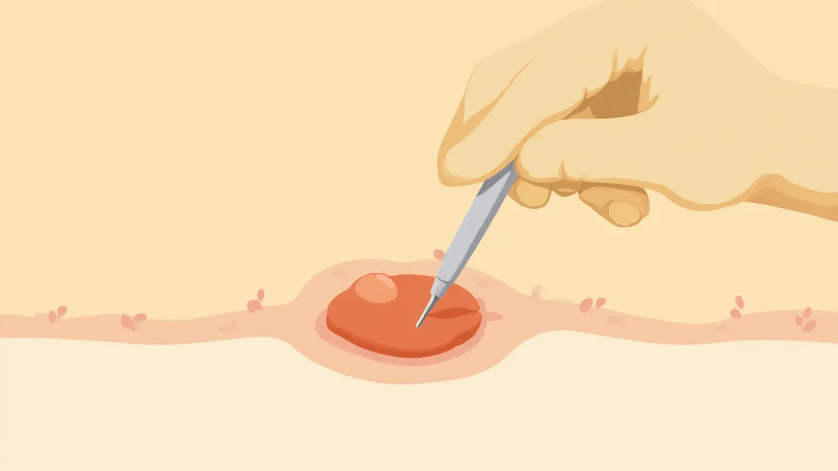
A fine-needle aspiration test being performed on a dog’s skin mass
A fine-needle aspirate of a skin mass is a primary diagnostic test in which cells are drawn from the lump using a thin needle and examined under a microscope. Its biggest advantage is that it can be performed immediately in an outpatient setting without anesthesia. It is the first test recommended when a lump is detected, and the results help determine whether to monitor the mass, proceed with a biopsy, or move on to surgical removal. The procedure is performed in the same way for both dogs and cats.

Which Lumps Require It?

While not every lump requires the same level of scrutiny, veterinary dermatology textbooks recommend attempting fine-needle aspiration (FNA) before proceeding to biopsy or surgical removal for most skin nodules (lumps) in dogs and cats. FNA is particularly helpful for diagnosis in the following types of lumps:
Firm, nodular lumps: FNA is most suitable for nodular lesions.
Lumps suspected to be tumors (neoplasms): Useful for differentiating neoplastic lesions.
Lumps accompanied by pus or granulomas: Helpful in cases of pyogranulomatous diseases.
Lumps suspected of infection: Can also identify infectious lesions such as those caused by bacteria or fungi.
If you feel a lump on your pet, it’s best to consult your veterinarian first to determine whether FNA is appropriate, regardless of the lump’s size or shape. Proper sampling technique is also important, as insufficient cell collection can lead to non-diagnostic results.

How Is the Procedure Performed?

The procedure typically takes 5 to 10 minutes and consists of three main steps.
Step 1: Preparation: The area around the lump is cleaned with medical-grade alcohol, and the lump is stabilized with a finger. Most animals tolerate the sample collection well while awake and without anesthesia.
Step 2: Sample Collection: A thin 21- to 23-gauge needle is attached to a 2- to 5-mL syringe and inserted into the lump. The needle is moved in various directions to collect cells, sometimes using suction by applying 2 to 3 mL of negative pressure with the syringe.
Step 3: Smear and Staining: The collected cells are spread thinly onto a slide, stained, and then examined under a microscope.
Unlike internal organs, skin masses that can be felt by hand can be aspirated directly without ultrasound guidance.
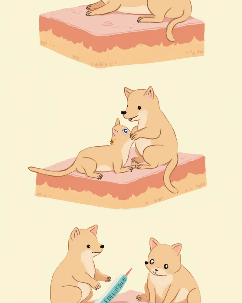
An illustration showing the three-step process of a skin mass fine-needle aspiration biopsy

Key Points to Know Before the Test

Cytology is a simple test, but it does have limitations. If too few cells are collected or if the lesion lies deep within a mass, the result may be “non-diagnostic.” In such cases, we may need to repeat the sampling or proceed to a biopsy. Also, please be sure to let your veterinarian know if your pet has a tendency to bleed easily. Mild swelling or bruising at the site is common after the procedure, but it typically resolves within one to two days.

How to Interpret the Results
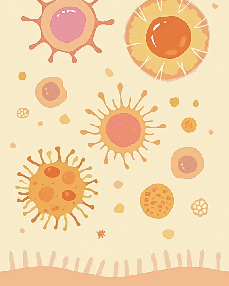
Cytology results are generally categorized into four main types.
Inflammatory: Bacterial or fungal infections, abscesses, granulomas, etc. — treated with antibiotics or antifungal medications.
Benign tumors: Lipomas, sebaceous adenomas, etc. — usually managed with monitoring or elective removal.
Suspected malignant tumors: Mast cell tumors, squamous cell carcinoma, lymphoma, etc. — require further testing and surgical removal.
Non-diagnostic: Insufficient or degenerated cells — requires re-sampling or a biopsy.
In particular, mast cell tumors are relatively easy to identify via cytology because mast cells are distinct. Therefore, it is recommended to perform cytology before surgically removing any skin mass. However, accurately determining the tumor grade (degree of malignancy) is difficult with cytology alone; a biopsy is necessary for precise grading and to determine the appropriate surgical margins.
Various cell types seen in a skin mass on cytology under the microscope

Fine-Needle Aspiration (FNA) vs. Biopsy: What’s the Difference?

ItemCytology (FNA)Biopsy
Sampling methodThin needleExcising part of the tissue with a scalpel/punch
AnesthesiaUsually not requiredLocal or general anesthesia required
Time required5–10 minutes20–40 minutes + recovery
Diagnostic accuracyVaries by type — favorable for round cell/epithelial tumors; nondiagnostic if the sample is scantMore favorable for definitive diagnosis, grading, and margin assessment
Result confirmationSame day–2 days3–7 days
InvasivenessVery lowModerate

Whether it is actually performed and the cost vary by lesion location and size. Decide in consultation with your attending veterinarian.

How to Care for Your Pet at Home After the Test

The sampling site heals quickly without any special treatment. However, please keep an eye on your pet for about 30 minutes, as persistent licking can lead to inflammation. Any bruising or small hematomas that may appear will typically resolve within one to two days.
Care Tips:
- Avoid bathing on the same day. - Consider using an Elizabethan collar to prevent licking. - Return to the clinic if you notice swelling, fever, or pus. If the results suggest malignancy, proceed with additional imaging tests (X-rays or ultrasound) or surgery as directed by the veterinary hospital.
A cat resting comfortably at home after a cytology test

When to Contact Your Veterinarian Immediately

Please contact your veterinarian immediately after a cytology test if you notice any of the following: the sampled area continues to grow or becomes firm, there is discharge of fluid or pus, your pet shows signs of severe pain or fever, or bruising does not subside and instead spreads after 24 hours. Since cats are adept at hiding pain, please also monitor for changes in their appetite and activity levels.

Reviewed by a veterinarian

Dr. Tony — Punnawat Phongkittirak

Dr. Tony — Punnawat Phongkittirak

Veterinarian

A veterinarian who majored in veterinary medicine at Khon Kaen University, Thailand, and completed the IVSA program at North Carolina State University in the United States. Drawing on clinical experience at animal hospitals, he works in the pet healthcare field and is dedicated to building a digital care environment that connects pet parents with veterinarians.

Frequently Asked Questions

How much does a cytology test cost?
Costs vary depending on the clinic, location, and number of lesions, but generally start at several tens of dollars for a single lesion, including the interpretation fee. For an exact quote, please contact the clinic directly during your visit.
Is it really okay to do it without anesthesia?
Yes, most skin masses are sampled with a very fine needle, so the discomfort is usually just a brief pinch. However, for particularly sensitive pets or when sampling delicate areas like the face or paw pads, we may use mild sedation.
The biopsy came back positive. Does it definitely need to be removed?
If the result is benign, most cases can be managed with routine monitoring. However, if the mass continues to grow or its location interferes with daily life, surgical removal may be considered. Please discuss the best course of action with your veterinarian.
Can a single test provide a definitive diagnosis?
Tumors with distinct characteristics, such as mast cell tumors, can be diagnosed through cytology alone. However, for certain tumors like soft tissue sarcomas, a biopsy is necessary to confirm the diagnosis.
Are the examination methods for dogs and cats different?
The basic principle is the same. However, since cats are more sensitive to stress and pain, extra care is taken during correction. In particular, because of the possibility of injection site sarcoma in cats, the interpretation of results is done cautiously.

Share

Related Guides

References

[1] Valenciano AC, Cowell RL. Small Animal Cytologic Diagnosis (Canine and Feline) — Chapter 1. Sample Acquisition and Preparation, 2nd Edition

[2] Jackson HA, Marsella R. BSAVA Manual of Canine and Feline Dermatology, 4th Edition — Diagnostic Techniques

[3] Karadsheh Z, Al-Haddad M. Fine-needle aspiration techniques: needle size and suction considerations, 2014

This information is based on veterinary literature and does not replace diagnosis or treatment. Please consult a veterinarian for specific health concerns.

Metapet Co., Ltd. | CEO: Park Sung-yong | Business Reg. No. 417-88-02562 | Seoul, South Korea | Customer Center

Do Dogs & Cats Need an FNA for a Skin Lump? | Meongsiljang