Like
Share
멍실장
망막박리 응급 대처와 수술

My Pet Suddenly Can’t See — Emergency Response and Surgical Timing for Retinal Detachment

Eye HealthQ&AMeongsiljang Veterinary Advisory Board

Retinal detachment is an emergency eye condition that can lead to rapid vision loss. Here’s a summary of the key points pet owners need to know, including symptoms, causes, surgery, and prognosis.

What Is Retinal Detachment?
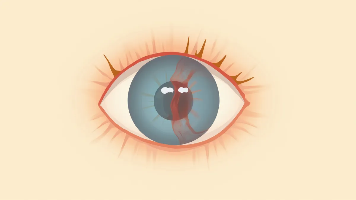
An illustration of a cross-section of a dog's eye showing the retina detached from the inner wall.
Retinal detachment is an emergency eye condition in which the retina, which should be attached to the inner wall of the eye, peels away, potentially causing sudden vision loss. Time is of the essence. According to veterinary textbooks, damage to retinal nerve cells can begin within hours of the detachment, so seeking treatment immediately upon noticing symptoms greatly improves the chances of vision recovery. If your pet suddenly starts bumping into things, has a cloudy appearance to the eye, or exhibits abnormally dilated pupils, take them to a veterinary clinic right away.

Why Does the Retina Detach? Main Causes

Retinal detachment is not caused by a single factor, but rather results from multiple overlapping conditions. In dogs, primary (bullous) retinal detachment is suspected to be immune-mediated, and veterinary ophthalmology textbooks identify German Shepherds, Australian Shepherds, and Labrador Retrievers as breeds at higher risk. Hypertension is also a common cause, and in cats, systemic hypertension is the most frequent cause of retinal detachment. It can also occur due to trauma, opacification of the anterior segment of the eye such as with cataracts, advanced uveitis, or adverse drug reactions, such as an overdose of phenylpropanolamine (PPA), a medication used for urinary incontinence.

Suspect Retinal Detachment If You See These Symptoms

Retinal detachment is often painless, so pet owners may not notice it until later. Be sure to check for the following signs: - Sudden vision loss: Bumping into walls or furniture, hesitating on stairs - Abnormal pupil size: One pupil is larger than the other, or reacts slowly to light - Bleeding inside the eye: The area around the pupil appears red - Cloudy or hazy appearance of the eye: Often accompanied by vitreous hemorrhage - White appearance inside the pupil (leukocoria): In cases of complete detachment, the eye may appear cloudy even from the outside
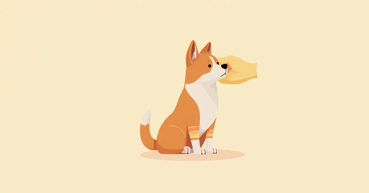
A puppy cautiously approaching furniture due to vision loss

When to Seek Immediate Veterinary Care

If any of the following signs are present, seek veterinary care within 24 hours, ideally at a clinic with a board-certified veterinary ophthalmologist. According to veterinary textbooks, damage to retinal nerve cells can begin within hours of detachment, and if left untreated for several weeks or more, widespread photoreceptor damage progresses, making recovery very difficult. - Sudden apparent vision loss in one or both eyes - Pupils that do not react to light - A red, hemorrhagic appearance inside the eye - A change in the eye’s condition immediately after trauma - Vision abnormalities in a cat diagnosed with chronic kidney disease or hypertension

How Is It Diagnosed at the Clinic?

Retinal detachment is difficult to confirm accurately with a routine eye exam alone. Typically, the following tests are performed in sequence: - Slit-lamp examination: To check for abnormalities in the cornea, lens, and anterior chamber - Fundus examination: Direct observation of the retina after pupil dilation - Ocular ultrasound: Essential when hemorrhage obscures the view of the retina - Blood pressure measurement: A mandatory test, especially for cats - Blood and urine tests: To assess for kidney failure, thyroid function, and infections Systemic hypertension is one of the most common causes of retinal detachment in cats and is emphasized as a key factor in veterinary textbooks. Blood pressure measurement is particularly important for senior cats or those with chronic kidney disease or hyperthyroidism.

Treatment Varies Depending on the Underlying Cause
The cornerstone of retinal detachment treatment is addressing the underlying cause while facilitating retinal reattachment. If hypertension is the culprit, blood pressure must be carefully managed using medications like amlodipine, strictly following your veterinarian’s prescription. In some cases, particularly in cats, the retina may spontaneously reattach depending on the chronicity and severity of the detachment. For inflammatory (steroid-responsive) retinal detachment, anti-inflammatory to immunosuppressive doses of steroids are the primary treatment. However, in cases of rhegmatogenous detachment caused by retinal tears, or severe detachments resulting from trauma or vitreous traction, microsurgical interventions such as vitrectomy, endoscopic retinopexy, or silicone oil exchange are necessary.
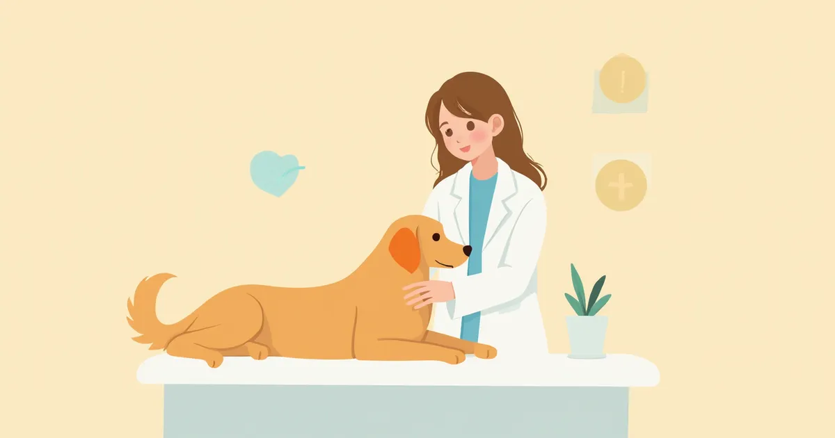
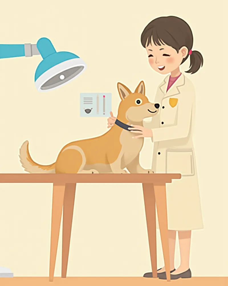
A veterinary ophthalmologist performing a fundus examination on a pet

Comparison of Treatments by Cause

ItemHypertensiveInflammatoryTraumatic·Tractional
Main causeChronic kidney disease, hyperthyroidism (cats)Uveitis, infectionCollision, after cataract surgery
First-line treatmentBlood pressure control medicationSteroids·immunosuppressantsSurgery (vitrectomy, etc.)
Need for surgeryLowModerateHigh
Likelihood of vision recoveryGood with early controlVaries depending on control of the underlying causeMay improve with early surgery

Compiled based on the veterinary ophthalmology textbook (Veterinary Ophthalmology, 5th ed.). Actual treatment is determined by the diagnosis of a veterinary ophthalmologist.

When Surgery Is Needed and Surgical Methods

Retinal surgery is complex and typically performed at university hospitals or specialized referral centers with veterinary ophthalmologists, rather than at general animal hospitals. - Vitrectomy: This procedure removes bleeding and scar tissue from inside the eye and reattaches the retina. - Silicone oil exchange and endoscopic retinopexy: These are surgical techniques used in conjunction with vitrectomy to ensure complete reattachment of the retina. - Retinal laser photocoagulation: In the early stages of partial retinal detachment, this method uses laser burns to seal torn areas and prevent further detachment. Preoperative blood work and cardiac evaluations are essential, and strict fasting before anesthesia must be observed.

Key Points for Post-Surgical Recovery Care

Surgery isn’t the end of the journey. For the retina to heal and reattach securely, your pet will need careful management for 2 to 4 weeks. - Rest: No jumping, stairs, or vigorous play for 2 weeks. - E-collar: Keep the cone on at all times to prevent irritation. - Eye drops: Administer exactly on schedule, 3 to 6 times daily. - Follow-up visits: Scheduled check-ups at 3 days, 1 week, and 1 month post-surgery. - Head position: Maintain the specific posture instructed by your vet (if gas was used during surgery). Contact the clinic immediately if you notice any redness returning or if your pet tries to scratch their eye.

Prognosis and Prevention of Recurrence

The prognosis for retinal detachment depends almost entirely on how quickly treatment is initiated. According to veterinary textbooks, retinal degeneration can begin within hours of detachment, and if left untreated for several weeks or more, widespread damage to photoreceptors will progress. The earlier treatment begins, the higher the likelihood of vision recovery; however, if the condition is left untreated for a long time or if the entire retina has detached, restoring vision may be difficult even with surgery. To prevent recurrence, it is essential to consistently manage underlying conditions (such as hypertension, kidney failure, or thyroid disease) and to have the other eye regularly examined with funduscopy. For dogs with cataracts, a thorough ophthalmic examination before surgery is mandatory.
An elderly cat resting comfortably after recovery

Reviewed by a veterinarian

Dr. Tony — Punnawat Phongkittirak

Dr. Tony — Punnawat Phongkittirak

Veterinarian

A veterinarian who majored in veterinary medicine at Khon Kaen University, Thailand, and completed the IVSA program at North Carolina State University in the United States. Drawing on clinical experience at animal hospitals, he works in the pet healthcare field and is dedicated to building a digital care environment that connects pet parents with veterinarians.

Frequently Asked Questions

Does retinal detachment cause pain?
The retina itself has very few pain-sensing nerves, so it’s usually painless. That’s why pet owners often don’t notice the problem until later. However, if uveitis or glaucoma develops alongside it, your pet may rub its eyes or squint.
Can it be treated with medication alone, without surgery?
The treatment approach depends on the underlying cause. In cases of hypertensive retinal detachment, blood pressure–lowering medications such as amlodipine may be sufficient to allow partial reattachment of the retina, particularly in cats. In contrast, detachments caused by vitreous traction or trauma typically require surgical intervention. A definitive diagnosis by a veterinary ophthalmologist is essential.
Is retinal detachment hereditary?
Certain breeds have a genetic predisposition. The veterinary ophthalmology textbook *Clinical Atlas of Canine and Feline Ophthalmic Disease* (2nd ed.) lists German Shepherds, Australian Shepherds, and Labrador Retrievers as breeds vulnerable to primary (immune-mediated) retinal detachment. We recommend genetic testing and ophthalmic examinations before breeding.
If only one eye has retinal detachment, is the other eye also at risk?
Yes, it is very dangerous. In particular, if the cause is hypertension, immune-mediated, or hereditary, the other eye is also at high risk of retinal detachment. It is essential to regularly have fundus examinations on the other eye while managing the underlying condition.
Can vision be restored through surgery even after blindness has occurred?
According to veterinary textbooks, retinal degeneration can begin within just a few hours after detachment. If the detachment persists for an extended period, leading to widespread damage to the retinal nerve cells, restoring vision becomes difficult. However, surgery to prevent complications such as pain and glaucoma may still be beneficial. Please consult with your veterinarian to make the best decision for your pet.

Share

Related Guides

References

[1] Gelatt KN, Gilger BC, Kern TJ. Veterinary Ophthalmology, 5th ed. Wiley-Blackwell, 2013 — Chapter: Diseases of the Retina

[2] Maggs DJ, Miller PE, Ofri R. Slatter's Fundamentals of Veterinary Ophthalmology, 6th ed. Elsevier, 2018

[3] Vainisi SJ, Wolfer JC. Canine retinal surgery. Vet Ophthalmol. 2004;7(5):291-306

[4] Grahn BH, Peiffer RL, Wilcock BP. Histologic Basis of Ocular Disease in Animals. Wiley-Blackwell, 2019

This information is based on veterinary literature and does not replace diagnosis or treatment. Please consult a veterinarian for specific health concerns.

Metapet Co., Ltd. | CEO: Park Sung-yong | Business Reg. No. 417-88-02562 | Seoul, South Korea | Customer Center

Sudden Blindness in Pets: Retinal Detachment Emergency Care | Meongsiljang