Like
Share
멍실장
안구 적출술 과정과 케어

What Is Canine Enucleation? — From Surgical Procedure to Recovery Care

Eye HealthQ&AMeongsiljang Veterinary Advisory Board

Enucleation is a surgical procedure performed to relieve pain and preserve quality of life in cases of difficult-to-treat eye conditions. Here’s an overview of the surgery and postoperative care.

What Is Enucleation?

Dogs and cats recovering comfortably after enucleation surgery
Enucleation is a surgical procedure in which one eye is removed to relieve pain and infection in cases of difficult-to-treat ophthalmic conditions. While pet owners are often initially concerned about the loss of an eye, the most important consideration is pain relief. An eye that has already lost vision and is left with chronic pain can significantly reduce a pet's quality of life, so when a veterinarian recommends enucleation, it is often the better choice for the animal's well-being.

When Is Enucleation Recommended?

Enucleation (eye removal) is recommended when the eye cannot be saved and the pet is experiencing severe pain. Common indications include: - End-stage glaucoma: Intraocular pressure remains uncontrolled despite medication, and vision is lost. - Severe ocular trauma: Irreversible damage such as corneal rupture or globe luxation. - Intraocular tumors: Malignant tumors, such as lymphoma or melanoma, requiring surgical removal. - Refractory uveitis: Chronic inflammation leading to loss of vision and structural integrity. - Severe ulcerative keratitis: Corneal perforation resulting in globe collapse. In these cases, enucleation is not about "giving up," but rather a form of pain management.

Comparison of Enucleation Surgical Methods

ItemTransconjunctival EnucleationTranspalpebral EnucleationEvisceration
Extent of RemovalEyeball + third eyelidEyeball + wider resection including eyelids and conjunctivaRemoval of inner eye contents only, preserving the outer coat
When Typically UsedStandard casesOcular tumors, severe infectionCosmetic purposes, prosthetic eye insertion
Surgery Time30–60 minutes60–90 minutes30–60 minutes
Recovery Period10–14 days14–21 days10–14 days
ScarringCovered by fur after suturingSunken appearanceLittle visible change

Surgery time and recovery period may vary depending on the patient's condition, and the final surgical technique is decided by the veterinarian based on the disease, breed, and eye condition.

How Is the Surgery Performed?
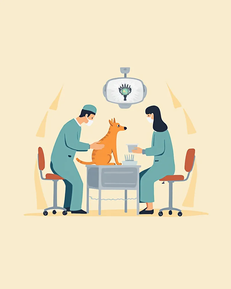
The surgery is performed under general anesthesia. Here’s the standard procedure for a routine enucleation: - Pre-anesthetic testing: Blood and cardiac evaluations to assess anesthesia risk - Induction of general anesthesia: A combination of injectable and inhalant anesthetics to maintain stability - Surgical site preparation: Hair around the eye is clipped, and antiseptic solution is applied - Conjunctival incision: The conjunctiva is incised in a 360-degree circle approximately 5 mm outside the limbus (the border of the cornea) - Handling of muscles and nerves: Extraocular muscles attached to the eyeball are detached close to the globe, and the optic nerve and blood vessels are severed with scissors - Removal of the eyeball and third eyelid: The eyeball and third eyelid (nictitating membrane) are removed together, and bleeding is controlled by applying pressure with gauze for 1–2 minutes - Suturing: Layered closure is performed from deep within the orbit, through the subcutaneous tissue, to the skin In many cases, the removed eyeball is sent for histopathological examination.
Image of the enucleation surgery process performed under general anesthesia

Essential Postoperative Precautions

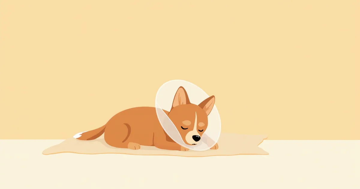
The 48 to 72 hours following surgery are a critical period with a heightened risk of bleeding, swelling, and infection. - Keep the e-collar on at all times: Leave it on 24/7 until the sutures are removed to prevent your pet from scratching the surgical site. - Monitor for bleeding: If blood continues to soak through the bandage or onto the skin, contact your veterinarian immediately. - Administer prescribed medications: Never stop antibiotics, pain relievers, or anti-inflammatory drugs without veterinary approval. - Prevent face rubbing: Ensure your pet does not rub its face against carpets or cushions. - No bathing or swimming: Keep the surgical site completely dry until the sutures are removed. Please contact us immediately if you notice any unusual discharge, severe swelling, or fever.

Care Points by Recovery Stage

Recovery typically takes 10 to 14 days, with specific care points varying by stage. - Days 0–3: Monitor the return of appetite and normal elimination, administer pain medication as prescribed, and apply cold compresses only if directed by your veterinarian. - Days 4–7: Check for reduced swelling, use warm compresses to aid recovery, gently disinfect the suture site, and keep activity to a minimum. - Days 10–14: Visit the clinic for suture removal and consult your vet about when it’s safe to remove the e-collar. - After 2 weeks: Gradually resume walks and play, and help your pet adjust to their new field of vision. Pets often adapt to having one eye much faster than owners expect. In many cases, it’s the owner who struggles with the adjustment longer than the pet.
A dog enjoying a walk after recovering from surgery

Living With One Eye: Is It Really Okay?

To put it simply, pets generally adapt quite well to living with one eye. Dogs and cats rely less on vision than humans do, as they gather a lot of information through their sense of smell and hearing. As long as you follow these guidelines, you’ll be doing great: - Keep furniture in place: Sudden changes in layout can lead to collisions. - Avoid approaching from the blind side: Coming up unexpectedly may startle them and trigger defensive aggression. - Be cautious with stairs and heights: Reduced depth perception increases the risk of falls. - Schedule regular check-ups for the remaining eye: It’s essential to monitor the healthy eye regularly. Protecting that remaining eye is the most important thing you can do.

Breed-Specific Additional Precautions

Surgical and recovery precautions vary by breed. - Brachycephalic breeds (Pugs, Bulldogs, Pekingese): Due to their prominent eyes, the unaffected eye is also at high risk of trauma, and anesthesia carries higher risks, making consultation with a specialist essential. - Small-breed dogs: Careful management of hypothermia and hypoglycemia during surgery is crucial. - Cats: Being sensitive to stress, they should recover quietly in an isolated space. - Senior animals: Anesthesia plans should be developed after evaluating heart and kidney function. Anesthesia protocols tailored to each breed’s characteristics ensure safety.

Reviewed by a veterinarian

Dr. Tony — Punnawat Phongkittirak

Dr. Tony — Punnawat Phongkittirak

Veterinarian

A veterinarian who majored in veterinary medicine at Khon Kaen University, Thailand, and completed the IVSA program at North Carolina State University in the United States. Drawing on clinical experience at animal hospitals, he works in the pet healthcare field and is dedicated to building a digital care environment that connects pet parents with veterinarians.

Frequently Asked Questions

Is it normal for my pet to seem depressed after surgery?
For the first few days after surgery, your pet may seem lethargic due to aftereffects of anesthesia and general discomfort. Keep a close eye on their appetite and energy levels as they gradually return to normal. If your pet remains unusually weak or refuses to eat even after the recovery period, please consult your veterinarian. Most pets bounce back to their usual selves once the sutures are removed.
Can a prosthetic eye be placed in the socket after enucleation?
In veterinary medicine, inserting an orbital implant (such as a PMMA spherical prosthesis) after enucleation is a documented and clinically used method to prevent postoperative orbital hollowing. Evisceration, which removes the eye’s contents while preserving the outer shape, is another option. However, whether to use an implant should be decided in close consultation with your veterinarian, taking into account the nature of the disease, presence of tumors, infection status, and other relevant factors.
How much does the surgery cost?
Costs can vary significantly depending on the clinic, location, anesthesia method, and scope of testing. It’s a good idea to get a written estimate in advance that covers all costs, including pre-anesthetic testing, surgery, hospitalization, and pathology exams.
Is it covered by insurance?
It depends on when you enroll your pet in insurance and the specific terms of the policy. Many policies include exclusions for pre-existing or congenital conditions, so it’s a good idea to request a pre-enrollment review from your chosen insurance provider.
Is it possible that both eyes will need to be removed?
In conditions like glaucoma or immune-mediated diseases, it is possible to lose vision in both eyes. Veterinary literature also documents cases where bilateral surgery is necessary for pain relief. Even with complete blindness, pets can adapt to life relying on their sense of smell and hearing, though this requires more time for environmental adjustments and training.

Share

Related Guides

References

[1] Maggs DJ, Miller PE, Ofri R. Slatter's Fundamentals of Veterinary Ophthalmology, 6th ed. Elsevier, 2018

[2] Gelatt KN, Gilger BC, Kern TJ. Veterinary Ophthalmology, 5th ed. Wiley-Blackwell, 2013

[3] Côté E. Clinical Veterinary Advisor: Dogs and Cats, 3rd ed. Elsevier Mosby, 2015

This information is based on veterinary literature and does not replace diagnosis or treatment. Please consult a veterinarian for specific health concerns.

Metapet Co., Ltd. | CEO: Park Sung-yong | Business Reg. No. 417-88-02562 | Seoul, South Korea | Customer Center

Canine Enucleation: Surgical Procedure to Recovery Care | Meongsiljang