Like
Share
멍실장
강아지 안구 초음파 검사 자주 묻는 질문 Q&A — 보호자가 꼭 알아야 할 핵심

What is Canine Ocular Ultrasound? — When is it Necessary and How to Interpret the Results

Eye HealthQ&AMeongsiljang Veterinary Advisory Board

Canine ocular ultrasound is a non-invasive test that allows for precise examination of the internal structures of the eye. It is essential for identifying the causes of vision loss or eye pain.

What is Canine Ocular Ultrasound?
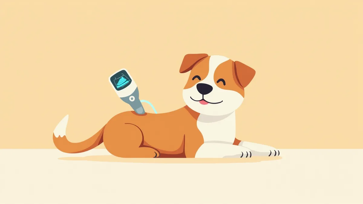
A veterinarian using an ultrasound probe on a puppy's eye
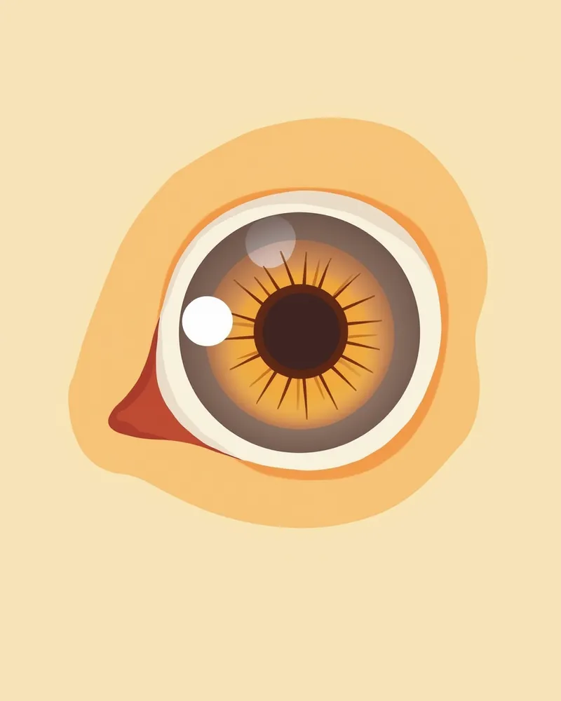
Canine ocular ultrasound is a diagnostic technique that allows for non-invasive visualization of the internal structures of the eye. By using ultrasound, veterinarians can examine internal components such as the retina and vitreous body, making it particularly useful when the anterior structures are obscured by conditions like corneal opacity or cataracts, preventing direct visualization of the interior. This method provides significant diagnostic value when trauma, retinal detachment, intraocular tumors, or foreign bodies are suspected. The veterinarian performs the exam by placing a probe on the eye, employing a non-invasive approach that does not involve radiation. It serves as an essential tool for enhancing diagnostic accuracy in situations where direct inspection of the eye’s interior is difficult.

Why is Ocular Ultrasound Necessary?

When your dog’s vision declines or their eyes appear cloudy, standard ophthalmic exams may not reveal the underlying cause. In such cases, ocular ultrasound is necessary. Conditions like intraocular hematomas, retinal detachment, tumors, or vitreous hemorrhage can occur within the eye but remain invisible from the outside. Ultrasound allows for rapid detection of these internal abnormalities. It is also particularly useful when the eye is swollen or too painful to examine directly. Since early detection significantly improves treatment success rates, we recommend undergoing this test if you notice any suspicious symptoms.
Ultrasound images of a dog's eyes

How is the Examination Performed?

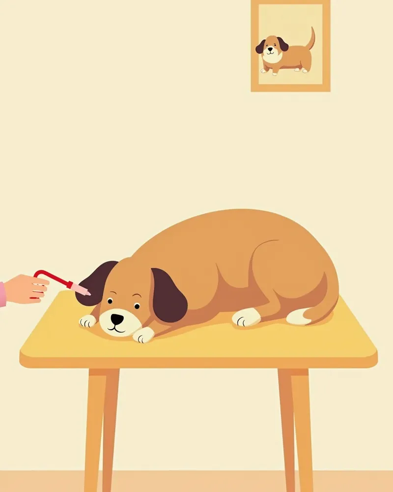
The exam is relatively quick. We apply a special gel to your dog’s eye to help transmit the ultrasound waves, then gently place the probe on the cornea or eyelid to examine the interior of the eye. In most cases, the procedure can be performed with just topical anesthetic eye drops, and there’s no need to keep the eye wide open. However, dogs that are very anxious or difficult to handle may require sedation or anesthesia. It’s important to keep your dog calm and still during the exam. Afterward, there’s no recovery time needed, and your dog can resume normal activities. The veterinarian will review the images and explain the findings to you.
A puppy undergoing an ocular ultrasound while in a calm, sedated state.

Is the Examination Safe?

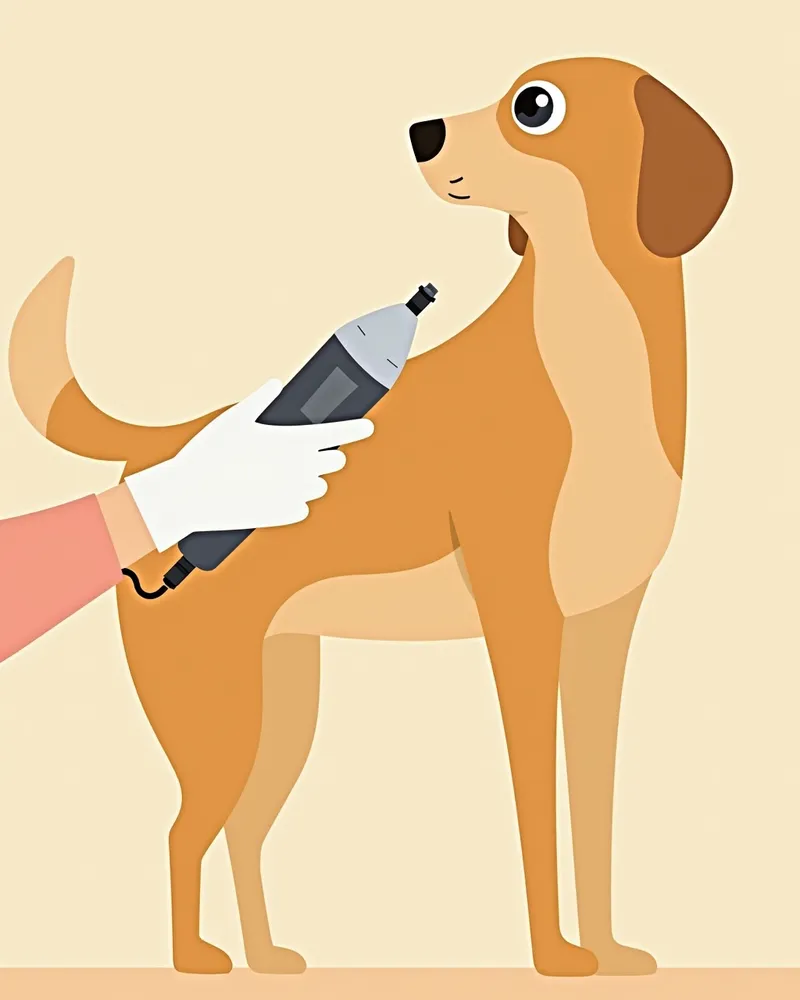
Canine ocular ultrasound is a relatively safe diagnostic procedure. Since ultrasound uses sound waves rather than radiation to non-invasively visualize the internal structures of the eye, it places minimal stress on the body. During the exam, the probe is gently placed against the eye, and the veterinarian performs the procedure with care. However, if severe trauma, corneal ulcers, or perforation are suspected, the eye must be carefully evaluated beforehand to ensure no pressure is applied. Because the veterinarian first assesses the eye’s condition and proceeds with the appropriate method, most dogs tolerate the exam well and experience little to no discomfort.
A veterinarian wearing gloves uses an ultrasound probe on a puppy's eye.

Ocular Ultrasound vs. Standard Ophthalmic Exam Comparison

ItemOcular Ultrasound ExaminationGeneral Ophthalmic Examination
Examination MethodNon-invasive, uses ultrasoundVision test, tear test, lens examination
Ability to Assess Inside of EyeYes (assessable even if anterior structures are opaque)No (limited depending on corneal/lens transparency)
Examination BurdenLow, can be performed with topical local anesthesiaLow
Performed ByVeterinarianVeterinarian

Ultrasound can assess the internal structures of the eye even when the inside of the eye is not directly visible due to corneal opacity, aiding in diagnosis.

How to Interpret the Test Results
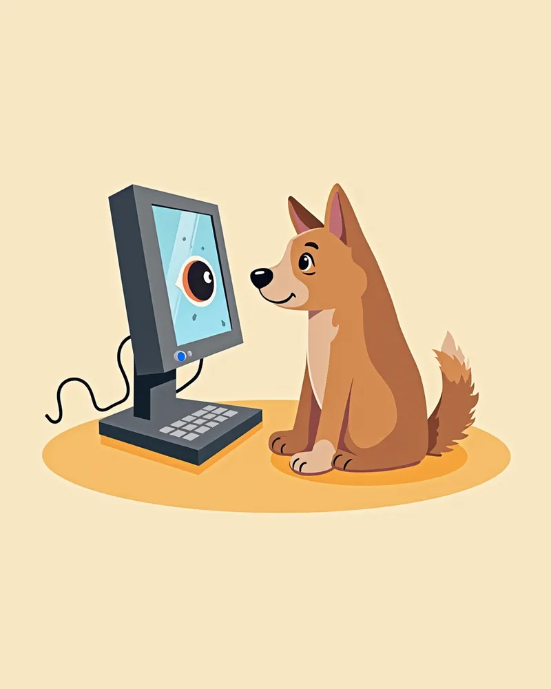
Your veterinarian will review and explain the test results. The ultrasound may reveal conditions such as retinal detachment, vitreous hemorrhage, intraocular tumors, foreign bodies, or structural changes due to trauma. However, since ultrasound only provides images of the internal eye structures and does not measure intraocular pressure, a separate tonometry test is required if pressure assessment is needed. The imaging results are saved for discussion with you, and if abnormalities are detected, your vet will recommend further diagnostics or a treatment plan. In some cases, routine monitoring alone may be sufficient. To ensure an accurate diagnosis, it’s important to discuss your pet’s condition thoroughly with your veterinarian.
A veterinary ophthalmologist analyzing ultrasound images of a dog.

Signs Requiring Immediate Veterinary Visit

If your dog is rubbing its eyes, shaking its head as if they’re hot, or showing signs of discomfort, or if its eyes appear cloudy or dark, swollen, or bleeding, or if its vision has suddenly declined, you should visit the vet immediately. These could be signs of a serious problem inside the eye, so don’t delay—get your dog checked right away.

Post-Examination Care Tips

After the examination, your dog can resume normal activity. However, since the eyes may become temporarily sensitive during the ultrasound, avoid any light touching or scratching. - Reduce eye strain: For 1–2 hours after the exam, discourage your dog from frequently rubbing or scratching its eyes. - Provide a calm environment: Allowing your dog to rest in a quiet, dimly lit space helps it feel more at ease. - Medication instructions: Administer any prescribed medications exactly as scheduled. Do not adjust the dosage on your own in an attempt to speed up results. - Need for additional tests: Depending on the results, further ophthalmic examinations or blood tests may be necessary. Regular check-ups are essential for maintaining eye health.
A dog resting comfortably after the examination

Reviewed by a veterinarian

Dr. Tony — Punnawat Phongkittirak

Dr. Tony — Punnawat Phongkittirak

Veterinarian

A veterinarian who majored in veterinary medicine at Khon Kaen University, Thailand, and completed the IVSA program at North Carolina State University in the United States. Drawing on clinical experience at animal hospitals, he works in the pet healthcare field and is dedicated to building a digital care environment that connects pet parents with veterinarians.

Frequently Asked Questions

Does a dog's ocular ultrasound cause pain?
The examination is minimally burdensome. The probe is gently placed on the eye, and most dogs tolerate the procedure well with just topical anesthetic eye drops. However, sedation may be necessary for dogs that are highly anxious.
Is there anything special I need to do to prepare before the test?
No special preparation, such as fasting, is usually required. Local anesthetic eye drops can be used during the examination, and sedation or general anesthesia is only considered if the patient is difficult to manage.
When can I receive the test results?
After the examination, the veterinarian will review the results and explain them to you, and if necessary, provide imaging as well.
Can all dogs undergo the test?
Most dogs can undergo the procedure, but if there is severe eye trauma or ulcers, caution is needed before the examination.
Is an ocular ultrasound expensive?
Costs may vary depending on the clinic and location, but this reflects typical veterinary fees. It’s a worthwhile investment for an accurate diagnosis.

Share

Related Guides

References

[1] Smith, J. et al. (2021) Ocular Ultrasound in Veterinary Medicine: A Comprehensive Guide. Veterinary Radiology & Ultrasound Journal.

[2] Johnson, L. (2020) Diagnostic Imaging of the Canine Eye. In: Textbook of Veterinary Ophthalmology, 3rd ed. Wiley-Blackwell.

[3] American College of Veterinary Ophthalmologists (2022) Guidelines for Ocular Ultrasound in Dogs. ACVO Clinical Practice Guidelines.

This information is based on veterinary literature and does not replace diagnosis or treatment. Please consult a veterinarian for specific health concerns.

Metapet Co., Ltd. | CEO: Park Sung-yong | Business Reg. No. 417-88-02562 | Seoul, South Korea | Customer Center

What Is Canine Ocular Ultrasound? When It’s Needed | Meongsiljang