
Like
Share
멍실장
강아지 비만세포종 등급별 치료법과 수술 후 관리 — 보호자가 꼭 알아야 할 단계별 가이드

Canine Mast Cell Tumor Treatment by Grade and Post-Operative Care — A Step-by-Step Guide for Pet Owners

Oncology/CancerQ&AMeongsiljang Veterinary Advisory Board

The prognosis and treatment for canine mast cell tumors vary significantly depending on the grade (Grade I–III). Here’s a comprehensive overview of the differences between grades, the diagnostic, surgical, and chemotherapy process, and key points for post-operative care.

Canine Mast Cell Tumors in a Nutshell

Examining a small lump on a puppy's chest
Mast cell tumors are the most common malignant tumors found in the skin and subcutaneous tissues of dogs. The most critical factor is the tumor's grade. Although they may appear similar as lumps, Grade I tumors can often be cured with surgical excision alone, whereas Grade III tumors typically require chemotherapy even after surgery. If you notice any small lump that persists for more than two weeks or changes in size, it is essential to have a cytology test performed.

Why Is the 'Grade' of a Mast Cell Tumor Important?

You can’t tell whether a mast cell tumor is benign or malignant just by looking at it. Even tumors of the same size and in the same location can behave very differently—varying widely in their risk of metastasis and recurrence—depending on how the cells appear under a microscope. That’s why veterinary pathologists assign a grade after a biopsy. Traditionally, the Patnaik 3-tier system (Grades I, II, III) was used, but the more simplified Kiupel 2-tier system (low-grade vs. high-grade) has now become the standard. Pet owners can check which grading system was used on their pathology report.

Comparison of Behavior and Prognosis by Grade

ItemGrade I (low)Grade II (intermediate)Grade III / high
Metastatic RiskLow — well-differentiated low grade usually follows a slow-growing benign courseDifficult to predict — assessed with additional indicators such as mitotic index, Ki-67, cKITHigh — invasive with a high metastatic rate and poor prognosis
Recommended Excision MarginWide local excision: 1-2cm laterally + deep to a clean fascial plane (mast cell tumors rarely penetrate fascia, so one layer is usually sufficient)Equally wide local excision + confirm margin adequacy by pathologyWide excision + due to high invasiveness, referral to a specialist (surgery/oncology) is recommended
Post-Surgical Additional TreatmentGenerally unnecessary if margins are clean; follow-up monitoringConsider radiation or chemotherapy if margins are insufficient or mitotic index is high (>5-7/HPF)High grade is indicated for chemotherapy (± targeted therapy)
1-Year Survival Rate (reference)About 95% (12-month survival with wide excision)Wide prognostic variability; determined by additional indicatorsPoor — high risk of metastasis/recurrence

Behavior by grade varies greatly between individuals, and prognosis differs depending on additional indicators such as mitotic index, Ki-67, and cKIT. Rather than specific survival/metastasis figures, the judgment of your attending veterinarian and pathology results takes priority.

What Is the Diagnostic Process?

If a mast cell tumor is suspected, the diagnostic process typically follows this sequence: fine-needle aspiration (FNA) → histopathological biopsy → staging. - Fine-needle aspiration (FNA): A needle is used to extract a small sample of cells from the mass for microscopic examination. This procedure can be performed without anesthesia, and the tumor type is usually diagnosed based on the characteristic metachromatic (metachromatic) purple granules of mast cells. However, since cytology alone cannot accurately determine the tumor grade, a histopathological biopsy is always required. - Histopathological biopsy: This is the key test for grading the tumor and evaluating surgical margins. Tissue samples are obtained either after surgical excision or via punch biopsy. - Staging: To check for metastasis, we perform fine-needle aspiration of regional lymph nodes, abdominal ultrasound (to assess lymph nodes, liver, and spleen), and thoracic radiographs. For tumors graded II or higher, fine-needle aspiration of regional lymph nodes is almost always essential before surgery.
A veterinary pathologist examining a mast cell tumor slide under a microscope

Do Not Delay Diagnosis If You See These Signs

If any of the following apply, it’s best to have a cytology test done within a week. - The lump repeatedly changes in size, growing larger or smaller suddenly (a sign of mast cell degranulation) - The surrounding skin becomes red and swollen when touched (Darier’s sign) - The pet constantly licks or scratches the lump, causing bleeding or ulceration - Loss of appetite, vomiting, or black, tarry stools occur (possible stomach ulcer due to excessive histamine release)

Standard Treatment by Grade — Surgery Is the First Priority
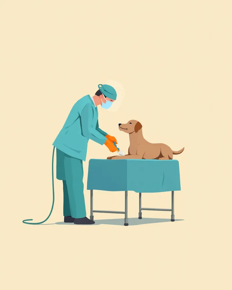
The first-line treatment for mast cell tumors is wide surgical excision. Additional therapies are determined based on the tumor grade. - Grade I / low-grade: In most cases, complete excision with adequate margins (1–2 cm plus fascia) results in a cure. If the surgical margins are clear after excision, no further treatment is needed, and regular follow-up monitoring is recommended. - Grade II: Treatment depends on the surgical outcome and pathology results. If risk factors are present—such as insufficient margins, a high mitotic index, or positive Ki-67 staining—radiation therapy or chemotherapy with vinblastine and lomustine may be added. - Grade III / high-grade: Surgery is combined with chemotherapy (vinblastine plus prednisolone) or targeted therapy (toceranib, masitinib). Testing for KIT gene mutations can help predict the response to targeted therapy.
The operating room prepared for a soft tissue tumor removal surgery in a puppy.

Post-Operative Recovery — The First Two Weeks Are Critical

Recovery after mast cell tumor removal is more delicate than after routine tumor surgeries. Surgical stimulation can cause mast cells to degranulate, leading to excessive histamine release. - Antihistamines (such as diphenhydramine) and gastroprotectants (famotidine, omeprazole) are typically prescribed for 7–14 days before and after surgery. - Wound care: Check the suture site twice daily. Contact us immediately if you notice swelling, discharge, or warmth. - Activity restriction: Until sutures are removed (usually in 10–14 days), shorten walk times and prohibit jumping or using stairs. - Preventing licking of the surgical site: Keep an Elizabethan collar or surgical suit on 24 hours a day. If the area is reached by the mouth, there is a risk of suture dehiscence and infection.

Schedule for Monitoring Recurrence and Metastasis

Regular follow-up is essential after surgery for mast cell tumors, with recommended schedules varying by grade: - Grade I: Palpate the surgical site and regional lymph nodes at 1, 3, 6, and 12 months post-surgery. - Grade II: Perform lymph node fine-needle aspiration at 1, 3, 6, 9, and 12 months post-surgery, plus abdominal ultrasound at 6 and 12 months. - Grade III: Conduct check-ups every 6–8 weeks during the first year, including regular thoracic radiographs, abdominal ultrasound, and complete blood counts (CBC). If a new mass appears, immediate cytology is required regardless of grade, as multiple occurrences are not uncommon.

Reviewed by a veterinarian

Dr. Tony — Punnawat Phongkittirak

Dr. Tony — Punnawat Phongkittirak

Veterinarian

A veterinarian who majored in veterinary medicine at Khon Kaen University, Thailand, and completed the IVSA program at North Carolina State University in the United States. Drawing on clinical experience at animal hospitals, he works in the pet healthcare field and is dedicated to building a digital care environment that connects pet parents with veterinarians.

Frequently Asked Questions by Pet Owners

Can mast cell tumors be cured with surgery alone?
Grade I tumors with clear surgical margins are often cured by surgery alone. However, for Grade II or higher tumors, or when margins are insufficient, combining radiation, chemotherapy, or targeted therapy is necessary to reduce the risk of recurrence.
Which breeds are most prone to mast cell tumors?
Breed predisposition is notably higher in Boxers, Bulldogs, Boston Terriers, Staffordshire Bull Terriers, and Labrador Retrievers. While this clear breed predisposition suggests a genetic tendency, exact incidence rates compared to other breeds are not yet definitively established in veterinary literature, making it difficult to provide precise figures. For these predisposed breeds, it is crucial to regularly palpate your dog’s entire body to detect any lumps early.
When is targeted therapy (toceranib) used?
This medication is used when the tumor cannot be surgically removed, is classified as Grade III, or when a KIT gene mutation is confirmed. It is administered as a pill every other day. Because it can cause side effects such as decreased appetite, diarrhea, and elevated liver enzymes, regular blood tests are necessary.
How should I manage my pet’s diet after surgery?
While a special prescription diet isn't always necessary after surgery, mast cell tumors can cause histamine release, leading to stomach and duodenal ulcers, vomiting, and loss of appetite. Therefore, a soft, gentle diet that minimizes gastrointestinal irritation can aid recovery. If you notice gastrointestinal symptoms such as vomiting or black, tarry stools, veterinary care and prescription gastrointestinal protectants should take priority over dietary adjustments.
Can it develop in other areas as well?
Yes, mast cell tumors can occur in multiple locations. Even after surgical removal of one tumor, new ones may develop elsewhere in some cases, so it’s helpful for pet owners to perform a full-body check on their pet every month.

Share

Related Guides

References

[1] Patnaik AK, Ehler WJ, MacEwen EG. Canine cutaneous mast cell tumor: morphologic grading and survival time in 83 dogs. Vet Pathol, 1984

[2] Kiupel M et al. Proposal of a 2-tier histologic grading system for canine cutaneous mast cell tumors to more accurately predict biological behavior. Vet Pathol, 2011

[3] Withrow SJ, Vail DM, Page RL. Withrow & MacEwen's Small Animal Clinical Oncology, 5th Edition, Elsevier

[4] Fossum TW. Small Animal Surgery, Elsevier — Preoperative and Perioperative Care

This information is based on veterinary literature and does not replace diagnosis or treatment. Please consult a veterinarian for specific health concerns.

Metapet Co., Ltd. | CEO: Park Sung-yong | Business Reg. No. 417-88-02562 | Seoul, South Korea | Customer Center

Canine Mast Cell Tumor: Treatment by Grade & Post-Op Care | Meongsiljang