
Like
Share
멍실장
강아지 각막 천공 자주 묻는 질문 Q&A — 보호자가 꼭 알아야 할 핵심

If You Suspect Your Dog’s Eye Has Perforated — Emergency Signs of Corneal Perforation and When to Seek Immediate Veterinary Care

Eye HealthQ&AMeongsiljang Veterinary Advisory Board

Canine corneal perforation is an emergency condition that can cause acute pain and vision loss. We’ve compiled the key information pet owners need to know in a Q&A format.

Corneal perforation in dogs is an emergency condition where the outermost layer of the eye is penetrated due to trauma or disease.

The eyes of a dog experiencing pain
A corneal perforation in dogs is a veterinary emergency in which the cornea is completely penetrated through all its layers, causing severe pain and vision loss. - Trauma: Puncture or scratch from a sharp object can cause immediate damage. - Infection: Viral (e.g., herpes) or bacterial infections can worsen corneal ulcers and increase the risk of perforation. - Dryness: Insufficient tear film (such as in dry eye disease) weakens the cornea and makes it more vulnerable. - Breed predisposition: Brachycephalic breeds like Shih Tzus, Pugs, Lhasa Apsos, and Boston Terriers are more prone to corneal ulcers and perforations because their central cornea is more exposed. Early detection and prompt surgical intervention are key to preserving vision. Delaying veterinary care can lead to serious complications such as iris prolapse.

The main causes of corneal perforation are trauma, infection, dryness, and genetic predisposition.

The most common cause of corneal perforation in dogs is trauma from objects piercing or scratching the eye, such as tree branches, eyelashes, toys, or foreign bodies. Infectious diseases can also worsen corneal ulcers and lead to perforation, with viral or bacterial infections posing a particularly high risk. A dry tear film or impaired tear production (keratoconjunctivitis sicca) weakens the cornea and increases the risk of perforation. Brachycephalic breeds like Shih Tzus and Pugs are more susceptible to ulcers and perforation because their central cornea is more easily exposed.
A dog’s eye with a corneal ulcer

The main symptoms of corneal perforation are excessive tearing, eye swelling, pain, and vision loss.

The most common sign of corneal perforation in dogs is excessive tearing. You may also notice swelling, redness, or cloudiness in the eye. Dogs may rub their eyes, keep them closed, or scratch and rub their heads against the floor. Severe pain can lead to loss of appetite and lethargy. If vision is impaired, your dog may bump into objects or fail to recognize familiar people. If you notice any of these symptoms, seek veterinary care immediately.
A puppy with its eyes closed

Emergency Signs Requiring Immediate Veterinary Visit

If your pet’s eyes become red and tear excessively, especially if they repeatedly squint or rub their eyes, seek veterinary care immediately. If the eyes appear cloudy or your pet behaves as though they have lost their vision, urgent medical attention is required. Corneal perforation can lead to vision loss, so do not delay—consult your veterinarian right away.

Diagnosis of corneal perforation involves ophthalmic examination and imaging.
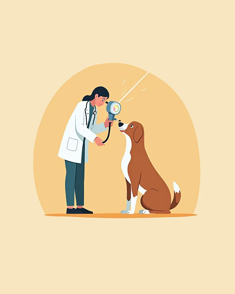
Diagnosing corneal perforation in dogs begins with a test in which the veterinarian applies a special dye (fluorescein) to the eye to identify the damaged area. This staining test allows us to determine the location and extent of the corneal defect. Because a perforated eye is structurally very fragile, it must be handled with extreme care to prevent further damage, such as iris prolapse or retinal detachment. As a general rule, avoid any forceful manipulation or attempts to remove foreign bodies before surgical repair. To minimize pain during the examination, topical anesthetic eye drops may be used. Accurate diagnosis is the foundation for developing a treatment plan, so prompt and precise testing is essential.
A veterinarian examining a dog's eyes with a slit lamp

Treatment for corneal perforation proceeds in stages with medication, surgery, and protective devices.

Treatment for corneal perforation begins with systemic antibiotics and anti-inflammatory medications to relieve pain and prevent infection. To prevent self-trauma from scratching or rubbing the eye, it is helpful to fit your dog with a protective Elizabethan collar. Please note that a third eyelid flap (nictitating membrane flap) alone is generally not recommended, as it does not provide sufficient support for the perforated area. Once a perforation is confirmed, surgical repair is necessary as soon as possible. Various techniques, such as corneal transplantation, conjunctival flaps, and suturing, are used to seal the perforation and aid recovery. Consistent medication and regular check-ups are essential after surgery. The success rate of treatment depends on early detection and appropriate management.
A dog wearing a protective cone

Home care focuses on pain relief and infection prevention.

Proper home care after a corneal perforation is crucial. It’s essential to ensure your dog takes the prescribed medications exactly as directed by your veterinarian. To prevent eye rubbing or scratching, fitting your dog with a protective cone is a must. Keep the environment clean and avoid dusty areas. If you notice your dog rubbing or scratching its eyes, stop the behavior immediately. Regular check-ups are necessary to monitor the healing process and adjust the treatment plan as needed.
A dog wearing a protective cone lying down comfortably.

Comparison of Corneal Perforation Treatments: Medication vs. Surgery

ItemWhen AppliedEffectPrecautions
Drug treatmentMild to moderate perforationPain relief, infection preventionCan promote recovery, non-invasive
Surgical treatmentSevere perforation, failure of drug treatmentDirect suturing of the damaged areaHigh possibility of preserving vision
Protective deviceUsed alongside all treatment stagesPrevents eye scratchingEnsures safety, reduces pain

The treatment method is decided by the veterinarian depending on the size, location, and cause of the perforation.

Reviewed by a veterinarian

Dr. Tony — Punnawat Phongkittirak

Dr. Tony — Punnawat Phongkittirak

Veterinarian

A veterinarian who majored in veterinary medicine at Khon Kaen University, Thailand, and completed the IVSA program at North Carolina State University in the United States. Drawing on clinical experience at animal hospitals, he works in the pet healthcare field and is dedicated to building a digital care environment that connects pet parents with veterinarians.

Frequently Asked Questions

My dog's eyes are red and watery. Could it be a corneal ulcer?
If your pet’s eyes become red and watery, there’s a high chance of corneal damage. Immediate veterinary care is essential. A veterinarian can determine whether the cornea has been perforated through a thorough examination.
Can vision return after treatment for a corneal perforation?
The potential for vision recovery depends on the timing of treatment and the severity of the perforation. Early diagnosis and appropriate treatment are crucial. In some cases, vision loss may persist, so regular check-ups are necessary.
My dog seems uncomfortable wearing an Elizabethan collar. What should I do?
At first, your dog may feel uncomfortable. Help them adjust gradually by creating positive experiences, such as offering treats and toys inside the cone. Be sure to prevent your dog from removing the cone.
Can corneal perforation recur?
There is a possibility of recurrence. Breeds with a genetic predisposition or pets with underlying conditions are at higher risk. Regular ophthalmic exams and proper care are essential.
How can corneal perforation be prevented?
To protect your pet’s eyes, provide a safe environment when going outdoors and be careful to prevent toys or objects from coming into contact with their eyes. Regular veterinary eye exams can help detect early signs of problems.

Share

Related Guides

References

[1] Fowler, L.B., et al. (2020). Ecological level analysis of primary lung tumors in dogs and cats and environmental radon activity. Journal of Veterinary Internal Medicine, 34(6), 2660–2670.

[2] American College of Veterinary Ophthalmologists. (2022). Canine Corneal Ulceration and Perforation: Diagnosis and Management Guidelines.

[3] Plumb's Veterinary Drug Handbook, 9th Edition. (2023). Wiley-Blackwell.

This information is based on veterinary literature and does not replace diagnosis or treatment. Please consult a veterinarian for specific health concerns.

Metapet Co., Ltd. | CEO: Park Sung-yong | Business Reg. No. 417-88-02562 | Seoul, South Korea | Customer Center

Is Your Dog's Eye Perforated? Corneal Perforation FAQ | Meongsiljang