Like
Share
멍실장
세포진 패턴 (염증·종양·증식) 구별법 완벽 가이드

The Ultimate Guide to Distinguishing Cytology Patterns (Inflammation, Neoplasia, and Hyperplasia)

ImmunityQ&AMeongsiljang Veterinary Advisory Board

We’ve broken down the three patterns seen in canine and feline cytology—inflammation, neoplasia, and hyperplasia—explaining their differences and what each means, all in pet-owner-friendly terms.

What Is a Cytology Pattern?

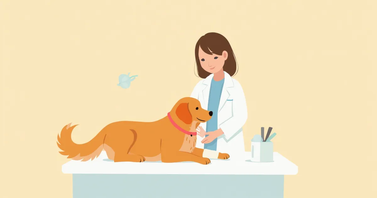
An image of a veterinary laboratory where a cytology slide is being examined under a microscope.
A cytology pattern refers to the three broad categories—inflammation, neoplasia, and hyperplasia—observed under a microscope when examining cells collected from a pet’s lumps, secretions, or body fluids. The most important first step is to identify the specific pattern name listed on the report. Even if two lumps look identical to the naked eye, their treatment plans can differ drastically depending on the underlying pattern. Understanding these basic concepts will help you grasp your veterinarian’s explanations much more quickly.

How Is Cytology Performed?

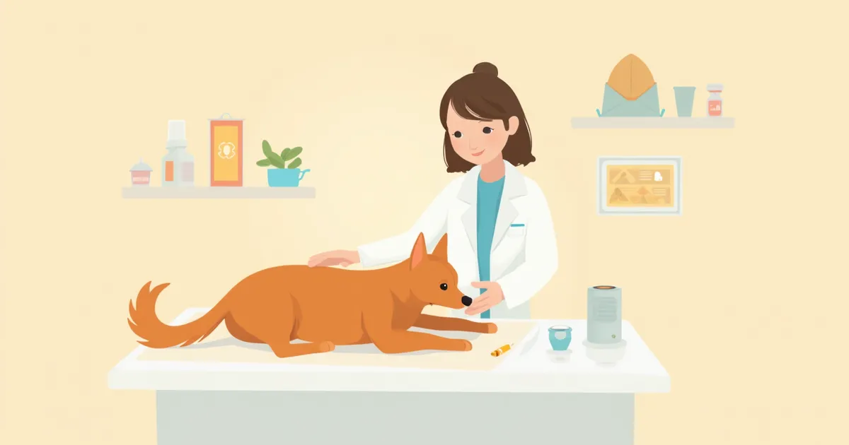
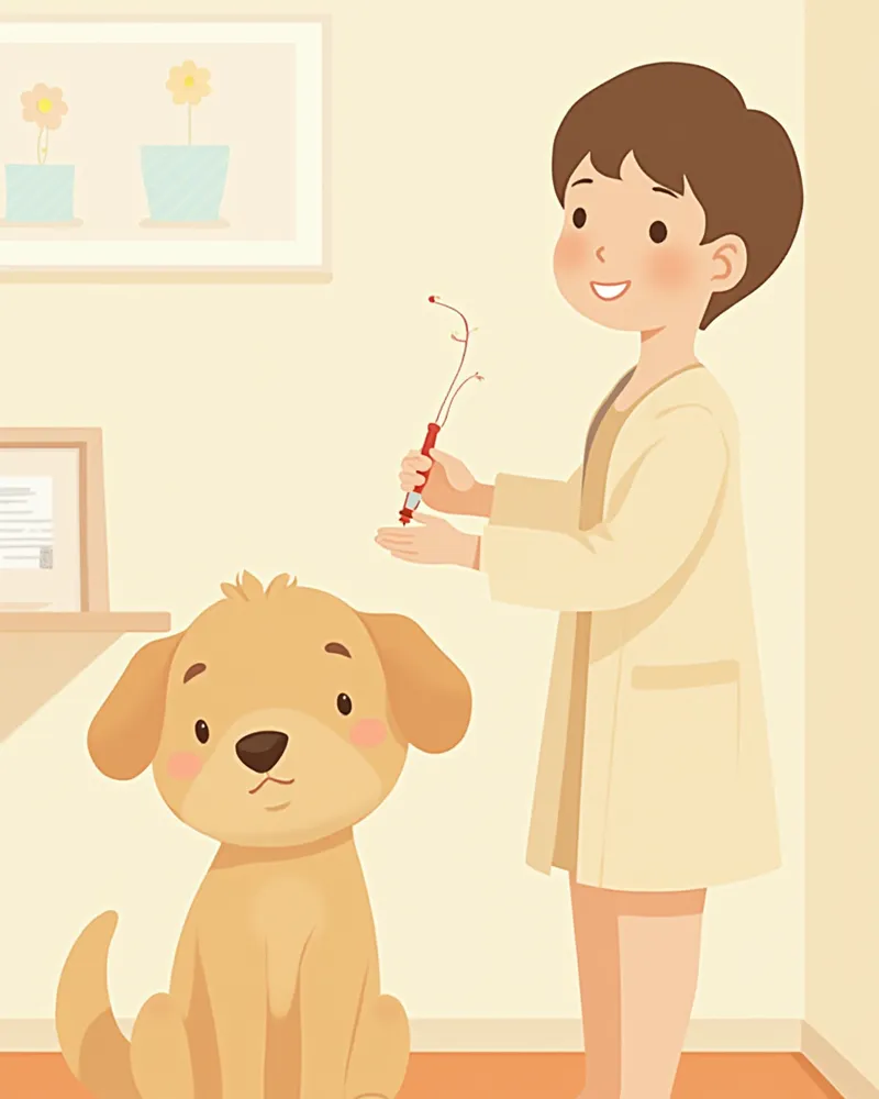
Cytology is most commonly performed using fine-needle aspiration (FNA). A thin needle is inserted into the lump or lymph node to extract cells, which are then smeared onto a slide, stained, and examined under a microscope. This procedure is minimally invasive for your pet, causing only slight bleeding, and can usually be done in the clinic without anesthesia. However, for lesions in internal organs, the procedure may be performed under ultrasound guidance.

Inflammation, Neoplasia, and Hyperplasia Patterns at a Glance

ItemInflammatory patternNeoplastic patternProliferative pattern
Predominant cellsNeutrophils/macrophages/lymphocytesNumerous abnormal cells of the same typeTissue cells close to normal
MeaningInfection/allergy/autoimmune reactionPossible benign/malignant tumorTissue proliferating due to irritation
UrgencyDepends on the causeModerate to highLow
Next stepCulture test/medical treatmentBiopsy/imaging testsMonitoring or removal

In practice, it is common for results to show a mix of two patterns.

Inflammatory Pattern: What Does It Mean?
An inflammatory pattern on cytology is characterized by a high presence of immune cells. The specific cause varies depending on which cell types are predominant and in what quantities. - Neutrophil predominance: Suggests a high likelihood of bacterial infection or abscess. - Macrophage predominance: May indicate chronic irritation or fungal infection. - Lymphocytes and plasma cells: Raises suspicion of autoimmune or allergic reactions. - Eosinophils: Suggests a possible reaction related to parasites or allergies. Not all inflammation is mild; many cases require culture tests or additional diagnostics to identify the underlying cause.
A veterinarian performing a fine-needle aspiration (FNA) on a dog.

Don’t Be Reassured by an Inflammatory Pattern

Even if the presentation looks like a routine inflammatory pattern, a bacterial culture must be performed whenever a bacterial infection is suspected. Switching antibiotics without proper guidance can lead to antibiotic resistance, and if the inflammation becomes chronic, it may ultimately require surgery. Don’t dismiss it with the assumption that “it’s just inflammation, so a topical ointment will suffice.”

Neoplastic Pattern: How to Distinguish Benign from Malignant?
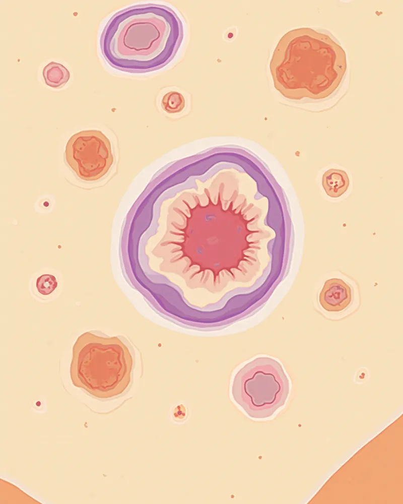
A tumor pattern refers to clusters of abnormal cells of the same type. Veterinarians make their assessment by evaluating multiple factors, including cell size, nuclear shape, nucleoli, and mitotic activity. - Benign indicators: Cells are uniform in size, nuclei are small and round, and mitotic figures are rare. - Malignant indicators: Cells vary significantly in size (pleomorphism), nuclei are enlarged and irregular (nuclear atypia and anisokaryosis), and multinucleated cells or abnormal mitotic figures are frequently observed. When three or more of these malignant indicators are present simultaneously, a diagnosis of malignancy can often be made based on cytology alone. However, if the findings are ambiguous, inflammation is present, or the tumor cells do not exfoliate well, a histopathological examination (biopsy) is required for definitive confirmation.
Microscopic image showing tumor cell patterns

Hyperplastic Pattern: Different from Neoplasia

Hyperplasia refers to a condition where the number of normal cells increases in response to stimulation, but the cells themselves remain normal and are not abnormal like tumor cells. Common examples include: - Sebaceous gland hyperplasia: Small bumps often seen in older dogs - Reactive lymph node hyperplasia: Enlargement of lymph nodes in response to nearby inflammation or infection - Gingival hyperplasia: Swollen or overgrown gums However, hyperplasia can often be difficult to confirm through cytology alone. In particular, reactive lymph node hyperplasia may resemble a mass and be mistaken for lymphoma, so a biopsy may be performed when the diagnosis is uncertain. In most cases, no immediate treatment is given and the condition is monitored, but if the size increases rapidly or the color changes, it is recommended to repeat cytology or perform a biopsy.

What Pet Owners Should Check When Receiving the Report

When you receive your cytology report, just check three key points: - Pattern name: Whether the findings indicate inflammation, a tumor, or hyperplasia. - Recommended additional tests: Whether culture, biopsy, or imaging studies are needed. - Recheck interval: When the next examination should be scheduled. If the report states that a definitive diagnosis is difficult or is labeled "non-diagnostic," it means an insufficient number of cells were collected. In such cases, consider repeating the test or proceeding with a biopsy. You may also find it helpful to read about symptoms and causes of swollen lymph nodes in dogs.

Cytology Is Not a Panacea

Cytology is a quick and minimally invasive test, but it cannot provide a definitive diagnosis for all lesions. In particular, fibrous tumors, hardened tissues, and deep-seated lesions in internal organs may yield unclear results because cells do not shed easily. In such cases, additional tests such as a biopsy (histopathology) or imaging studies like CT scans or ultrasounds are necessary.

Reviewed by a veterinarian

Dr. Tony — Punnawat Phongkittirak

Dr. Tony — Punnawat Phongkittirak

Veterinarian

A veterinarian who majored in veterinary medicine at Khon Kaen University, Thailand, and completed the IVSA program at North Carolina State University in the United States. Drawing on clinical experience at animal hospitals, he works in the pet healthcare field and is dedicated to building a digital care environment that connects pet parents with veterinarians.

Frequently Asked Questions

Will the biopsy procedure cause my pet a lot of pain?
Because we use a very fine needle, the discomfort is comparable to that of a routine vaccination. Most procedures are completed in just a few minutes in the clinic without anesthesia. However, sedation or brief general anesthesia may be required for lesions located deep within internal organs.
If the cytology results show "inflammation," does that completely rule out the possibility of a tumor?
Not necessarily. Inflammation often develops around tumors, so if the mass continues to grow or changes shape, it’s safest to perform a biopsy for further evaluation.
How long does it take to get the cytology results?
If the test is performed in-house, results are typically available within 30 minutes to an hour. If we send the sample to an external specialized pathology lab, it usually takes 3 to 7 days. More complex cases may take even longer.
Do cats also frequently undergo cytology tests?
Yes, cytology is commonly used in cats to evaluate skin masses, enlarged lymph nodes, and abdominal lesions. However, because cats are more sensitive to stress, they often require sedation more frequently than dogs.
What should I do if the cytology result is reported as “non-diagnostic”?
This happens when there aren’t enough cells collected or when the sample is heavily contaminated with blood. You can discuss with your veterinarian whether to repeat the test in one to two weeks or proceed directly to a tissue biopsy.

Share

Related Guides

References

[1] Raskin, R.E., Meyer, D.J., Small Animal Cytologic Diagnosis: Canine and Feline Disease, 2nd Edition, Chapter 1. Sample Acquisition and Preparation

[2] Hnilica, K.A., Patterson, A.P., BSAVA Manual of Canine and Feline Dermatology, 4th Edition, Chapter on Cytology

[3] Valenciano, A.C., Cowell, R.L., Cowell and Tyler's Diagnostic Cytology and Hematology of the Dog and Cat, 5th Edition

This information is based on veterinary literature and does not replace diagnosis or treatment. Please consult a veterinarian for specific health concerns.

Metapet Co., Ltd. | CEO: Park Sung-yong | Business Reg. No. 417-88-02562 | Seoul, South Korea | Customer Center

Cytology Patterns: Inflammation vs Neoplasia vs Hyperplasia | Meongsiljang