Like
Share
멍실장
강아지 백내장수술 과정과 회복 완벽 가이드

The Ultimate Guide to Cataract Surgery and Recovery in Dogs

Eye HealthQ&AMeongsiljang Veterinary Advisory Board

Cataract surgery in dogs is a specialized ophthalmic procedure that removes the clouded natural lens and replaces it with an artificial one. We’ll walk you through the surgical process and post-operative care in detail.

What Is Cataract Surgery in Dogs?
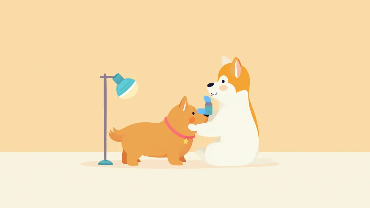
A puppy undergoing an eye examination.
Cataract surgery in dogs is a specialized ophthalmic procedure that restores vision by using ultrasound to emulsify and aspirate the clouded natural lens, followed by the implantation of an artificial intraocular lens. Timing is critical. If the cataract progresses beyond the mature stage to the hypermature stage, surgical success rates drop significantly, and the risk of complications such as glaucoma and uveitis increases. If you suspect vision problems, seek a comprehensive examination from a veterinary ophthalmologist as soon as possible.

Indications for Surgery and Causes

Cataracts occur when proteins in the lens degenerate, causing it to become cloudy and opaque. They are classified into genetic (onset at a young age), diabetic, senile, and traumatic types. Notably, dogs with diabetes tend to experience a more rapid progression of cataracts. When vision impairment arises due to lens opacity, surgery should be considered; performing the procedure at an appropriate time before the cataracts advance to the hypermature stage, which carries a high risk of complications, is crucial for a favorable prognosis. However, normal retinal function is required to expect vision recovery after surgery.

Characteristics of Cataract Progression Stages

Cataracts are classified into four stages based on their progression, which serves as a key criterion for determining the appropriate timing for surgery.

Cataract Progression Stages and Optimal Timing for Surgery

ItemIncipientImmatureMatureHypermature
Lens OpacityLess than 10%10–99%100%100% + lens liquefaction
VisionNormalPartially reducedAlmost noneComplete loss
Optimal Surgery TimingMonitor progressionOptimal timingSurgery possibleIncreased complication risk
Complication RiskLowLowModerateHigh (uveitis, glaucoma)

Based on the veterinary ophthalmology textbook standard (E7), the immature to early mature stage is the optimal time for surgery.

Essential Preoperative Tests
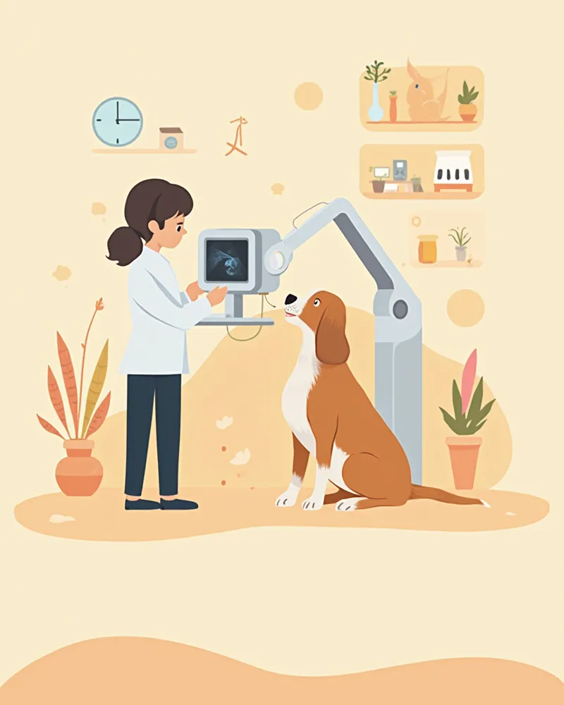
Cataract surgery requires general anesthesia and precise surgical technique, making preoperative testing essential.
Electroretinography (ERG): Assesses whether the retina is still functional—this is the most critical test.
Ocular Ultrasound: Evaluates structures behind the lens and checks for retinal detachment.
Intraocular Pressure Measurement: Screens for concurrent glaucoma.
Blood Work and Electrocardiogram (ECG): Evaluates safety for general anesthesia.
Comprehensive Diabetes Testing: If the cataract is diabetes-related, this confirms blood sugar control status.
The decision to proceed with surgery depends on these test results, so it’s important to complete them thoroughly.
A scene of a detailed ophthalmic examination for a puppy

Contraindications for Surgery

If retinal detachment has already progressed or if the retina shows no response on an electroretinogram, it is difficult to expect vision recovery even with surgery. Additionally, surgery may be challenging due to the high risks associated with anesthesia and the procedure itself in cases of uncontrolled diabetes, severe heart disease, or active uveitis. Your veterinarian will make a comprehensive assessment.

Step-by-Step Explanation of the Surgical Procedure

The actual surgery is a precise ophthalmic procedure performed under general anesthesia using an operating microscope. Whether both eyes are operated on simultaneously depends on the dog’s overall health and is determined by the veterinarian. Step 1: General Anesthesia – Anesthesia is carefully induced using an anesthetic agent tailored to the dog’s weight and health status, ensuring a stable anesthetic state. Step 2: Corneal Incision – A tiny incision is made in the cornea to create a surgical access pathway. Step 3: Anterior Capsulotomy – The front portion of the lens capsule is opened with a circular incision. Step 4: Phacoemulsification – The clouded lens is broken down into small fragments using ultrasound and then suctioned out (Phacoemulsification). Step 5: Intraocular Lens Implantation – A foldable artificial lens is inserted into the lens capsule and securely positioned. Step 6: Suturing – The incision site is closed with fine sutures to complete the procedure.

Postoperative Recovery Period and Care
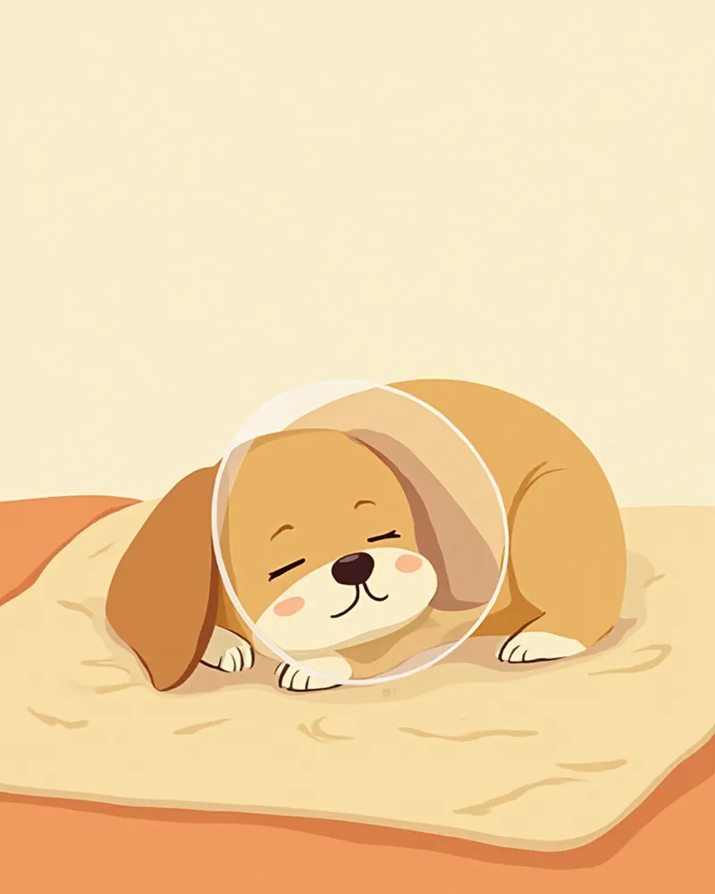
Intensive care is required from immediately after surgery for a certain period, and the intensity of care is adjusted according to the stage of recovery.
Day of surgery to 1 week: Eye drops are administered frequently as prescribed by the veterinarian, whether the pet is hospitalized or returns home the same day.
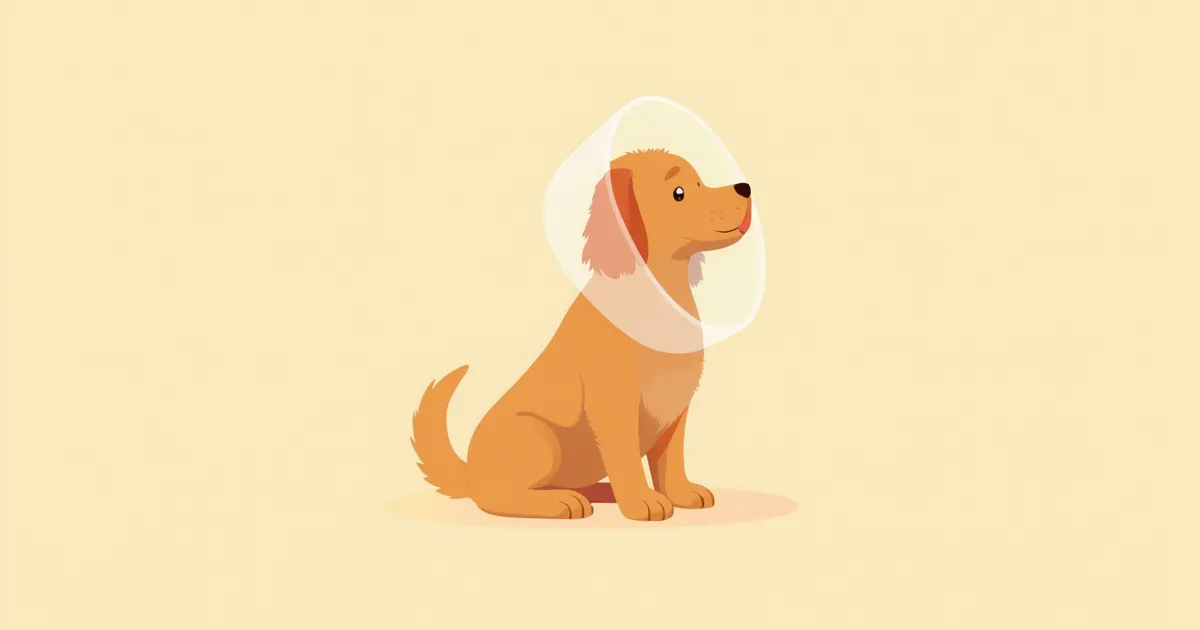
1 to 2 weeks: An e-collar must be worn to protect the eyes, strenuous activity should be avoided, and eye drop administration continues.
2 to 4 weeks: The frequency of eye drops is gradually reduced while monitoring progress, and follow-up visits are made at scheduled intervals for check-ups.
Subsequent monitoring period: Eye drops are continued, and complications such as inflammation, glaucoma, and retinal detachment are consistently monitored.
If the eye drop schedule is missed, inflammation may recur or the artificial lens may not remain securely in place, so it is essential to strictly adhere to the eye drop schedule set by the veterinarian.
A puppy recovering after surgery while wearing a neck collar.

Signs Requiring Immediate Veterinary Attention After Surgery

If you notice any of the following symptoms, it could be an emergency, so please take your pet to the vet immediately: severe eye rubbing or scratching with the paws, bloody or purulent discharge from the eyes, a cloudy and swollen cornea, inability to open the eyes accompanied by signs of pain, or loss of appetite and lethargy lasting more than 48 hours. Glaucoma and retinal detachment are the most serious postoperative complications, so prompt action is crucial.

Surgical Success Rates and Prognosis

According to veterinary ophthalmology research, cataract surgery in dogs (phacoemulsification and intraocular lens implantation) generally yields good visual recovery when performed at the appropriate time. However, long-term maintenance of successful vision may decline over time due to the development of secondary complications such as glaucoma and retinal detachment. Preoperative examinations and owner compliance significantly influence the prognosis. The following conditions help improve success rates:
Timing of surgery: During the immature to early mature stages of cataract development
Normal retina: Confirmation of retinal function through preoperative testing
Diabetes control: Stable management of blood glucose levels
Owner compliance: Strict adherence to eye drop administration and follow-up visit schedules
If you are also concerned about your dog’s joint health, please refer to our Dog Joint Supplement Guide.

Reviewed by a veterinarian

Dr. Tony — Punnawat Phongkittirak

Dr. Tony — Punnawat Phongkittirak

Veterinarian

A veterinarian who majored in veterinary medicine at Khon Kaen University, Thailand, and completed the IVSA program at North Carolina State University in the United States. Drawing on clinical experience at animal hospitals, he works in the pet healthcare field and is dedicated to building a digital care environment that connects pet parents with veterinarians.

Frequently Asked Questions

How much does cataract surgery for dogs cost?
Costs vary widely depending on the clinic and the scope of testing, but in general, the total expense—including surgery, diagnostics, and hospitalization for one eye—can run into thousands of dollars. Please consult with your veterinarian for an accurate estimate.
Can both eyes be operated on at the same time?
Yes, surgery on both eyes at the same time is generally recommended. This approach minimizes the number of anesthesia sessions, reducing stress on your dog, and helps prevent visual imbalance. However, the final decision will be made by your veterinarian based on your dog’s overall health.
Should I continue applying eye drops after surgery?
The first two to three months require intensive eye drop administration, and long-term use is often recommended afterward to prevent inflammation and glaucoma. Always consult your veterinarian before stopping eye drops.
Can diabetic dogs undergo surgery?
If blood sugar levels are well controlled, surgery is possible. In fact, diabetic cataracts can progress rapidly, and once they reach the hypermature stage, the risk of complications increases significantly, so early surgery is recommended.
Can senior dogs undergo surgery?
Overall health matters more than age. If heart and kidney function are good and retinal function is intact, many dogs over 10 years old can undergo surgery successfully. Preoperative testing is essential.

Share

Related Guides

References

[1] Gelatt KN, Gilger BC, Kern TJ, Veterinary Ophthalmology, 5th Edition, Wiley-Blackwell, 2013

[2] Maggs DJ, Miller PE, Ofri R, Slatter's Fundamentals of Veterinary Ophthalmology, 6th Edition, Elsevier, 2018

[3] Fossum TW, Small Animal Surgery, 5th Edition, Elsevier, 2019

This information is based on veterinary literature and does not replace diagnosis or treatment. Please consult a veterinarian for specific health concerns.

Metapet Co., Ltd. | CEO: Park Sung-yong | Business Reg. No. 417-88-02562 | Seoul, South Korea | Customer Center

Cataract Surgery in Dogs: The Ultimate Guide to Recovery | Meongsiljang