Like
Share
멍실장
고양이 종양 생검·세침흡인검사 자주 묻는 질문 Q&A — 보호자가 꼭 알아야 할 핵심

Cat Tumor Biopsy and Fine-Needle Aspiration FAQ — Key Information for Pet Owners

Oncology/CancerQ&AMeongsiljang Veterinary Advisory Board

We’ve compiled essential information for pet owners about biopsies and fine-needle aspiration tests used to diagnose tumors in cats. These procedures are the first step toward accurate diagnosis and effective treatment.

What Are Cat Tumor Biopsy and Fine-Needle Aspiration?
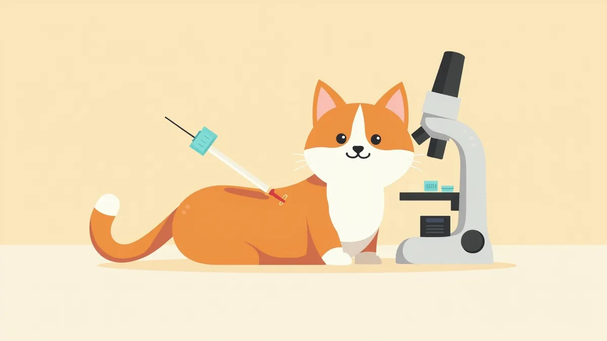
A veterinarian performing a fine-needle aspiration biopsy on a tumor located on a cat’s neck.
To accurately determine the nature of a cat’s tumor, we rely on two key diagnostic methods: biopsy and fine-needle aspiration. A biopsy involves surgically removing part or all of the tumor to collect tissue samples, while fine-needle aspiration uses a thin needle to extract cells from within the tumor. - Biopsy: This method analyzes the entire tissue structure, offering high diagnostic accuracy. However, it requires surgery and a recovery period. - Fine-needle aspiration: This procedure is less painful and provides faster results, but it may be difficult to obtain a definitive diagnosis for certain types of tumors. Both methods involve microscopic examination to determine whether the tumor is benign or malignant and to identify its specific type. Without an accurate diagnosis, we cannot develop an effective treatment plan. Therefore, we ask that you understand the importance of these tests and cooperate actively in your pet’s care.

Why Are Biopsy or Fine-Needle Aspiration Necessary?

You can’t tell whether a cat’s tumor is benign or malignant just by looking at it. Tumors that look similar can be either benign or malignant, and this distinction significantly affects treatment options and prognosis. Biopsy or fine-needle aspiration: These procedures allow for histological analysis to determine the true nature of the tumor, which is essential for developing a treatment plan and predicting outcomes. Ignoring this step can allow malignant tumors to spread rapidly and delay treatment. An accurate diagnosis directly impacts the chances of early intervention and recovery, making testing a crucial first step.
A veterinarian examining feline tumor cells under a microscope

What Is the Difference Between Biopsy and Fine-Needle Aspiration?
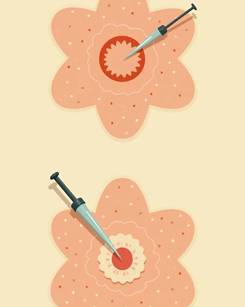
- Biopsy: This involves surgically removing the entire tumor or a portion of it to obtain tissue samples. It offers high accuracy, allowing for a clear determination of whether the tumor is malignant and its specific type. However, it requires surgery and a longer recovery period. - Fine-needle aspiration (FNA): This test uses a thin needle to extract cells from inside the tumor. It causes minimal discomfort and provides immediate results. However, for some tumors, cell samples alone may not be sufficient for an accurate diagnosis. Your veterinarian will select the most appropriate test based on the tumor’s size, location, and characteristics. Both methods are essential procedures for achieving an accurate diagnosis.
Comparison diagram of fine-needle aspiration and biopsy: cell collection with a needle vs. tissue removal through surgery

What Is the Examination Process Like?

Whether anesthesia is required depends on the type of diagnostic procedure. Fine-needle aspiration is generally well-tolerated by most cats while conscious, so it is often performed without anesthesia. In contrast, biopsies involve removing a tissue sample, so they are routinely performed under anesthesia. After disinfecting the site with an antiseptic, a needle or instrument is inserted to collect cells or tissue. Fine-needle aspiration is quick, typically taking only one to two minutes, and most cats adapt rapidly. The collected cells are examined under a microscope, allowing for relatively rapid diagnostic information, whereas histopathological analysis of tissue samples may take several days to yield results. Mild bleeding or swelling may occur after the procedure, but most cats recover naturally. Please follow your veterinarian’s instructions for post-procedure care.
After a fine-needle aspiration, a cotton swab is gently pressed against the cat’s neck to stop the bleeding.

Signs Requiring Immediate Veterinary Visit

If the tumor grows rapidly, or if you notice bleeding, ulceration, pain, loss of appetite, or weight loss, seek veterinary care immediately. These could be signs of a malignant tumor, making early diagnosis and treatment crucial.

How Are Test Results Interpreted?
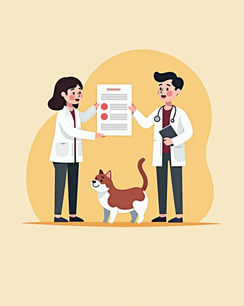
Test results are categorized into benign tumors, malignant tumors, inflammation, and congenital abnormalities. - Benign tumors: These are generally easier to treat and have a low likelihood of recurrence. - Malignant tumors: Because they can spread rapidly, early treatment is crucial. Pathologists make a diagnosis by analyzing cell morphology, arrangement, and division rate. In some cases, additional tests or imaging may be required. The veterinarian will explain the results in detail to the pet owner and discuss the treatment plan together.
A veterinarian explaining the pathology test results to a cat owner.

Comparison Table: Biopsy vs. Fine-Needle Aspiration

ItemFine-Needle AspirationBiopsy
Level of PainVery lowModerate
Time Required1–2 minutes10–30 minutes
Need for AnesthesiaNot neededGenerally needed
AccuracyModerateHigh
Recovery TimeImmediateSeveral days

The veterinarian selects the appropriate test based on the tumor's location, size, and nature.

Post-Examination Care Points

- Monitor the examination site: Check daily for any bleeding, swelling, or pus. - Limit activity: Reduce physical activity and allow for rest for about a day after the examination. - Watch for signs of infection: Contact us immediately if you notice an odor or significant swelling. - Regular check-ups: Continue to periodically inspect the examination site even after diagnosis. - Nutritional support: Ensure your pet receives adequate nutrition during treatment. - Consult your veterinarian: Feel free to reach out anytime if you notice unusual symptoms or have any questions.
The cat resting comfortably after a fine-needle aspiration test.

Reviewed by a veterinarian

Dr. Tony — Punnawat Phongkittirak

Dr. Tony — Punnawat Phongkittirak

Veterinarian

A veterinarian who majored in veterinary medicine at Khon Kaen University, Thailand, and completed the IVSA program at North Carolina State University in the United States. Drawing on clinical experience at animal hospitals, he works in the pet healthcare field and is dedicated to building a digital care environment that connects pet parents with veterinarians.

Frequently Asked Questions

Is a biopsy painful for cats?
Since a biopsy is performed under anesthesia, your pet will feel little to no pain during the procedure. There may be some mild discomfort afterward, but your veterinarian will prescribe appropriate medication to keep your pet comfortable.
Are the results of the fine-needle aspiration test accurate?
In most cases, an accurate diagnosis is possible. However, some tumors can be difficult to distinguish based on cells alone. If necessary, additional tests will be conducted.
What should I do if my cat becomes lethargic or sleepy after the examination?
It’s normal for your pet to feel drowsy or less active for a short time after anesthesia. They should recover within 1–2 hours. If the symptoms persist, please contact your veterinarian.
Should I still have a biopsy done if the tumor is small?
Yes, regardless of the tumor's size, an accurate diagnosis is essential. The smaller it is, the earlier it can be detected, which increases the chances of successful treatment.
Can the tumor grow back after the procedure?
In most cases, diagnostic procedures do not cause tumors to recur or spread. However, for certain types of tumors, such as splenic tumors, there have been reports of cancer cells spreading along the needle track. Therefore, veterinarians carefully select the safest diagnostic method based on the tumor type and location. After the procedure, regular monitoring is recommended.

Share

Related Guides

References

[1] Ryseff J.K., Bohn A.A. (2012) Detection of alkaline phosphatase in canine cells previously stained with Wright-Giemsa and its utility in differentiating osteosarcoma from other mesenchymal tumors. Vet Clin Pathol 41(3):391–395.

[2] Cotter, S.M. (1981) Oral pharyngeal neoplasms in the cat. J Am Anim Hosp Assoc 17:917–920.

[3] Hostetter S.J. (2023) Oral cavity, gastrointestinal tract, and associated structures. In: Canine and Feline Cytology: A Color Atlas and Interpretation Guide, 2nd edn. Saunders Elsevier, St. Louis, pp. 287–296.

This information is based on veterinary literature and does not replace diagnosis or treatment. Please consult a veterinarian for specific health concerns.

Metapet Co., Ltd. | CEO: Park Sung-yong | Business Reg. No. 417-88-02562 | Seoul, South Korea | Customer Center

Cat Tumor Biopsy & Fine-Needle Aspiration: Owner FAQ | Meongsiljang