

Feline Tumor Fine-Needle Aspiration (FNA) — Procedure, Cost, and Accuracy Guide
We’ve put together a clear, easy-to-understand guide for pet owners on the fine-needle aspiration (FNA) test—the first step when a tumor is found in a cat. Learn about the procedure, cost range, accuracy, and limitations, as well as what to expect before and after the test, how results are interpreted, and when additional biopsies may be needed.
What Is Feline Tumor Fine-Needle Aspiration (FNA)? One-Line Summary
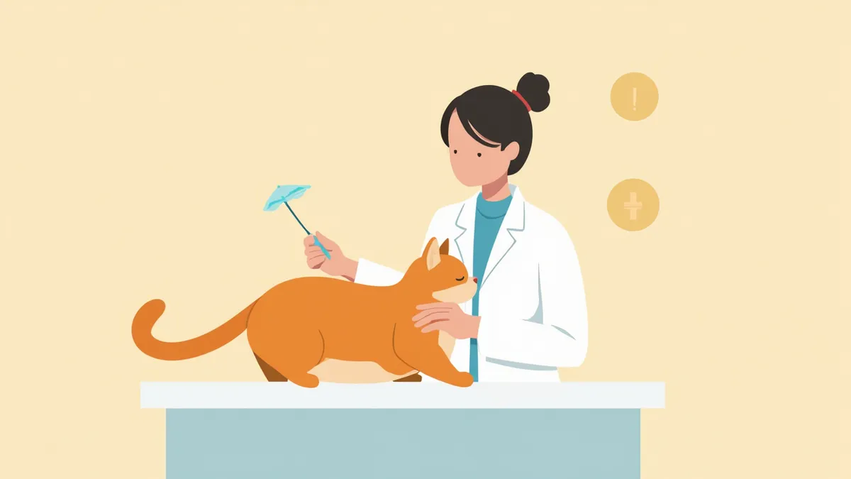
When Is Fine-Needle Aspiration Recommended?
Fine-Needle Aspiration vs. Core Biopsy vs. Excisional Biopsy — At a Glance
| Item | Fine-needle aspiration (FNA) | Core biopsy | Excisional biopsy |
|---|---|---|---|
| Sample amount | Cell-level (small amount) | Portion of tissue (core) | Entire mass |
| Anesthesia | Usually no sedation | Sedation or local anesthesia | General anesthesia required |
| Time required | 5–15 minutes | 15–30 minutes | 30 minutes to several hours |
| Diagnostic accuracy | About 80–89% depending on reports | Generally higher than fine-needle aspiration | Highest (evaluation of entire mass) |
| Cost burden | Low | Medium | Highest |
| Main use | Primary screening | Definitive diagnosis of tumors other than lymphoma | Both treatment and definitive diagnosis |
Accuracy varies depending on tumor type, location, and degree of inflammation. Some reports indicate the diagnostic yield of fine-needle aspiration for liver/spleen tumors is as low as 14–33% (Wang et al., 2004).
Procedure — From Sampling to Interpretation
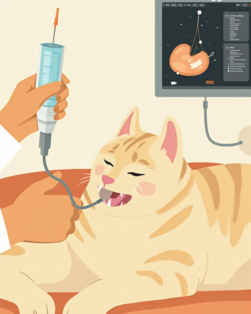
Key Pre-Procedure Checks
Fine-needle aspiration is a relatively safe procedure, but cats with bleeding tendencies or low platelet counts are at risk of post-procedural bleeding or hematoma. To ensure safety, basic blood tests and coagulation times (BMBT or PT/aPTT) should be checked before the procedure. Additionally, because intrathoracic masses carry a risk of pneumothorax and splenic masses may cause intra-abdominal hemorrhage, cats should be monitored for 30 to 60 minutes after the procedure. For pregnant cats or those in a state of extreme agitation, it is safer to perform the procedure after sedation.
Feline Tumor Testing Costs — Ranges by Site and Guidance

Accuracy and Limitations — When Additional Testing Is Needed
Notes on Interpreting Results
Even if a fine-needle aspiration report reads "negative" or "inflammatory changes," it cannot rule out a tumor with 100% certainty. This is because normal tissue may be present within the mass, or the sampled area may not represent the center of the tumor. Therefore, if clinical suspicion for a tumor remains high despite ambiguous results, a follow-up test should be performed in 2–4 weeks, or a core needle biopsy or excisional biopsy should be conducted. Please note that the phrase "consistent with" on the report does not indicate a definitive diagnosis, but rather suggests a high likelihood.
Reviewed by a veterinarian

Dr. Tony — Punnawat Phongkittirak
VeterinarianA veterinarian who majored in veterinary medicine at Khon Kaen University, Thailand, and completed the IVSA program at North Carolina State University in the United States. Drawing on clinical experience at animal hospitals, he works in the pet healthcare field and is dedicated to building a digital care environment that connects pet parents with veterinarians.
Frequently Asked Questions
Doesn't my cat hurt a lot during the examination?
What should I do if the test result comes back as “nonspecific”?
Does a needle aspiration cause the tumor to grow faster or spread?
Does a negative result mean I can be completely reassured?
How long does it take to get the results?
Share
Related Guides

7 Factors Determining Prognosis and Life Expectancy in Dogs with Tumors, According to Veterinarians

7 Key Points of Canine Tumor Immunotherapy Selected by Veterinarians — From Principles to Selection Criteria

7 Criteria Veterinarians Recommend for Choosing Canine Tumor Biopsy and Fine-Needle Aspiration

Cat Suddenly Biting and Scratching — Causes and Correction of Play Aggression

My Cat Keeps Licking Themselves — Is Excessive Grooming Normal?

Cats Keep Fighting — Causes and Intervention Timing for Multi-Cat Household Conflicts

What is feline cognitive dysfunction? — Early signs of feline dementia

Why Is My Cat Suddenly Aggressive? — Causes and Management of Fear-Based Aggression

My Cat Keeps Meowing — Is Excessive Meowing Normal?

If Your Cat Has Changed After the Move — Is Environmental Change Stress Normal?
References
[1] Raskin RE, Meyer DJ. Canine and Feline Cytology: A Color Atlas and Interpretation Guide, 3rd Edition. Elsevier, 2016
[2] Valenciano AC, Cowell RL. Small Animal Cytologic Diagnosis: Canine and Feline Disease, 2nd Edition
[3] Kudnig ST, Séguin B. Veterinary Surgical Oncology, 2nd Edition. Wiley-Blackwell
[4] Wang KY et al., Accuracy of ultrasound-guided fine-needle aspiration of the liver and cytologic findings in dogs and cats: 97 cases (1990-2000). JAVMA, 2004
[5] Jasik A et al., BSAVA Manual of Canine and Feline Dermatology, 4th Edition (Chapter 34: Neoplastic and paraneoplastic syndromes affecting the skin)