
Like
Share
멍실장
강아지 심낭삼출·심낭압전 증상과 원인, 진단·치료와 관리 포인트 총정리

If Your Dog Collapses Suddenly — Emergency Signs and Response for Pericardial Effusion and Cardiac Tamponade

HeartDiseasesMeongsiljang Veterinary Advisory Board

Canine pericardial effusion and cardiac tamponade are acute conditions in which fluid accumulates around the heart, impairing its function. Early diagnosis and treatment are critical for survival.

Pericardial Effusion and Cardiac Tamponade in Dogs Are Acute Conditions That Threaten Heart Function
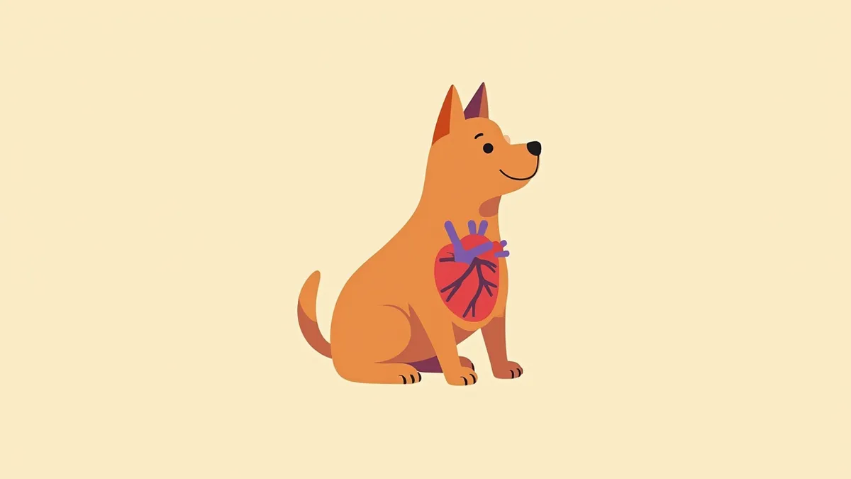
An anatomical diagram of a dog’s heart showing fluid accumulation around the heart
Pericardial effusion and cardiac tamponade in dogs are acute conditions in which abnormal fluid accumulates in the pericardium surrounding the heart, compressing the organ. As fluid builds up, the heart cannot contract properly, impairing blood circulation and potentially leading to acute heart failure. Because onset is often sudden and life-threatening, immediate diagnosis and treatment are essential. Pericardial effusion refers to the abnormal accumulation of fluid around the heart, causing severe symptoms such as weakened heartbeats and low blood pressure. Cardiac tamponade is a medical emergency in which excessive fluid accumulation severely compresses the heart, potentially causing it to stop beating. Early detection and rapid intervention are key to improving survival rates.

The Main Causes of Pericardial Effusion Include Tumors, Infection, Trauma, and Heart Disease
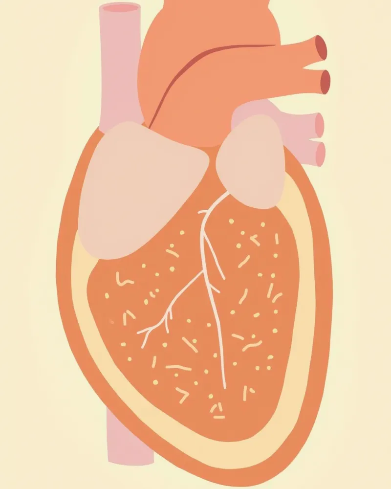
The causes of pericardial effusion are varied. In dogs, the most common causes are idiopathic (of unknown origin) and neoplastic. Among tumors, hemangiosarcoma is the most frequent cause of pericardial effusion, while cardiac base tumors (chemodectomas) and mesotheliomas can also be responsible. These tumors may lodge in the pericardium or cause bleeding, leading to continuous fluid accumulation. Other, less common causes include bleeding due to coagulopathies or anticoagulant rodenticide toxicity, trauma, and left atrial rupture in dogs with severe mitral valve regurgitation. Inflammatory conditions such as infectious pericarditis can also result in fluid accumulation. Accurately identifying the underlying cause is essential for determining the treatment approach and prognosis. - Neoplastic and idiopathic causes: Tumors such as hemangiosarcoma and idiopathic (unknown cause) factors are the most common causes of pericardial effusion in dogs. - Other causes: Coagulopathies, anticoagulant toxicity, trauma, left atrial rupture, and infectious pericarditis can all lead to fluid accumulation.
Comparison diagram of a normal pericardium and pericardial effusion

Key Symptoms of Cardiac Tamponade Include Difficulty Breathing, Shock, and Decreased Heart Rate

Cardiac tamponade is a condition in which fluid accumulation compresses the heart, preventing it from filling properly and reducing cardiac output. Key symptoms include difficulty breathing, lethargy, exercise intolerance, shock, a weak and rapid pulse, pale gums, abdominal distension (ascites) due to systemic venous congestion, jugular vein distension, and collapse. A characteristic sign may be pulsus paradoxus, where the femoral arterial pulse weakens during inspiration. If your pet suddenly develops rapid breathing or altered consciousness, seek immediate veterinary care. These are signs of a rapid decline in heart function, and prompt intervention is critical for survival. Early detection and treatment significantly improve the chances of recovery. - Difficulty breathing and shock: Reduced cardiac output and low blood pressure can lead to labored breathing, pale gums, and decreased consciousness. - Signs of venous congestion: Abdominal distension (ascites), jugular vein distension, and a weak femoral pulse may be observed.
A dog experiencing difficulty breathing due to pericardial compression

Cardiac Tamponade Is a Life-Threatening Emergency

This is an acute condition in which the heart is compressed, potentially causing the heartbeat to stop. If your pet shows signs of difficulty breathing, shock, lethargy, or collapse, they must be transported to a veterinary clinic immediately. Because delays can be fatal, prompt action by pet owners is crucial.

Diagnosis Is Accurately Confirmed via Echocardiogram and Thoracic Radiographs
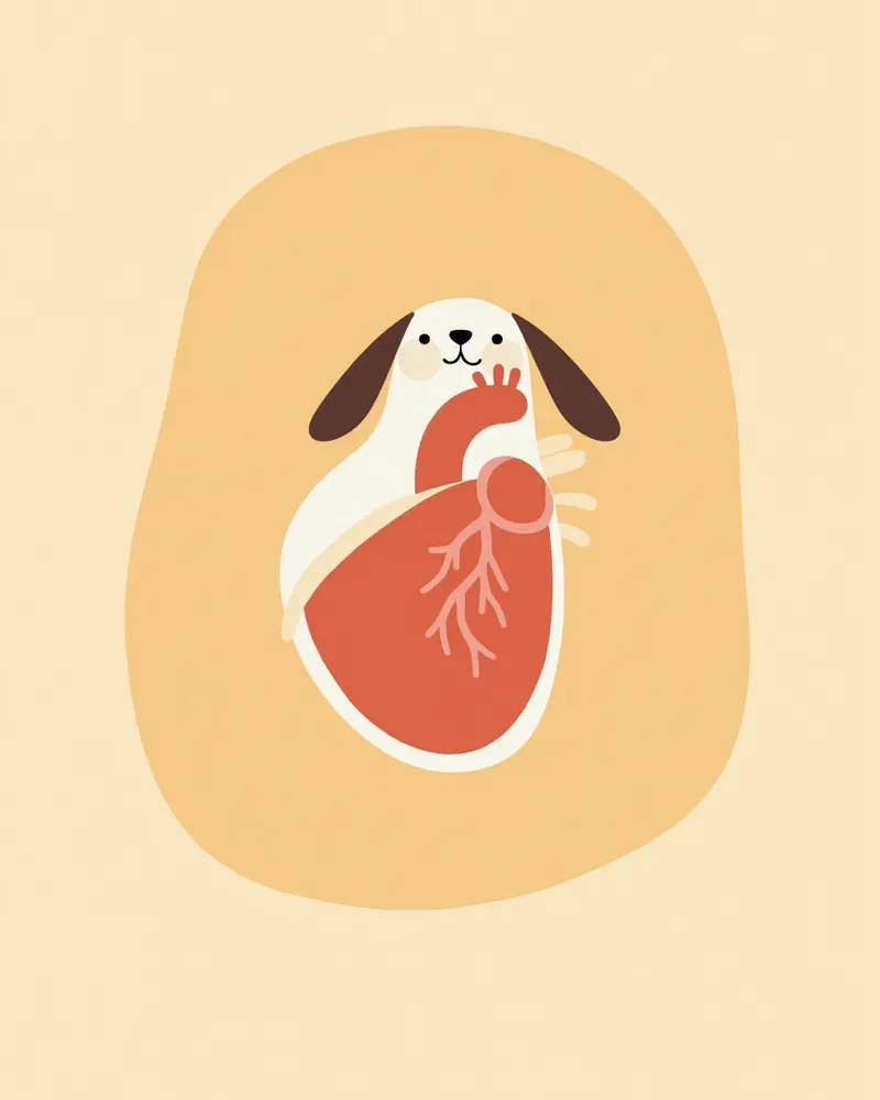
Pericardial effusion is most accurately diagnosed with an echocardiogram (cardiac ultrasound). This test allows real-time visualization of fluid accumulation around the heart and compression or collapse of the right atrium, which is a characteristic sign of cardiac tamponade. While thoracic radiographs may show an enlarged, globoid cardiac silhouette, acute and rapid fluid accumulation can sometimes result in a normal-appearing heart size, so caution is warranted. Blood tests and cytological analysis of pericardial fluid are used to identify underlying causes, helping to determine whether tumors or infections are present. In some cases, pulsus paradoxus—a characteristic weakening of the femoral pulse during inspiration—may also be observed. - Echocardiography: The key diagnostic tool for accurately assessing fluid accumulation and the degree of right atrial compression. - Thoracic Radiographs: Helpful for evaluating changes in the cardiac silhouette, though the heart may appear normal in size with acute effusion.
An image showing pericardial effusion detected on an echocardiogram

Treatment Is Divided Into Pericardiocentesis and Treatment of the Underlying Cause

The most urgent priority in treating cardiac tamponade is the immediate removal of fluid. Pericardiocentesis, which involves draining the fluid surrounding the heart with a needle, is the fastest method. This procedure is performed precisely by a veterinarian while monitoring real-time ultrasound. After fluid removal, treatments such as chemotherapy, antibiotics, or immunosuppressants are prescribed depending on the underlying cause. If a tumor is the cause, surgery or radiation therapy may also be considered. Ongoing management is necessary after treatment to prevent recurrence. - Pericardiocentesis: A critical treatment that restores heart function by draining fluid in emergency situations. - Treating the underlying cause: Addressing the root cause, such as tumors or infections, is key to long-term recovery.
A veterinarian performing pericardiocentesis while viewing an echocardiogram.

Home Care Focuses on Maintaining Heart Function and Preventing Recurrence

After treatment, ongoing care to maintain heart function and prevent recurrence (reaccumulation of fluid) is essential. Following pericardiocentesis, complications such as reaccumulation of pericardial fluid, bleeding, or ventricular arrhythmias may occur, so close monitoring under your veterinarian’s guidance is crucial. Unlike other forms of fluid retention, diuretics and vasodilators are contraindicated in pericardial effusion; therefore, medications must never be administered without a veterinary prescription. Avoid excessive exercise, regulate activity levels, and monitor for recurrence and overall cardiac status through regular cardiac ultrasound examinations. Neoplastic and idiopathic effusions carry a high risk of recurrence, and surgical intervention may be necessary to avoid repeated pericardiocentesis, making consistent long-term management vital. - Recurrence monitoring: Follow-up observations are essential to promptly detect reaccumulation of fluid, bleeding, or arrhythmias after pericardiocentesis. - Regular check-ups: Routine cardiac ultrasound examinations are recommended to identify recurrence and assess heart condition at an early stage.
A dog resting for heart health

Certain Breeds Are at Higher Risk for Pericardial Effusion

Pericardial effusion is the most common pericardial disease in dogs, and it is usually caused by a tumor or is idiopathic (of unknown origin). Among tumors, hemangiosarcoma is the most frequent cause of pericardial effusion, while tumors at the base of the heart and mesotheliomas can also be responsible. Because it is difficult to definitively link the condition to specific breeds or ages based solely on textbook evidence, regular cardiac check-ups are key to early detection and improving treatment success rates.

Reviewed by a veterinarian

Dr. Tony — Punnawat Phongkittirak

Dr. Tony — Punnawat Phongkittirak

Veterinarian

A veterinarian who majored in veterinary medicine at Khon Kaen University, Thailand, and completed the IVSA program at North Carolina State University in the United States. Drawing on clinical experience at animal hospitals, he works in the pet healthcare field and is dedicated to building a digital care environment that connects pet parents with veterinarians.

Frequently Asked Questions

Can cardiac tamponade be completely cured?
It depends on the underlying cause. If the issue is caused by a tumor—particularly hemangiosarcoma—a complete cure is unlikely, but symptoms can be managed through procedures such as aspiration or surgery. In cases resulting from infection or trauma, the likelihood of recovery after treatment is generally higher.
Is pericardiocentesis dangerous?
When performed under precise ultrasound guidance, the risk is low. However, because complications such as heart wall injury, bleeding, or ventricular arrhythmias can occur, the procedure should be carried out by a specialist veterinarian, and your pet should be closely monitored afterward.
Can pericardial effusion recur?
Yes, whether the effusion is due to a tumor or is idiopathic, fluid tends to reaccumulate after aspiration in most cases. Regular cardiac ultrasound screenings and close monitoring are essential, and surgical intervention may be considered if the condition recurs repeatedly.
What are the early symptoms of pericardial compression?
Signs include difficulty breathing, lethargy, shock, a weak and rapid pulse, pale gums, abdominal distension, and collapse. If these symptoms appear suddenly, immediate veterinary care is essential.
Can senior dogs also develop pericardial tamponade?
Yes, it can occur. While neoplastic pericardial effusion is more commonly reported in older dogs, there’s no strict age cutoff; what matters most is early detection through regular cardiac check-ups and prompt veterinary care if symptoms like difficulty breathing or shock appear.

Share

Related Guides

References

[1] Clinical Medicine of the Dog and Cat, 4th Ed. 2020, Elsevier

[2] Textbook of Cardiovascular Medicine in Dogs and Cats, 2018, Wiley-Blackwell

[3] Small Animal Critical Care Medicine, 3rd Ed. 2021, Elsevier

This information is based on veterinary literature and does not replace diagnosis or treatment. Please consult a veterinarian for specific health concerns.

Metapet Co., Ltd. | CEO: Park Sung-yong | Business Reg. No. 417-88-02562 | Seoul, South Korea | Customer Center

Dog Collapses? Pericardial Effusion & Tamponade Emergency | Meongsiljang