Like
Share
멍실장
강아지 안구 초음파 검사 증상과 원인, 진단·치료와 관리 포인트 총정리

Canine Ocular Ultrasound Examination — When Is It Needed and What Can It Detect?

Eye HealthDiseasesMeongsiljang Veterinary Advisory Board

Canine ocular ultrasound is a diagnostic method that allows for non-invasive visualization of the internal structures of the eye. It is essential for the early detection of ocular injuries, diseases, and tumors.

What Is a Canine Ocular Ultrasound Examination?
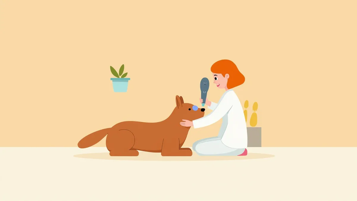
A veterinarian using an ultrasound probe on a puppy's eye.
Canine ocular ultrasound is a key diagnostic technique for non-invasively examining the internal structures of the eye. - Ocular Ultrasound Examination: This method uses ultrasound to observe the internal condition of the eye in real time through its transparent tissues. It is particularly useful when there is corneal clouding or vision impairment following trauma. - Non-Invasive Procedure: The exam can be performed without anesthesia, minimizing stress for your dog, and typically takes about 5 to 10 minutes. - Real-Time Imaging: Results appear immediately on the screen during the procedure, allowing for precise analysis of any abnormalities. - Foundation for Accurate Diagnosis: This test is essential for the early detection of various eye conditions, including hemorrhage, tumors, and retinal detachment. Immediate availability of results helps facilitate timely treatment planning.

Main Reasons for an Ocular Ultrasound Examination
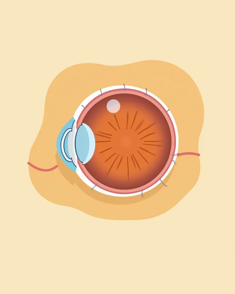
The main reason your dog may need an ocular ultrasound is to detect abnormalities inside the eye at an early stage. - Abnormalities after trauma: If bleeding, vitreous degeneration, or opacification is suspected following eye injury, an ultrasound is necessary to examine the internal structures. - Suspected retinal detachment: If your dog shows vision loss or repeatedly exhibits dilated pupils with a sluggish response to light, it is important to check for retinal detachment. Ultrasound can be helpful when direct visualization via fundus examination is difficult. - Suspected intraocular tumors: If a new growth inside the eye is suspected, an ultrasound is recommended to evaluate the presence of a tumor. - Corneal opacity preventing fundus examination: When the cornea is cloudy and prevents direct visualization of the interior of the eye, ultrasound helps assess the internal structures. - Evaluation of foreign bodies or trauma: If a foreign body or trauma inside the eye is suspected, it is essential to determine the internal condition of the eye. Early diagnosis through ultrasound can significantly aid in preserving vision.
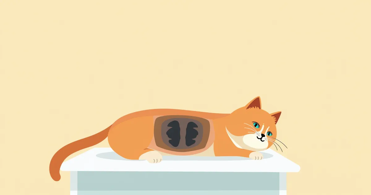
The anatomical structure of a dog’s eye and the propagation of ultrasound waves

Main Symptoms and Signs

The main symptoms that indicate your dog may need an ocular ultrasound include frequent blinking or eye rubbing, persistent tearing, collisions due to vision loss, swelling of the eye or prominent blood vessels in the sclera, corneal clouding, and dilated pupils with a diminished light reflex. In particular, if abnormal symptoms appear shortly after trauma, immediate examination is necessary.
Increased tear production: Persistent tearing, which can signal inflammation or trauma.
Vision loss: Repeated behaviors such as bumping into walls or difficulty recognizing objects.
Corneal opacity: The eye appears cloudy or loses its transparency.
Ocular swelling: Swelling around the eye or a sensation that the eye looks enlarged.
Pupil abnormalities: Dilated pupils or a sluggish response to light.
A puppy rubbing its eyes and producing excessive tears.

Criteria for Immediate Veterinary Visit

If your dog suddenly squints, experiences excessive tearing, suffers a rapid decline in vision, or shows signs of eye swelling and pain, immediate veterinary attention is necessary. These symptoms may indicate intraocular hemorrhage, retinal detachment, or infection. Because delays can lead to permanent vision loss, prompt diagnosis is essential.

Diagnostic Procedure and Method
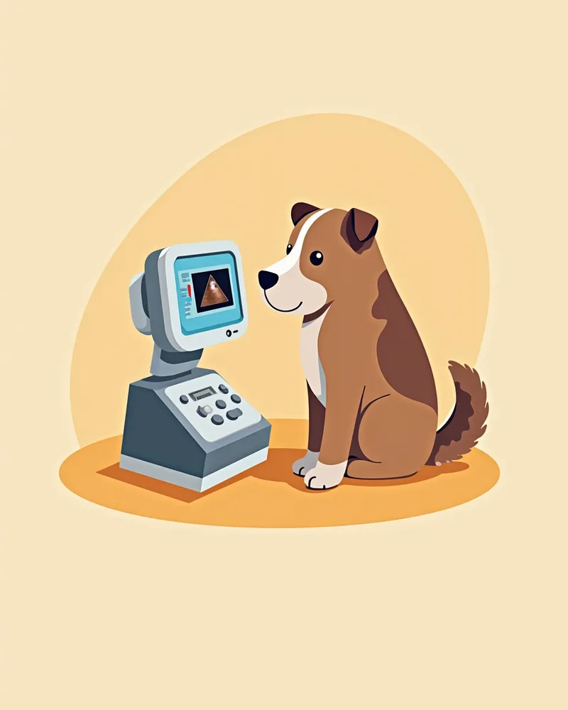
Ocular ultrasound in dogs is a safe procedure that can be performed without anesthesia. - Preparation: The veterinarian applies a gel to the eye and gently presses the ultrasound probe against it. - Real-time imaging: Internal eye structures are displayed on the screen in real time during the exam, allowing any abnormalities to be magnified and analyzed. - Duration: The procedure typically takes 5–10 minutes. If the dog becomes anxious, some gentle behavioral guidance may be needed. - Accurate diagnosis: This test enables precise detection of various conditions, including vitreous opacity, hemorrhage, tumors, and retinal detachment. - Immediate results: Results are available right away at the clinic, allowing for quick decisions on treatment direction. The findings serve as essential baseline information for planning any additional tests or therapies.
A veterinarian using ocular ultrasound equipment, with the resulting image displayed on the screen.

Treatment Methods and Step-by-Step Management

The treatment plan is determined based on the results of the ocular ultrasound. If inflammation is confirmed, we prescribe local or systemic anti-inflammatory medications (steroids or non-steroidal), and antibiotics may be used concurrently if necessary. If a tumor is suspected, a biopsy (histopathological examination) is performed for definitive diagnosis, and additional treatments such as surgical excision are considered depending on the type of tumor. After trauma, an Elizabethan collar (protective collar) can be used to prevent the dog from scratching or irritating its eye. During the treatment period, it is important to minimize irritation and support recovery in a quiet environment.
Anti-inflammatory treatment: Steroid or non-steroidal medications are used to reduce eye inflammation.
Biopsy and surgical treatment: If a tumor is suspected, a biopsy confirms the diagnosis, and excision is performed if necessary.
Use of a protective collar: An Elizabethan collar is used after trauma to prevent the dog from touching its eye.
Medication management: Administer prescribed medications at the exact scheduled times.
Regular check-ups: Continue periodic examinations even after recovery.
A dog wearing a protective mask

Home Care and Daily Care

Proper home care after an ocular ultrasound is essential to improving treatment success. - Medication adherence: Administer prescribed medications exactly as directed and on schedule. - Prevent eye contact: Keep your pet from scratching or touching their eyes. - Environmental management: Provide a quiet, well-lit space for comfortable rest. - Nutritional support: Offer a balanced diet and fresh, clean water consistently. - Follow-up appointment: Schedule a recheck within one week after the examination. - Close monitoring: Regularly observe your pet’s eyes for any changes, and contact your veterinarian immediately if abnormalities arise.
An indoor environment where your dog can rest comfortably

Prevention of Recurrence and Breed Precautions

Certain breeds are more prone to eye conditions. For example, Shih Tzus, Bichon Frises, Italian Greyhounds, Yorkshire Terriers, Jack Russell Terriers, and Labrador Retrievers are known to have a higher risk of internal eye disorders such as vitreous degeneration and retinal detachment. Additionally, because recurrent eye problems can lead to vision loss, regular check-ups and preventive care are essential. Early detection significantly improves treatment outcomes.

Reviewed by a veterinarian

Dr. Tony — Punnawat Phongkittirak

Dr. Tony — Punnawat Phongkittirak

Veterinarian

A veterinarian who majored in veterinary medicine at Khon Kaen University, Thailand, and completed the IVSA program at North Carolina State University in the United States. Drawing on clinical experience at animal hospitals, he works in the pet healthcare field and is dedicated to building a digital care environment that connects pet parents with veterinarians.

Frequently Asked Questions

Is an ocular ultrasound painful?
It won’t hurt. The probe will only lightly touch the eye. Some dogs may feel a bit tense, but the veterinarian will calmly guide them through the process.
Are there any special precautions I should take after the test?
Administer the medication exactly as prescribed, and prevent your pet from touching or scratching their eyes. Additionally, keep the environment quiet and minimize any potential irritants.
Does a normal test result mean everything is okay?
Even if the test results are normal, persistent symptoms warrant further investigation to rule out other underlying causes. We recommend consulting with your veterinarian for a thorough evaluation.
Which breeds are more susceptible to eye diseases?
Breeds such as Shih Tzus, Bichon Frises, Italian Greyhounds, Yorkshire Terriers, Jack Russell Terriers, and Labrador Retrievers are at a higher risk for vitreous degeneration and retinal detachment due to their genetic predispositions. They require careful monitoring and management.
What should I do if my pet’s vision worsens after the examination?
Please return to the clinic immediately for a recheck. Vision loss can be a sign of internal eye abnormalities, so prompt action is crucial.

Share

Related Guides

References

[1] Textbook of Cardiovascular Medicine in Dogs and Cats, 2021

[2] Small Animal Critical Care Medicine, 3rd Ed, 2020

[3] Feline Emergency and Critical Care Medicine, 2nd Ed, 2019

This information is based on veterinary literature and does not replace diagnosis or treatment. Please consult a veterinarian for specific health concerns.

Metapet Co., Ltd. | CEO: Park Sung-yong | Business Reg. No. 417-88-02562 | Seoul, South Korea | Customer Center

Canine Ocular Ultrasound: When It’s Needed and Why | Meongsiljang