Like
Share
멍실장
강아지 안구 돌출·프로프토시스 증상과 원인, 진단·치료와 관리 포인트 총정리

If Your Dog’s Eyeball Suddenly Protrudes — Emergency First Aid for Globe Prolapse and the Golden Time

Eye HealthDiseasesMeongsiljang Veterinary Advisory Board

Canine globe prolapse is an emergency condition in which the eyeball protrudes from the socket, and it occurs especially frequently in brachycephalic (flat-faced) breeds. Immediate veterinary care is essential.

What Is Canine Globe Prolapse (Proptosis)?
A medical illustration showing a dog’s eye protruding from the eye socket
Canine proptosis, or eye bulging, is an acute emergency condition in which the eye partially or completely protrudes from its normal position within the orbit (eye socket). If left untreated, it can lead to vision loss or permanent eye damage. - Proptosis: The eye protrudes beyond the eyelids and out of the orbit, most commonly due to trauma. It is also frequently caused by severely swollen tissues around the eye that prevent the eyeball from returning to its socket, trapping it in place. - High-risk breeds: This condition occurs most often in brachycephalic (flat-faced) breeds such as Shih Tzus, Pugs, and Lhasa Apsos. - Emergency nature: Because proptosis can be accompanied by other life-threatening injuries from trauma, stabilizing the patient’s overall condition is the first priority. - Immediate action required: If your dog’s eye is protruding, seek veterinary care immediately. The sooner professional treatment is administered, the higher the chance of saving the eye and preserving vision. This is not merely a discomfort—it is a true emergency.

What Are the Main Causes?

The primary cause of proptosis in dogs is trauma. - Trauma: Blunt force to the eye, such as from being hit by a car or fighting with another dog, can cause the eyeball to protrude. - Structural vulnerability: Brachycephalic breeds with flat faces have shallow eye sockets, making their eyeballs more prone to slipping out. - Periocular tissue swelling: After proptosis occurs, severe swelling of the tissues around the eye often prevents the eyeball from returning to its normal position. - Breed predisposition: This condition is most commonly seen in brachycephalic breeds such as Shih Tzus, Pugs, and Lhasa Apsos. - Prevention is key: Managing the environment to ensure safety during walks and play helps reduce the risk of trauma, which directly aids in preventing further injury.
A medical illustration highlighting the shallow eye socket structure characteristic of flat-faced breeds.

What Are the Main Symptoms and Signs?
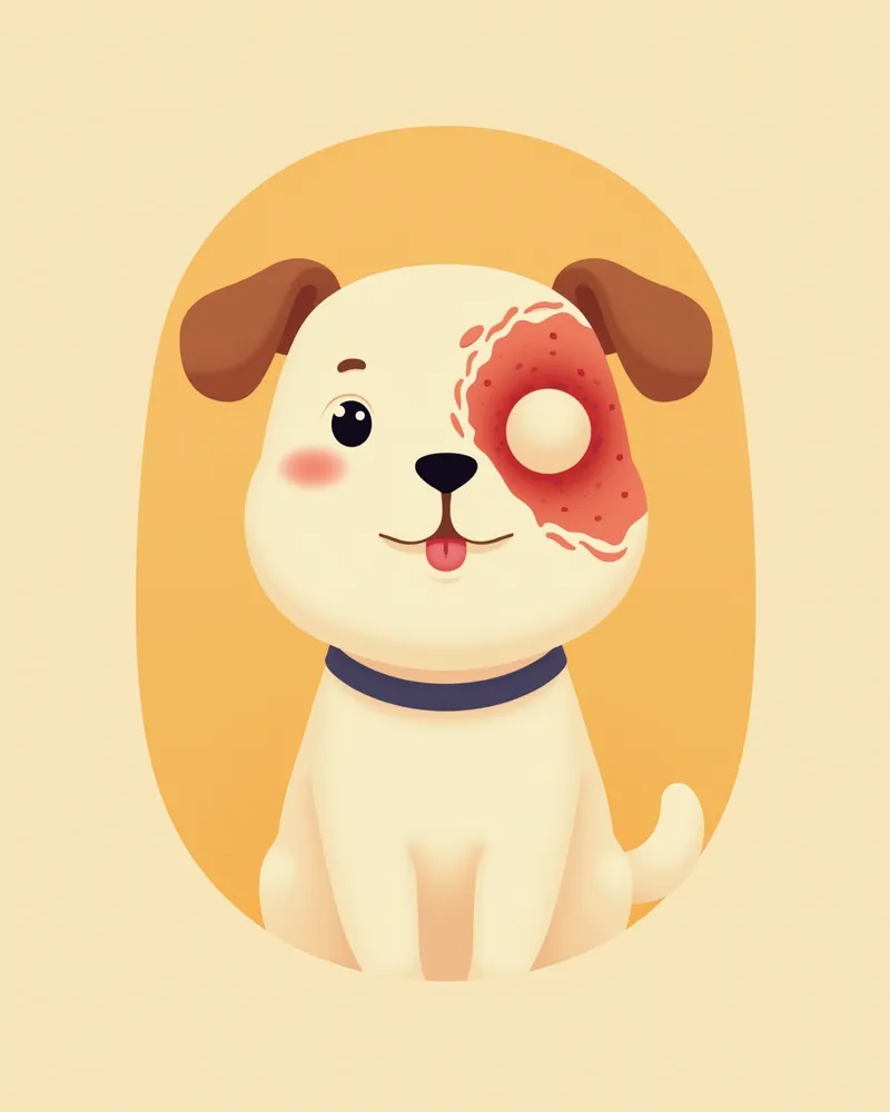
The most obvious sign of suspected proptosis in dogs is when the eyeball protrudes from the socket and bulges in front of the eyelids. - Change in eye position: The eye appears displaced outward from its normal position. - Bleeding and congestion: Subconjunctival hemorrhage, bleeding around the sclera, or anterior chamber hemorrhage (bleeding inside the eye) may occur. - Corneal exposure and damage: Inadequate eyelid closure can lead to corneal drying and exposure keratopathy. - Swelling: Severe swelling of the tissues surrounding the eye can prevent the eyeball from returning to the socket, causing it to become trapped. - Pupil response check: A constricted pupil or a remaining light reflex (pupillary light reflex) is a positive prognostic sign. If you notice these symptoms, immediate veterinary care is required.
A photo of a dog with completely protruding eyeballs, appearing red and swollen.

Emergency Situation Requiring Immediate Veterinary Visit

When the eyeball protrudes from the socket, the sooner professional treatment is received, the higher the chances of saving the eye and preserving vision. Delaying treatment can worsen the prognosis, so it’s essential to reach the clinic immediately. During transport, cover the eye with a clean, damp cloth to prevent drying and further damage.

How Is It Diagnosed?
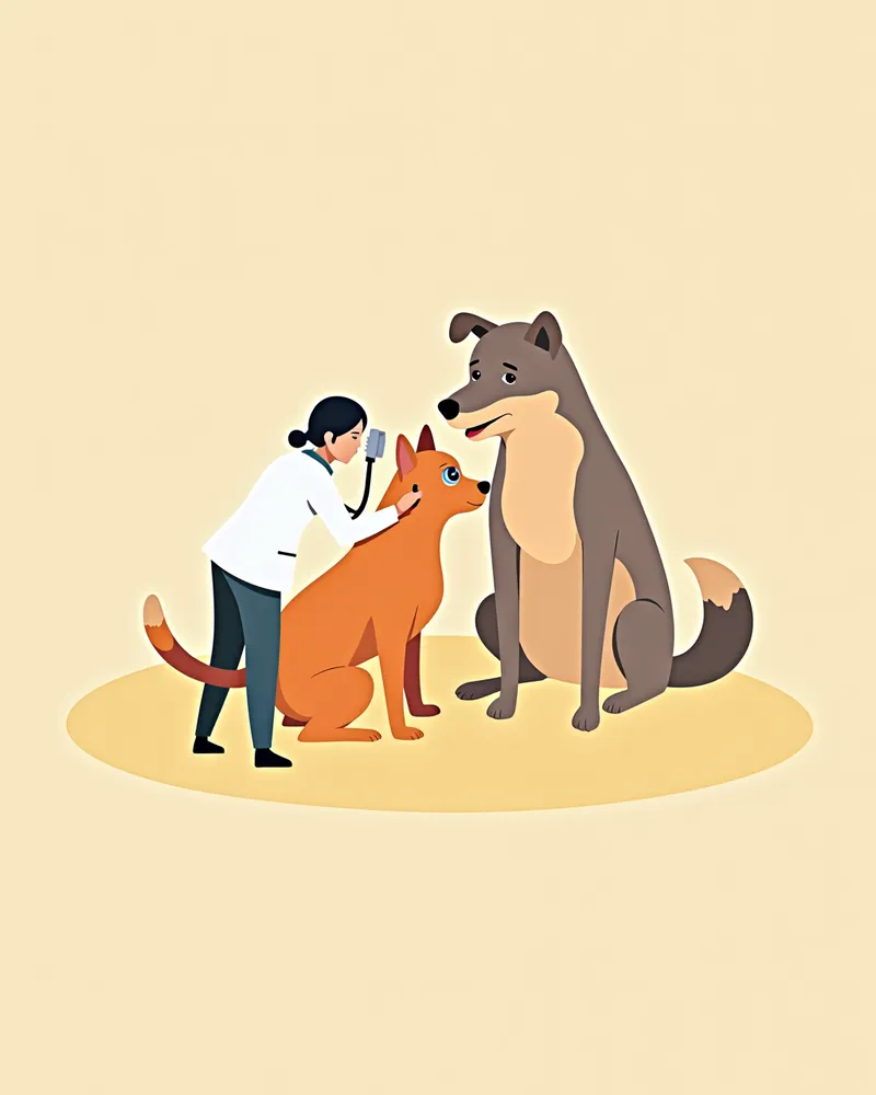
The veterinarian will first directly examine the eye, assessing pupil size, the pupillary light reflex (PLR), and any corneal damage. - Pupillary Light Reflex Assessment: A preserved pupillary light reflex or pupil constriction (miosis) is a relatively positive prognostic indicator. - Corneal Examination: A special fluorescent dye is used to check for exposed corneal damage. - Check for Hemorrhage: Bleeding inside or around the eye, such as hyphema (anterior chamber hemorrhage), is a negative prognostic factor. - Evaluation of Extraocular Muscle and Scleral Damage: The prognosis worsens if multiple extraocular muscles are ruptured or if there is a rupture of the cornea or sclera. - Systemic Evaluation: We also check for life-threatening injuries associated with the trauma to develop the optimal treatment plan.
A photo of a veterinarian examining a puppy’s protruding eyes.

Treatment Methods and Step-by-Step Management

The cornerstone of treatment is a reduction surgery performed under general anesthesia to return the protruding eyeball to its normal position. - Preoperative preparation: First, life-threatening injuries are addressed to stabilize the patient, and the ocular surface is kept moist with physiological saline to prevent drying. - Surgical procedure: If severe eyelid swelling makes reduction difficult, a canthotomy (lateral canthotomy) is performed to create space, after which the eyeball is repositioned into the orbit. - Temporary tarsorrhaphy: After reduction, temporary tarsorrhaphy sutures are typically maintained for 10–14 days. - Severe damage: If the eyeball is severely damaged and unlikely to be saved, enucleation (eye removal) may be considered. - Postoperative care: Avoid corneal abrasions and schedule regular check-ups with your veterinarian until the swelling subsides.
A photo of a dog wearing a spectacle-style protective cone after eye proptosis surgery.

Home Care and Recovery Points

After surgery, it is most important to protect the sutured eyelids and cornea. - Maintain temporary sutures: Temporary tarsorrhaphy sutures are typically kept in place for 10–14 days and should not be removed prematurely. - Medication administration: Administer medications exactly as directed by your veterinarian and complete the full prescribed course. - Maintain cleanliness: Keep the area around the eye clean to prevent bacterial infection. - Prevent scratching: Use an Elizabethan collar or similar protective device to prevent your dog from scratching or rubbing its eye. - Regular check-ups: Restrict activity during the recovery period to avoid straining the eye, and schedule regular veterinary visits to monitor healing progress.
A photo of a puppy lying quietly after eye proptosis surgery

Certain Breeds Have a Higher Risk of Recurrence

Brachycephalic breeds with flat faces, such as Shih Tzus, Pugs, and Lhasa Apsos, have shallow eye sockets, making them prone to globe protrusion. Even after surgery, it’s essential for these breeds to consistently maintain habits that protect their eyes, and it’s important to manage their environment to prevent injuries during outdoor activities or play.

Reviewed by a veterinarian

Dr. Tony — Punnawat Phongkittirak

Dr. Tony — Punnawat Phongkittirak

Veterinarian

A veterinarian who majored in veterinary medicine at Khon Kaen University, Thailand, and completed the IVSA program at North Carolina State University in the United States. Drawing on clinical experience at animal hospitals, he works in the pet healthcare field and is dedicated to building a digital care environment that connects pet parents with veterinarians.

Frequently Asked Questions

Can vision return after an eye has been displaced from the socket?
The sooner your pet receives professional treatment, the higher the chances of saving the eye and preserving vision. Delaying treatment can worsen the prognosis, while a retained pupillary light reflex is a relatively positive sign.
Can exophthalmos be completely cured?
If the injury isn’t severe, the eye can usually be repositioned and recover. However, if the damage is extensive, removal of the eye may be necessary.
Can we prevent proptosis?
Brachycephalic breeds are structurally predisposed to certain conditions, making complete prevention difficult. However, it’s important to manage their environment to prevent injuries during outdoor activities or play.
Can the eye bulge out again after it has been displaced?
Since trauma can cause complications, it’s important to consistently protect your pet’s eyes and prevent injuries even after surgery.
Which breeds are most commonly affected by exophthalmos?
It most commonly occurs in brachycephalic breeds with flat faces, such as Shih Tzus, Pugs, and Lhasa Apsos.

Share

Related Guides

References

[1] Clinical Atlas of Canine and Feline Ophthalmic Disease, 2nd Ed. 2023.

[2] The Dog Care Handbook, Things I Wish My Vet Had Told Me. 2021.

[3] Gilger BC et al. Traumatic ocular proptoses in dogs and cats: 84 cases (1980-1993). J Am Vet Med Assoc. 1995;206(8):11.

This information is based on veterinary literature and does not replace diagnosis or treatment. Please consult a veterinarian for specific health concerns.

Metapet Co., Ltd. | CEO: Park Sung-yong | Business Reg. No. 417-88-02562 | Seoul, South Korea | Customer Center

Dog Eye Popped Out: Emergency First Aid and the Critical Golden Hour | Meongsiljang