
Like
Share
멍실장
강아지 각막 색소침착 증상과 원인, 진단·치료와 관리 포인트 총정리

If Your Dog’s Pupil Has Turned Dark — Causes of Corneal Pigmentation and Warning Signs of Vision Loss

Eye HealthDiseasesMeongsiljang Veterinary Advisory Board

Corneal pigmentation in dogs is a condition characterized by the appearance of black or brown spots on the eye, primarily caused by genetic factors and age. Early detection and regular check-ups are essential.

Canine Corneal Pigmentation Is a Genetic Condition Characterized by Black Spots on the Eye

A condition in which pigment deposits have appeared on the cornea of a dog's eye.
Canine corneal pigmentation, also known as pigmentary keratitis, is a condition characterized by the gradual deposition of brown pigment on the surfaces of the cornea and conjunctiva, appearing as smooth or irregular brown opacities. Rather than being a single-gene disorder, it results from multiple contributing factors, including eyelid structures that fail to adequately cover the eye (exposure), chronic or severe keratitis, abnormalities in the quantity or quality of the tear film, chronic irritation from eyelashes or distichiasis, and genetic predisposition. It is particularly common in breeds such as the Pug. While small pigmented spots may appear initially, the area of deposition can expand over time. - Multiple Contributing Factors: Exposure due to eyelid structure, chronic keratitis, tear film abnormalities, eyelash irritation, and genetic predisposition all play a role. - Progression: The course varies among individuals, and inflammatory keratitis accompanied by neovascularization may also occur. - Asymptomatic Period: In the early stages, there is often no noticeable discomfort, making it easy for owners to overlook; therefore, regular monitoring is essential.

The Main Causes Are Genetic Predisposition and Age, with Inflammation Also Playing a Role

Corneal pigmentation in dogs isn’t caused by a single factor, but rather by a combination of several contributing elements. These include eyelid structures that fail to fully cover the cornea (common in brachycephalic breeds and associated with exposure), chronic or severe keratitis, abnormalities in the quantity or quality of the tear film (such as dry eye), chronic irritation from conditions like distichiasis or trichiasis, and genetic predisposition. It is frequently observed in certain breeds, such as Pugs. - Structural factors: If the eyes are not adequately closed, the cornea remains chronically exposed, which can promote pigmentation. - Chronic irritation: Eyelashes or ectopic cilia can continuously irritate the cornea. - Inflammatory impact: Chronic or severe keratitis can influence the development of corneal pigmentation. - Genetic predisposition: Certain breeds have a higher susceptibility, making early monitoring beneficial.
A scene of an eye examination for a Carener Terrier

The Main Symptom Is the Appearance of Black Spots on the Eye, and Vision Loss May Occur
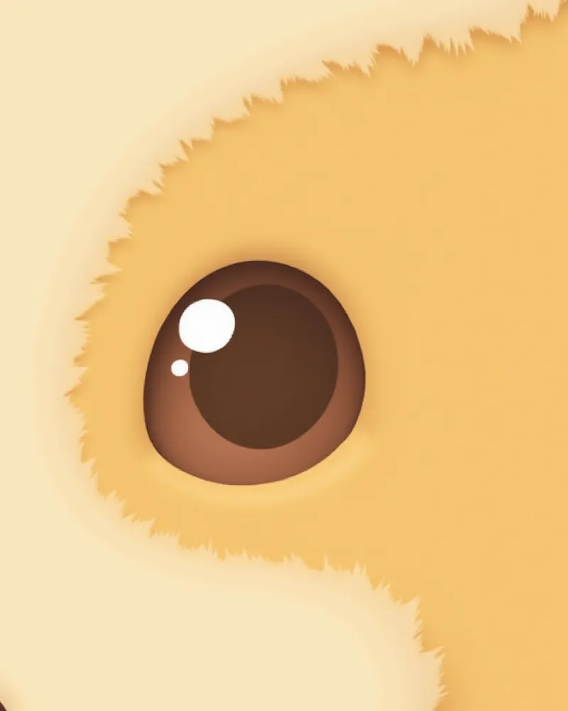
The primary sign of corneal pigmentation in dogs is the appearance of smooth or irregular brown opacities (pigment deposits) on the surface of the cornea. The extent of pigmentation varies; it may start small but can gradually expand over time. It can occur alongside inflammatory keratitis with neovascularization, and in severe cases, it may obstruct the visual field, leading to vision impairment or functional blindness. - Progression of pigmentation: The brown opacity gradually spreads, potentially reducing corneal transparency. - Changes in vision: If the pigmentation covers a large area of the central cornea, it can reduce light transmission and affect vision. - Accompanying inflammation: When neovascularization and inflammation are present, the eye may appear red or uncomfortable. - Early detection: Monitor for symptoms from the earliest stages, and visit a veterinary clinic immediately if any abnormal signs are observed.
A dog's eye with progressive pigment deposition on the cornea

Immediate Veterinary Visit Is Necessary If Vision Loss Is Severe or the Eye Is Red

If you notice rapid progression of pigmentation, along with corneal redness, new blood vessel growth, and declining vision, consult your veterinarian immediately. This is because pigmentation can occur alongside other conditions such as glaucoma (pigmentary glaucoma) or keratitis. Since glaucoma involves increased intraocular pressure, early diagnosis and pressure assessment are crucial for preserving vision.

Diagnosis Is Made Through Specialized Ophthalmic Examination and Intraocular Pressure Measurement
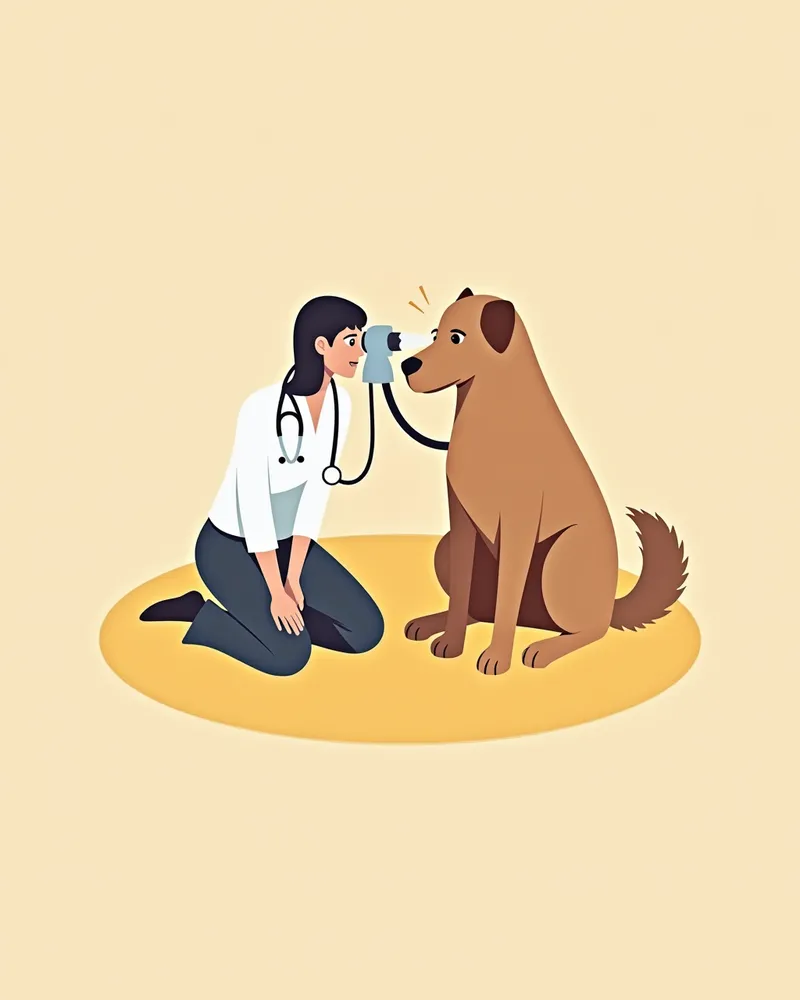
Corneal pigmentation in dogs is diagnosed based on clinical findings during an ophthalmic examination. The extent and location of brownish pigmentary opacity are directly observed, and additional tests are performed to differentiate it from other corneal diseases. - Intraocular Pressure Measurement: Measured using applanation (e.g., Tono-Pen) or rebound (e.g., Tono-Vet) tonometers. Normal intraocular pressure is approximately 15–25 mmHg. This measurement is essential to rule out pigmentary glaucoma. - Fluorescein Staining: Sodium fluorescein staining is used to check for the presence of corneal epithelial defects (ulcers). - Disease Differentiation: Since corneal pigmentation can overlap with other corneal conditions, accurate differentiation is the foundation for appropriate treatment. - Specialized Examination: An ophthalmic examination is necessary for an accurate assessment.
A veterinarian examining a dog's eyes with a slit lamp.

Treatment Varies Depending on the Cause, and There Is Rarely Specific Medication

For corneal pigmentation in dogs, we focus on addressing the underlying cause rather than trying to eliminate the pigmentation directly with medication. If the pigmentation is linked to glaucoma or inflammatory keratitis, we treat the underlying condition alongside managing the pigmentation. - Intraocular pressure control: If pigmented glaucoma is causing elevated intraocular pressure, we lower it using medication or surgery. - Anti-inflammatory treatment: If inflammatory keratitis is present, we use anti-inflammatory medications to reduce inflammation. - Correcting the root cause: If the issue stems from an exposed eyelid structure or eyelash irritation, surgical correction may be considered. - Treatment goal: The primary objective is to slow progression and preserve vision as much as possible.
A veterinarian administering eye drops to a puppy

Home Care Centers on Regular Ophthalmic Exams and Eye Protection

Managing corneal pigmentation in dogs requires consistent monitoring of its progression. Regular ophthalmic exams are essential to track the extent of pigmentation and screen for concurrent conditions such as glaucoma or keratitis. It is also helpful to minimize chronic irritants like eyelashes, prevent the dog from scratching its eyes, and avoid exposure to eye-irritating dust and chemicals. - Regular Exams: Once corneal pigmentation is identified, schedule regular ophthalmic check-ups based on the severity of the condition. - Environmental Management: Reduce factors that irritate the eyes and provide a safe environment. - Owner Observation: Frequently check for changes in the pigmented area, redness, neovascularization, or signs of vision loss. - Immediate Veterinary Visit: Seek prompt veterinary care if any abnormal symptoms appear.
A dog wearing protective glasses

Carener Terriers, Shar-Peis, and Cocker Spaniels Have a High Genetic Risk and Require Attention

Brachycephalic breeds like Pugs are prone to corneal pigmentation due to their exposed eye structure, which often prevents the eyelids from closing completely. If you have a dog of such a breed, it’s a good idea to monitor the condition through regular veterinary eye exams. Since corneal pigmentation results from multiple contributing factors, managing potential irritants and detecting the issue early are more important than treatment.

Reviewed by a veterinarian

Dr. Tony — Punnawat Phongkittirak

Dr. Tony — Punnawat Phongkittirak

Veterinarian

A veterinarian who majored in veterinary medicine at Khon Kaen University, Thailand, and completed the IVSA program at North Carolina State University in the United States. Drawing on clinical experience at animal hospitals, he works in the pet healthcare field and is dedicated to building a digital care environment that connects pet parents with veterinarians.

Frequently Asked Questions

Should you always take your dog to the vet if you notice brown or black spots on its eyes?
It’s a good idea to have your pet examined by a veterinarian as soon as you notice any pigmentation. This is because it may not be simple pigment deposition but another type of corneal disease, so an accurate diagnosis based on clinical findings is essential.
Can corneal pigmentation be treated?
There are limited medication options that directly eliminate the pigment itself. However, if there is an underlying condition such as glaucoma or inflammatory keratitis, we will treat that condition concurrently. In cases of structural issues like exposure blepharitis, surgical correction may be considered.
What should I do if the pigmentation is progressing rapidly?
Because rapid progression can impair vision, you should consult a veterinarian immediately. Ophthalmic examinations, such as intraocular pressure measurement and fluorescein staining, are necessary to identify the underlying cause and any concurrent conditions.
Are Pugs prone to corneal pigmentation?
Yes, brachycephalic breeds like Pugs are commonly prone to corneal pigmentation. It’s important to monitor its progression through regular ophthalmic examinations.
Can pigmentation in the eyes lead to vision loss?
When pigment spreads widely across the center of the cornea, it can reduce light transmission and impair vision. If glaucoma is also present, vision loss may become even more severe. Early monitoring is essential.

Share

Related Guides

References

[1] Petersen-Jones SM, Mentzer AL, Dubielzig RR, et al. Ocular melanosis in the Cairn Terrier: histopathological description of the condition, and immunohistological and ultrastructural analysis. Vet Ophthalmol. 2007;10 Suppl 1:63–69.

[2] Hanselman BA. Melanocytic glaucoma in a cairn terrier. Can Vet J. 2002;43(4):296–298.

[3] Clinical Atlas of Canine and Feline Ophthalmic Disease, 2nd Ed. Wiley-Blackwell, 2018.

This information is based on veterinary literature and does not replace diagnosis or treatment. Please consult a veterinarian for specific health concerns.

Metapet Co., Ltd. | CEO: Park Sung-yong | Business Reg. No. 417-88-02562 | Seoul, South Korea | Customer Center

Canine Corneal Pigmentation: Causes & Vision Loss Signs | Meongsiljang