Like
Share
멍실장
고양이 안구 초음파 검사 증상과 원인, 진단·치료와 관리 포인트 총정리

Feline Ocular Ultrasound — Key Points for Diagnosing Cataracts, Tumors, and Retinal Detachment

Eye HealthDiseasesMeongsiljang Veterinary Advisory Board

Feline ocular ultrasound is a non-invasive diagnostic method that precisely evaluates abnormalities within the internal structures of the eye. Your veterinarian may recommend this test if your cat exhibits symptoms such as vision loss or eye pain.

What is Feline Ocular Ultrasound?
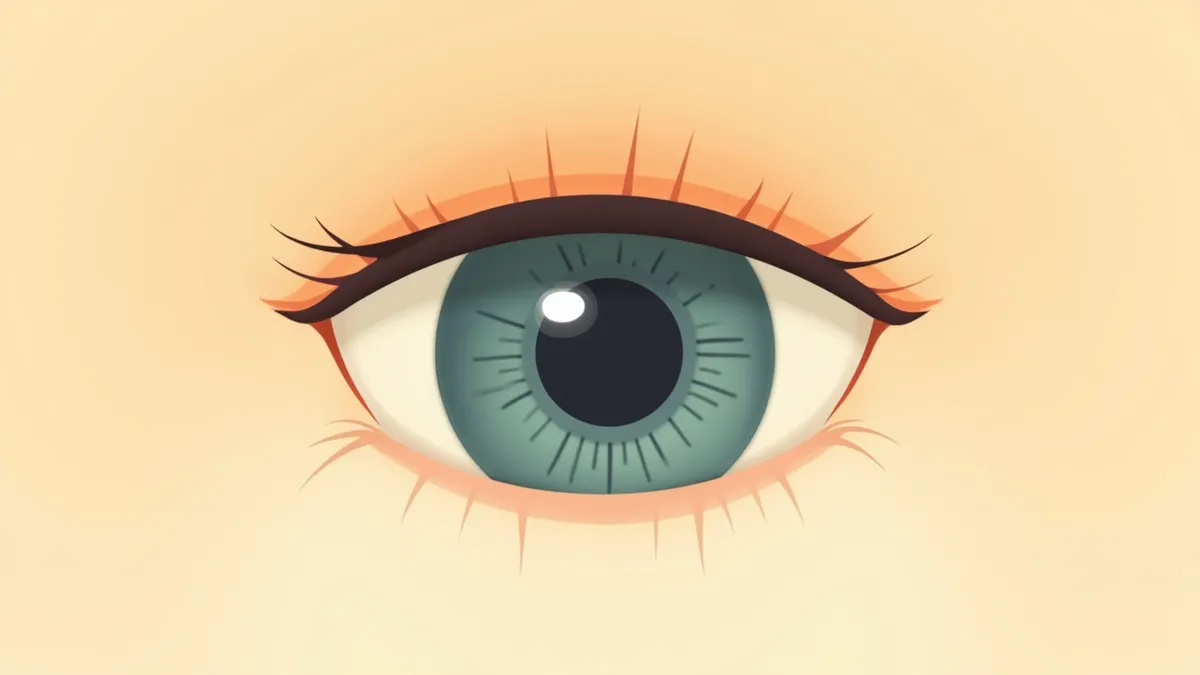
An ultrasound image of the internal structures of a cat's eye
Feline ocular ultrasound is a diagnostic method that allows non-invasive visualization of the internal structures of the eye. It plays a crucial role in detecting abnormalities within the eye and formulating treatment plans, especially when direct visualization is difficult due to corneal opacity or anterior segment lesions.
Detection of internal ocular abnormalities: Identifies structural issues such as retinal detachment, intraocular hemorrhage, and tumors.
Assessment of structural changes due to abnormal intraocular pressure: While intraocular pressure is measured with a tonometer, ultrasound is used to examine structural changes within the eye caused by pressure abnormalities.
Confirmation of substance accumulation: Evaluates for vitreous opacity or inflammatory changes.
Timely diagnosis: Helps identify the cause early when vision loss or pain occurs.
This procedure is typically performed quickly while the cat is awake, using gentle restraint and topical local anesthesia, without the need for general anesthesia.

Main Causes and Pathogenesis

There are several key reasons why a cat may need an ocular ultrasound. This test is essential for accurately identifying the underlying cause when direct visualization of the interior of the eye is difficult.
Retinal detachment: This condition involves the separation of the retina from the inner wall of the eye, and ultrasound is particularly useful for diagnosis. Complete detachment can result in a poor prognosis for vision.
Intraocular hemorrhage: Bleeding into the vitreous humor or retina, often caused by trauma or vascular disease, can be detected via ultrasound.
Ocular tumors: Tumors developing in the vitreous, retina, or other intraocular structures may appear as masses on ultrasound imaging.
Inflammatory diseases: Intraocular inflammation, such as uveitis, may occur in association with systemic diseases.
Abnormal intraocular pressure: Ultrasound can help assess structural changes in the eye associated with abnormal intraocular pressure, such as high intraocular pressure.
Because these conditions directly affect a cat’s vision and ocular health, early diagnosis is crucial.
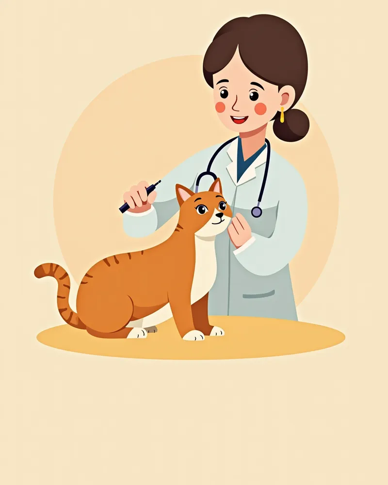
A veterinarian performing an ocular ultrasound examination on a cat

Main Symptoms and Signs

There are several key symptoms that may indicate your cat needs an ocular ultrasound. It’s important for pet owners to observe carefully.
Decreased vision: You may notice your cat misjudging distances or bumping into walls more often.
Increased eye closure: Your cat may frequently squint or keep one eye closed, or show sensitivity to light.
Excessive tearing: There may be an abnormal increase in tears or the presence of sticky discharge.
Eye redness: The area around the eye may appear red, with visible blood vessel dilation.
Eye swelling: Swelling around the eye or irregular bulging of the eyeball itself may occur.
Abnormal eye movements: The eyes may tremble or move in unusual directions.
If these symptoms persist, there may be an underlying issue inside the eye, so it's best to consult with a veterinarian.
A cat’s eye showing redness and swelling.

When to Visit the Vet Immediately

If your cat suddenly loses vision, squints in pain, or frequently shows its third eyelid (nictitating membrane), you should visit a vet immediately. This could be a sign of an acute issue inside the eye.

Diagnostic Procedure and Methods
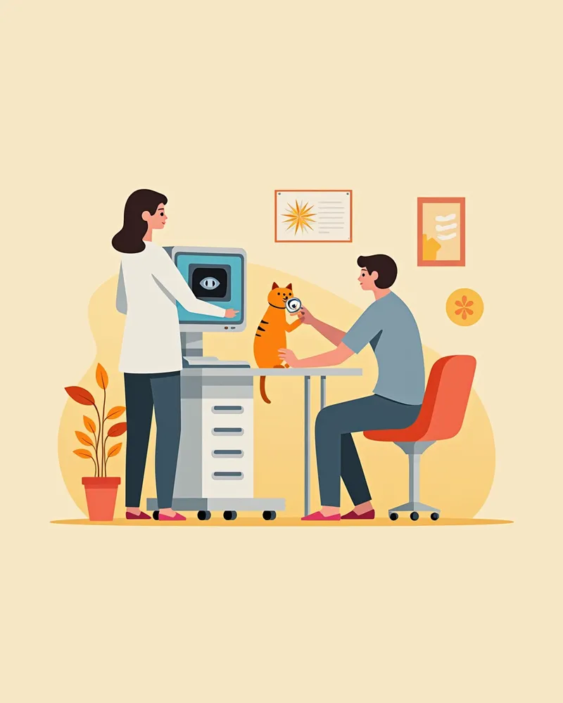
Feline ocular ultrasound is performed through a specialized procedure, with the veterinarian carefully preparing to ensure an accurate diagnosis.
Ultrasound Equipment Setup: High-resolution ultrasound equipment suitable for a cat’s eye is used.
Stable Positioning: The cat is gently restrained to minimize stress and maintain a stable position.
Periocular Cleaning: The area around the eye is thoroughly cleaned before the examination.
Topical Anesthesia and Gel Application: To reduce discomfort, a local anesthetic eye drop is administered, followed by the application of ultrasound gel to the contact area before beginning the scan.
Multi-angle Imaging: Images are captured from various angles, including front, side, and top views.
Image Analysis: The veterinarian analyzes the captured images to identify any abnormalities.
This process typically takes about 10–15 minutes and places minimal burden on the cat.
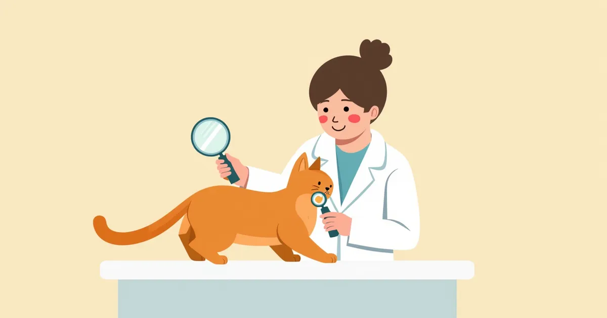
A veterinarian reviewing real-time ocular ultrasound images of a cat.

Treatment Methods and Step-by-Step Management
The treatment plan is determined based on the results of the ocular ultrasound examination, and the specific approach depends on the underlying cause.
Medication: If inflammation (uveitis) is present, topical and/or systemic anti-inflammatory medications are prescribed.
Surgical treatment: If an intraocular tumor is identified, surgical options such as enucleation (eye removal) may be considered.
Intraocular pressure management: If elevated intraocular pressure is detected, medications to lower eye pressure are used.
Management of underlying conditions: For retinal detachment, underlying causes such as systemic hypertension are treated concurrently.
Regular follow-up examinations: Ultrasound monitoring continues after treatment to track the condition.
Environmental adjustments: Provide a comfortable environment for your cat.
Throughout the treatment period, maintain regular communication with your veterinarian and keep a record of any changes in symptoms.
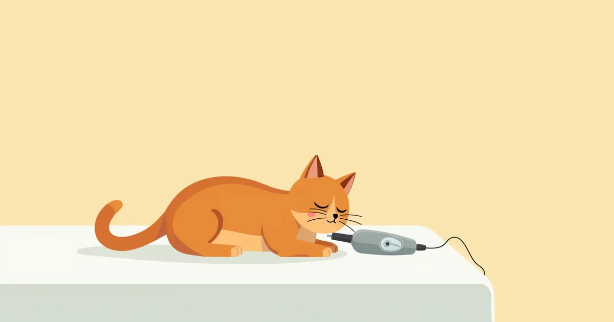
A cat undergoing treatment resting comfortably

Home Care Points

Regular care remains essential even after treatment. There are several steps you can take to protect your cat’s eye health.
Medication management: Administer medication precisely at the scheduled times.
Maintain eye hygiene: Gently wipe around the eyes with a soft cloth.
Avoid stimulating environments: Keep your cat away from areas with bright light or excessive noise.
Regulate activity levels: Avoid strenuous exercise and maintain a calm, comfortable environment.
Dietary management: Choose food that provides balanced nutrition.
Regular check-ups: Schedule appointments with your veterinarian for routine examinations.
These care practices significantly support vision recovery and help prevent recurrence.
A caregiver gently cleaning a cat's eyes.

Breed-Specific Precautions and Prevention of Recurrence

Certain breeds may be more susceptible to eye diseases. For example, brachycephalic (short-nosed) cats are known to be more prone to ocular surface issues such as corneal sequestrum. Please regularly check your cat's eye health according to their breed characteristics.

Reviewed by a veterinarian

Dr. Tony — Punnawat Phongkittirak

Dr. Tony — Punnawat Phongkittirak

Veterinarian

A veterinarian who majored in veterinary medicine at Khon Kaen University, Thailand, and completed the IVSA program at North Carolina State University in the United States. Drawing on clinical experience at animal hospitals, he works in the pet healthcare field and is dedicated to building a digital care environment that connects pet parents with veterinarians.

Frequently Asked Questions

Does an ocular ultrasound hurt cats?
No. Cats typically don’t experience significant pain during the procedure. They’re gently restrained during the exam, and most cats simply feel a bit tense.
Is fasting required before the test?
No special preparation is needed. The examination is performed using only topical eye drops for local anesthesia, without general anesthesia, and can be safely done without fasting.
How soon will the test results be available?
A veterinarian can analyze the images on the spot and provide you with the results. The official report will be ready within 1 to 2 hours.
Are there any special precautions to take after an ocular ultrasound?
After the examination, please avoid touching the area around your cat’s eyes and maintain a quiet environment to help keep them calm and relaxed.
Is this test necessary for all cats?
No. We recommend consulting a veterinarian if you notice symptoms such as vision loss, eye pain, or redness.

Share

Related Guides

References

[1] Textbook of Cardiovascular Medicine in Dogs and Cats, 2022, Chapter 11: Echocardiography in Feline Cardiac Disease

[2] Small Animal Critical Care Medicine, 3rd Ed, 2021, Section 43.1: Indications for Echocardiography

[3] Feline Emergency and Critical Care Medicine, 2nd Ed, 2020, Figure 14.1: Clinical Approach to Dyspneic Feline Patients

This information is based on veterinary literature and does not replace diagnosis or treatment. Please consult a veterinarian for specific health concerns.

Metapet Co., Ltd. | CEO: Park Sung-yong | Business Reg. No. 417-88-02562 | Seoul, South Korea | Customer Center

Feline Ocular Ultrasound: Cataracts, Tumors & Retinal Detachment | Meongsiljang