Like
Share
멍실장
시바이누 고관절·팔꿈치 검진 — 일본 견종 호발 가이드

Shiba Inu Hip and Elbow Dysplasia — Complete Guide to Screening Timelines and Common Conditions

JointBreed HealthMeongsiljang Veterinary Advisory Board

Shiba Inus are a Japanese breed that is relatively prone to hip and elbow dysplasia. We’ve outlined the recommended veterinary screenings for each life stage, from four months of age through eight years and beyond.

What Is Shiba Inu Joint Health?

A Shiba Inu sitting on the examination table
The Shiba Inu is a native Japanese medium-sized breed. Hip and elbow dysplasia are developmental disorders that occur during joint formation. While they are more common in large and giant breeds, they can also affect medium and small breeds. Therefore, it is important for Shiba Inus to undergo early imaging screenings to assess joint health in advance. Dysplasia can progress to osteoarthritis over time, but clinical symptoms often remain subtle until pain develops in middle age. By the time owners notice limping or difficulty getting up in the morning, osteoarthritis has often already advanced significantly. Thus, the key is to regularly monitor joint health throughout the growth and adult stages, even in the absence of symptoms.

Three Joint Conditions Common in Shiba Inus

Here’s a summary of joint conditions that Shiba Inu owners should be aware of: - Hip Dysplasia: A developmental disorder in which the muscles, connective tissues, and ligaments supporting the joint become lax, preventing the femoral head from sitting properly in the pelvic socket. - Elbow Dysplasia: A condition in which the bones forming the elbow grow unevenly, leading to cartilage damage. - Patellar Luxation: A disorder in which the kneecap (patella) dislocates from its normal position; it is best diagnosed through orthopedic palpation (physical examination) rather than X-rays. These conditions are known to result from a combination of genetic predisposition and management during the growth period. They typically arise not from a single cause, but from the interplay of inherent constitution and environmental factors during development.

Why Hip Dysplasia Is Common in Shiba Inus
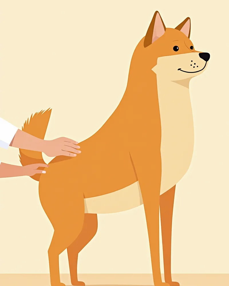
Hip dysplasia is a condition in which the ligaments, muscles, and connective tissues that support the joint become lax, causing the femoral head to repeatedly slip in and out of the pelvic socket (subluxation). This process leads to abnormal wear of the joint surfaces and progression to osteoarthritis. As the condition advances, the pelvic socket becomes shallow and wide, and the femoral head flattens. Non-genetic factors such as nutrition, growth rate, body size, exercise level, and muscle mass also influence the development of the disease. In particular, excessive jumping or rough play during the growth period can place undue stress on the joints. Clinical signs are often first noticed in middle-aged dogs or older, typically presenting as hind limb pain or difficulty standing up.
A veterinarian gently palpating the joints of a Shiba Inu named Fuji

See These Signs? Seek Immediate Orthopedic Evaluation

If your dog exhibits "bunny hopping" (jumping with both hind legs together), suddenly refuses to climb stairs, shows hind leg instability when standing up, or takes longer to sit down after walks, these could be signs of hip pain. If these symptoms persist for more than 24 to 48 hours, it's important to seek orthopedic care and X-ray imaging rather than general veterinary treatment. Shiba Inus tend to hide pain well, so even mild symptoms should not be overlooked.

Shiba Inu Joint Screening Schedule and Items by Age

Item4–6 months12 months3–5 years6 years and older
Basic physical examRequiredRequiredRequiredRequired
Hip X-rayRecommended (baseline)Required (evaluation)RecommendedRequired
Elbow X-rayOptionalRecommendedRecommendedRequired
Gait examinationRecommendedRecommendedRequiredRequired
Weight and muscle mass assessmentRequiredRequiredRequiredRequired

Standard recommendations by growth, adult, and senior stages. If parent dogs have a history of dysplasia, increasing frequency is recommended.

Elbow Dysplasia Screening — What Do We Look For?
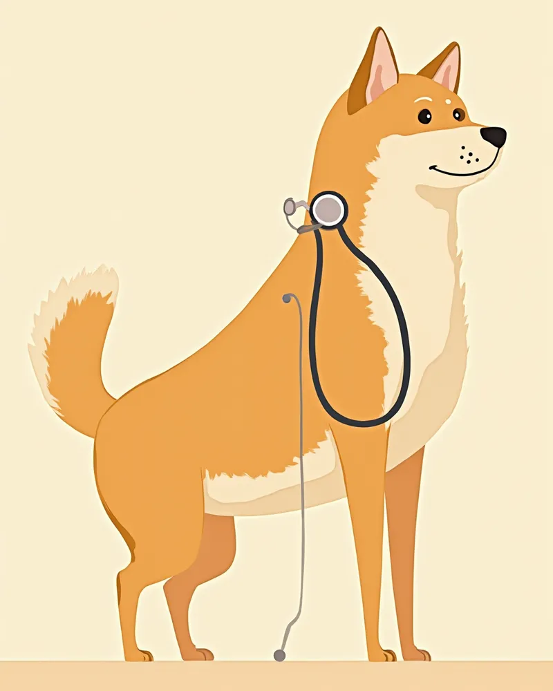
Elbow dysplasia is a condition in which the cartilage becomes damaged due to uneven growth of the three bones that make up the elbow joint (the humerus, radius, and ulna). Changes caused by this developmental abnormality often first appear as limping in young, growing dogs. The initial diagnostic step is a standard X-ray, but a flexed lateral view is particularly effective at sensitively revealing osteoarthritic changes such as bone spurs. If a more definitive diagnosis or a detailed assessment of the extent of cartilage damage is needed, a CT scan is recommended because it is more sensitive than X-rays and allows for viewing the joint in multiple cross-sections. Signs for pet owners to watch for include front leg limping, dragging the front paw after walks, and walking with the front legs splayed outward.
A veterinarian examining the elbow joint of a Shiba Inu's front leg

How Owners Can Understand Screening Results

Hip X-ray reports typically include assessments of joint congruency, femoral head shape, acetabular depth, and the presence of osteoarthritic changes. Normal findings are usually described as "mild" or "within normal limits," while abnormalities are classified as "mild," "moderate," or "severe." Even with mild findings, Shiba Inus are prone to rapid progression due to their high activity levels, so follow-up imaging every six to twelve months is recommended. If terms such as "subluxation," "flattening," or "osteophyte formation" appear on the report, consultation with a veterinary orthopedic specialist is necessary.

Key Management Points for Protecting Shiba Inu Joints

Along with regular check-ups, daily home care is just as important. - Weight management: Obesity can place excessive stress on joints like the hips and worsen osteoarthritis, so it’s best to keep your pet at a lean body condition. - Limit jumping: Reduce jumping on and off sofas and beds from puppyhood, and use stairs or ramps instead. - Prevent slipping: Use non-slip mats on hardwood floors or apply paw pads to improve traction. - Consistent, low-impact exercise: During growth, controlled leash walks are better than rough, free play, as they help strengthen the muscles and supportive structures around the joints. - Nutrition during growth: Overfeeding, rapid growth, and excess calcium or vitamin D can increase the risk of developmental joint diseases. Feed appropriate amounts based on the correct calcium-to-energy ratio. Maintaining a lean body condition during growth is known to lower the risk of hip dysplasia.
A Shiba Inu taking a leisurely walk with its owner

Checking Parents’ Screening History Is the Most Powerful Prevention

Hip and elbow dysplasia are conditions with a strong genetic predisposition. The most effective preventive measure when acquiring a Shiba Inu is to request the parents’ hip and elbow evaluation reports (such as OFA, PennHIP, or JAHD). If you already have a Shiba Inu at home, ask the breeder for the parents’ health history; if that information is unavailable, be sure to have an X-ray evaluation done when your dog is one year old. Having baseline data early on allows you to compare the extent of changes as your dog ages.

Reviewed by a veterinarian

Dr. Tony — Punnawat Phongkittirak

Dr. Tony — Punnawat Phongkittirak

Veterinarian

A veterinarian who majored in veterinary medicine at Khon Kaen University, Thailand, and completed the IVSA program at North Carolina State University in the United States. Drawing on clinical experience at animal hospitals, he works in the pet healthcare field and is dedicated to building a digital care environment that connects pet parents with veterinarians.

Frequently Asked Questions

At what age should a Shiba Inu first undergo a hip joint screening?
A basic evaluation is typically recommended once between 4 and 6 months of age, while a comprehensive assessment is standard at 12 months (after the growth plates have closed). If there is a known family history of joint issues, we recommend a more thorough examination at the 4-month mark.
Can hip dysplasia be diagnosed using only X-rays?
In most cases, an X-ray is sufficient for diagnosis. However, to assess subtle ligament laxity (subluxation), specialized imaging such as PennHIP or a CT scan may be necessary.
Is an exam necessary even if there are no symptoms at all?
Yes, Shiba Inus are notoriously good at hiding pain. By the time symptoms become apparent, osteoarthritis is often already advanced, which makes imaging during the asymptomatic stage the most valuable diagnostic tool.
When should I start giving my pet joint supplements (glucosamine and chondroitin)?
If minor abnormalities are detected during a checkup, you can start as early as one year of age. If the results are normal and there are no symptoms, it’s common to begin after six years of age as a preventive measure against aging.
How much does a check-up usually cost?
While costs vary by clinic, this is the standard baseline for a general physical exam plus hip and elbow X-rays. For the most accurate pricing, we recommend contacting a veterinary clinic that offers orthopedic services directly.

Share

Related Guides

References

[1] Textbook of Veterinary Orthopaedic Surgery, Chapter 9: Hip Dysplasia

[2] Clinical Medicine of the Dog and Cat, 4th Edition — Hip Dysplasia

[3] The Dog Care Handbook — Things I Wish My Vet Had Told Me

This information is based on veterinary literature and does not replace diagnosis or treatment. Please consult a veterinarian for specific health concerns.

Metapet Co., Ltd. | CEO: Park Sung-yong | Business Reg. No. 417-88-02562 | Seoul, South Korea | Customer Center

Shiba Inu Hip & Elbow Dysplasia: Screening & Care Guide | Meongsiljang