
Border Collie CEA (Collie Eye Anomaly) — A Complete Guide from Screening Timelines to Genetic Management
We’ve put together a comprehensive guide covering the definition of CEA, a common hereditary eye condition in Border Collies, as well as screening timelines, the differences between fundus and genetic testing, and important considerations for breeding.
Understanding Border Collie CEA at a Glance
Why Does It Occur Frequently in Border Collies?
Signs Owners Can Notice
The Golden Time for Screening Is 6–8 Weeks of Age
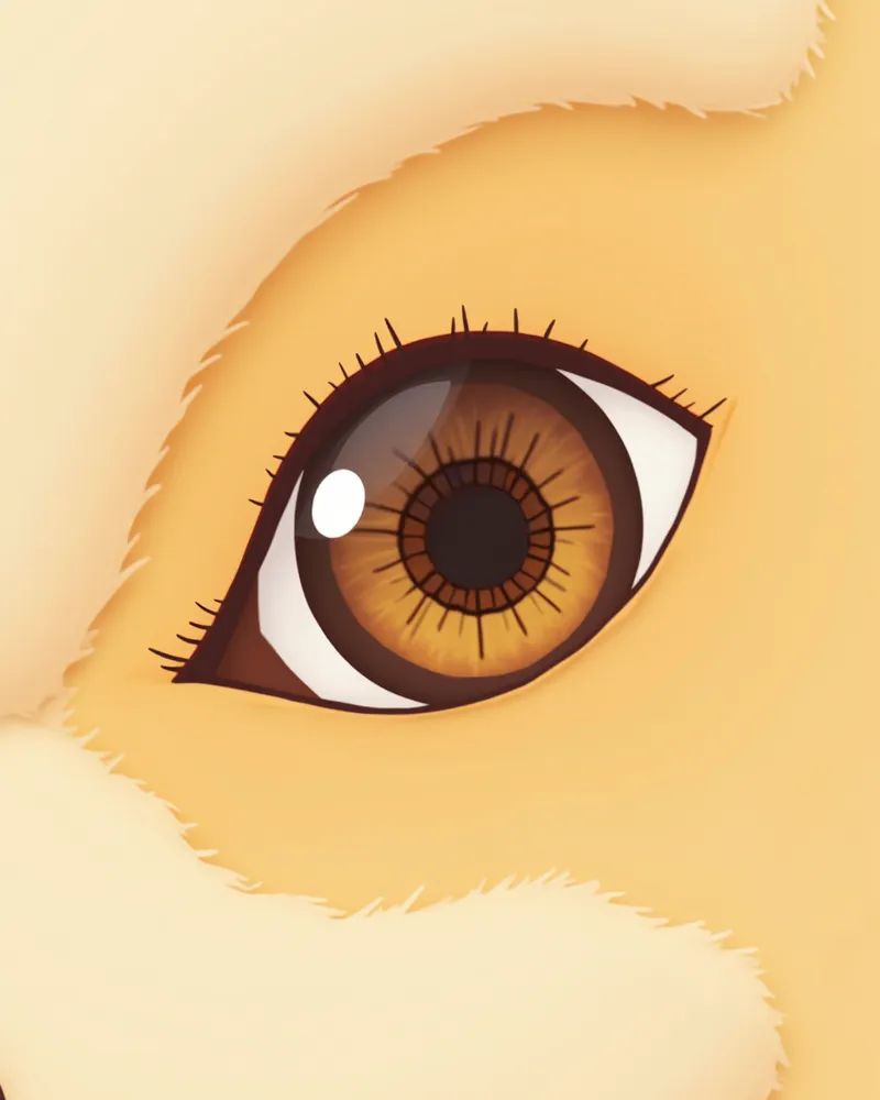
CEA fundus examinations are most recommended at around 6 to 8 weeks of age. As time passes, some mild lesions or retinal folds may no longer be visible on follow-up exams between 6 months and 1 year of age. Due to this "Go Normal" phenomenon, puppies with CEA may actually be classified as normal. Therefore, the initial exam results from early in life are crucial. If diagnosis through fundus examination alone is difficult, a separate genetic test should be performed to more accurately determine carrier status. If you are considering acquiring a puppy, be sure to request the health screening results for both parent dogs from the breeder.
How Is It Tested?
Fundus Examination vs. Genetic Testing
| Item | Dilated fundus exam | Genetic test (NHEJ1) |
|---|---|---|
| What it checks | Presence of actual retinal lesions | Genotype (normal/carrier/affected) |
| Recommended timing | 6–8 weeks of age | Possible at any age |
| Test method | Fundus observation after pupil dilation | Buccal swab or blood draw |
| Advantages | Directly confirms lesion severity | Not affected by 'Go Normal' |
| Limitations | Mild lesions may appear to disappear after 6 months to 1 year of age | Cannot determine lesion severity |
| For breeding decisions | Supplementary data | Essential data |
Doing both tests together is the most accurate.
How to Interpret Screening Results
Be Sure to Verify Before Breeding or Rehoming
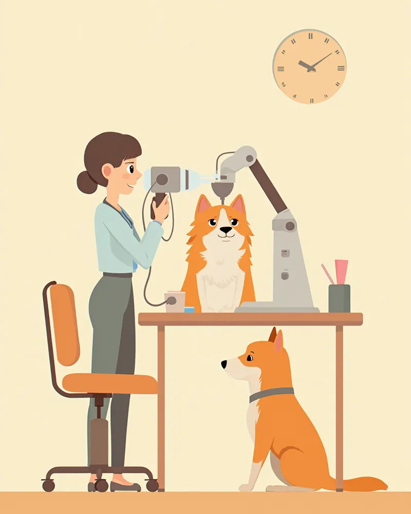
CEA is a genetic disorder, so it’s essential to verify the screening results of both parent dogs before breeding. Mating two carriers can result in 25% of their puppies being affected. If you’re considering getting a Border Collie, ask for an ophthalmic examination certificate from the American College of Veterinary Ophthalmologists (ACVO) or an equivalent accredited institution in Korea, along with the NHEJ1 gene test results. It’s wise to carefully reconsider any breeder who cannot provide both documents.
Lifelong Management Points After Screening
Reviewed by a veterinarian

Dr. Tony — Punnawat Phongkittirak
VeterinarianA veterinarian who majored in veterinary medicine at Khon Kaen University, Thailand, and completed the IVSA program at North Carolina State University in the United States. Drawing on clinical experience at animal hospitals, he works in the pet healthcare field and is dedicated to building a digital care environment that connects pet parents with veterinarians.
Reviewed 2026-04-30
Frequently Asked Questions
How much does the checkup cost?
My dog is already an adult. Should I still get him tested now?
Can a dog with CEA still live a normal daily life?
Can it be confused with other eye diseases?
Is there a way for pet owners to check this at home in advance?
Share
Related Guides

Top 5 Dog Tear Supplements Recommended for 2026

What is Lutein? Effects on Dog Eye Health

The Best Canine Uveitis Management Products and Selection Criteria: A Comprehensive Guide
Dachshund Grooming by Coat Type (Smooth, Longhaired, Wirehaired)
Cocker Spaniel Grooming Guide
Cocker Spaniel Ear Care (Prone to Otitis Externa)

7 Hereditary Conditions in Shih Tzus: Symptoms, Causes, and Lifelong Care Guide

The Ultimate Guide to Shih Tzu Long-Hair Grooming — From Brushing to Grooming Schedules
Golden Retriever Regular Check-up Schedule
Golden Retriever Genetic Condition Checklist — Essential Guide for Owners
References
[1] Esson DW, Calvarese S. Clinical Atlas of Canine and Feline Ophthalmic Disease, 2nd Edition, Chapter 134 Collie Eye Anomaly. John Wiley & Sons, 2022
[2] Lowe JK, Kukekova AV, Kirkness EF, et al. Linkage mapping of the primary disease locus for collie eye anomaly. Genomics. 2003;82(1):86–95
[3] Parker HG, Kukekova AV, Akey DT, et al. Breed relationships facilitate fine-mapping studies: a 7.8-kb deletion cosegregates with Collie eye anomaly. Genome Research, 2007
[4] Munyard KA, Sherry CR, Sherry L. A retrospective evaluation of congenital ocular defects in Australian Shepherd dogs in Australia. Vet Ophthalmol. 2007;10(1):19–22