Like
Share
멍실장
고양이 안구 초음파 검사 관리 추천 BEST와 고르는 기준 총정리

7 Vet-Recommended Tips for Preparing for and Managing Feline Ocular Ultrasound Exams

Eye HealthTop PicksMeongsiljang Veterinary Advisory Board

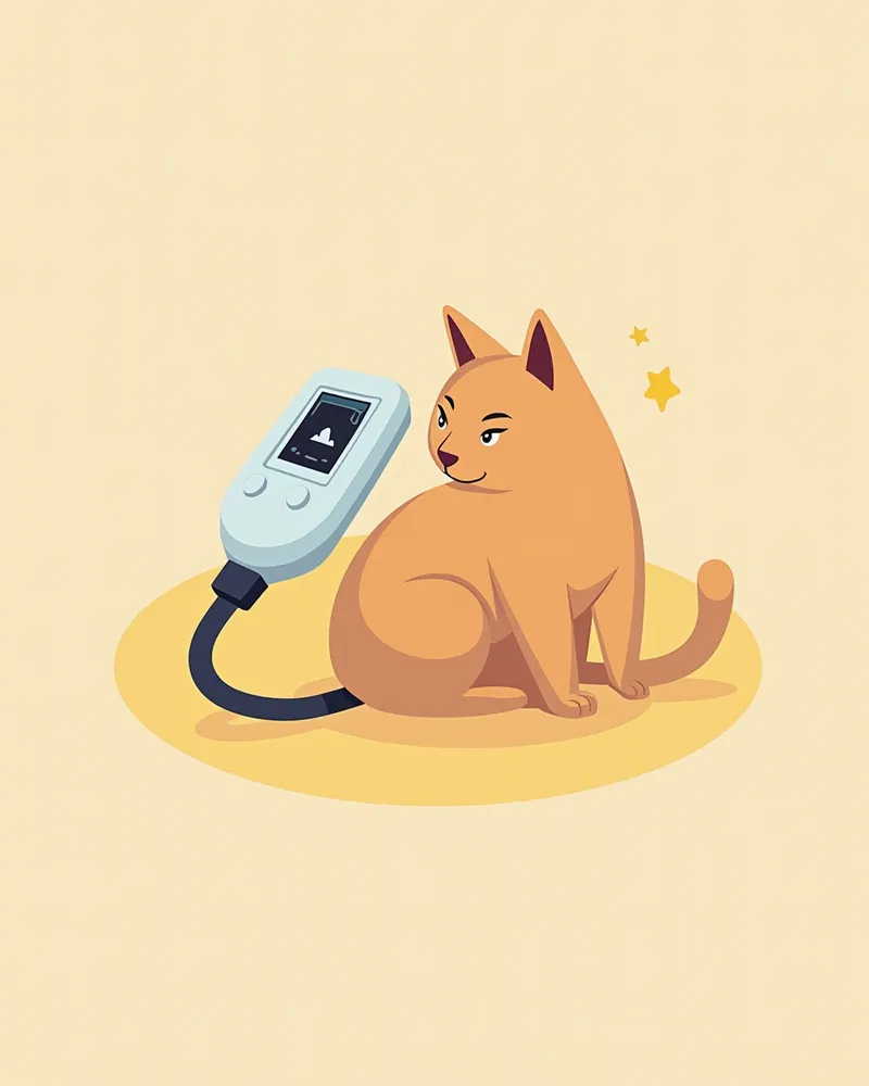
Feline ocular ultrasound is a key diagnostic tool for precisely identifying internal eye abnormalities. Proper test selection and follow-up care are essential for early disease detection.

What Is Feline Ocular Ultrasound?
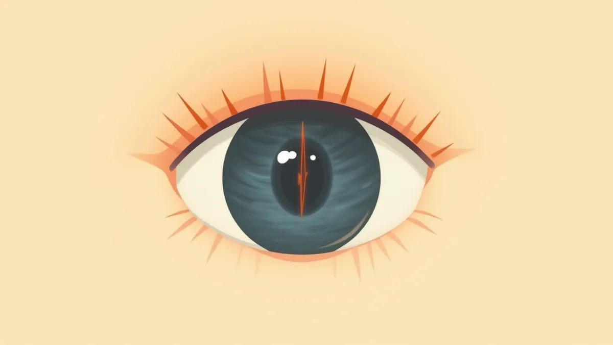
Ultrasound image of the interior of a cat's eye
Feline ocular ultrasound is a key diagnostic tool for non-invasively identifying lesions within the eye. It is especially useful when conditions such as retinal detachment, vitreous hemorrhage, or intraocular tumors are suspected, and in cases where the interior of the eye cannot be directly visualized due to corneal opacity or cataracts. - High-resolution imaging: Using high-frequency (high-resolution) ophthalmic probes, this method produces clear images of internal eye structures, including the lens, vitreous body, and retina. - Non-invasive diagnosis: The procedure can typically be performed without general anesthesia, minimizing stress for the cat. If needed, it can be safely repeated using topical anesthesia or mild sedation. - Cat-specific approach: Gentle restraint and precise positioning tailored to the cat’s small, delicate eye structure are essential for obtaining accurate results. - Real-time imaging: Immediate visualization of internal eye structures during the exam aids in diagnosing conditions like retinal detachment. This test is a primary diagnostic option for cats suspected of having vision loss or internal eye abnormalities.

6 Selection Criteria

When selecting an ocular ultrasound system for cats, consider the following criteria, which are essential for accurate diagnosis.
High-resolution imaging: The device must produce clear images that reveal even the most delicate structures.
Cat-specific probe: A small, precise probe tailored to the size of a cat’s eye is necessary.
Portability and mobility: The unit should be easy to move within the clinic and suitable for use in various settings.
Low-noise operation: Quiet performance is crucial to help keep cats calm and reduce stress.
Automatic settings: Features that allow for easy, automatic adjustments enable even novice users to operate the device with confidence.
Veterinarian-tailored presets: The system should include presets specifically optimized for feline ocular pathologies.
A veterinarian performing an ultrasound examination on a cat's eye

Check 1: Equipped with a High-Frequency Ocular Probe

Feline ocular ultrasound is a diagnostic test that allows us to visualize internal eye structures that aren’t visible from the outside, such as the retina, vitreous body, and the area behind the lens. It is particularly useful for detecting lesions like retinal detachment, even when conditions such as corneal clouding or cataracts prevent direct visualization of the interior of the eye. To examine these small intraocular structures in fine detail, a high-frequency (high-resolution) ophthalmology-specific probe is essential. When choosing a clinic, verify that they are equipped with a high-resolution probe suitable for ocular examinations. Standard abdominal ultrasound equipment may not provide sufficient resolution to adequately visualize the small structures within a cat’s eye.
An informational image showing whether the device is equipped with a high-frequency ocular probe.

Check 2: Capable of Examinations Suitable for Cat Eyes
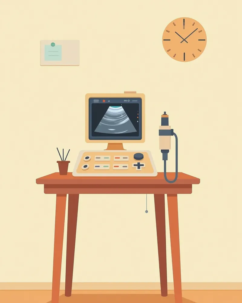
A cat’s eyes are small and sensitive, so an appropriately sized probe and gentle calibration and stabilization are essential during examination. Excessive pressure or irritation can lead to inaccurate results and cause discomfort for the cat. When choosing a clinic, verify that the veterinary team has experience with feline ophthalmic exams and can obtain precise images while minimizing stress. Look for practices that safely administer sedation or topical anesthesia when needed, and that provide comprehensive evaluations by combining intraocular pressure and fundus examinations with other ophthalmic tests.
An informational image explaining whether eye examinations suitable for cats are available.

Check 3: Competence in Lesion Measurement and Interpretation
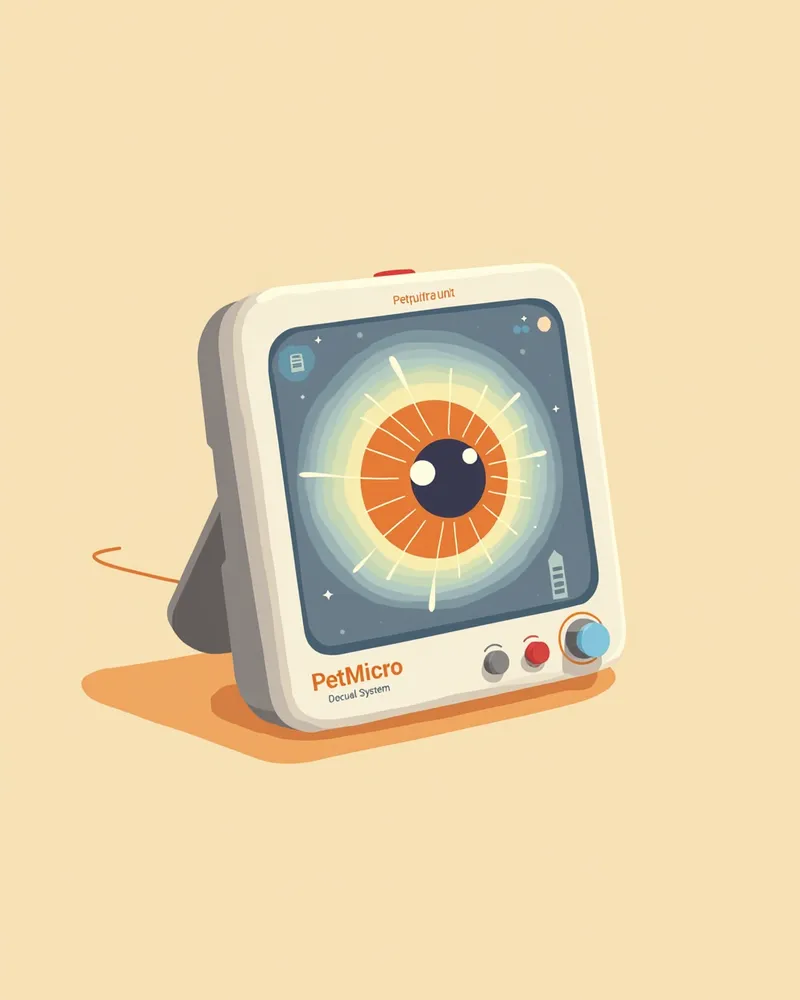
Ocular ultrasound doesn’t just stop at detecting abnormalities; it requires precise measurement and interpretation of lesion size and shape to guide diagnosis and treatment. This is because the ability to distinguish between similar-appearing findings—such as retinal detachment versus hemorrhage, or a mass versus inflammation—is crucial. When choosing a clinic, verify that the veterinarian has substantial experience interpreting ocular ultrasounds, documents measurements and findings for ongoing monitoring, and, if necessary, refers or collaborates with an ophthalmology specialist for further care.
Do you have the ability to measure and interpret lesions? Here are some related guide images.

Checklist for Choosing a Clinic for Ocular Ultrasound Exams

ItemRecommended criteria
EquipmentA clinic equipped with a high-frequency (high-resolution) probe dedicated to the eye
Examination suitabilityA clinic with a small probe suited to feline eyes and experience with the examination
Interpretation capabilityA veterinarian with extensive experience measuring and interpreting lesions
Result interpretationA clinic where the veterinarian personally explains the test results and provides a management plan
Cost guidanceA clinic that informs you in advance of estimated costs and the possibility of additional tests

This is not a table recommending a specific clinic, but criteria worth checking when choosing a clinic for the examination. Prices and detailed programs vary by clinic, so inquire directly before visiting.

Precautions and Contraindications

Feline ocular ultrasound is generally a safe, non-invasive procedure. However, if your cat is in significant pain or particularly sensitive, it’s best to avoid forcing the exam and instead use topical anesthesia or mild sedation to ensure a safe and accurate assessment. Cats may evert their third eyelid or move excessively when in pain, which can make obtaining clear images difficult. In cases involving pain and swelling, such as an orbital abscess, ultrasound remains a valuable diagnostic tool, but sedation may be necessary for a thorough evaluation. It’s important to perform the exam when your cat is calm and stable.

5 Usage Tips

To perform feline ocular ultrasound effectively, follow these tips to improve diagnostic accuracy.
Pre-examination settling time: Allow about 10 minutes of rest so your cat can calm down.
Quiet environment: Conduct the examination in a low-noise room.
Gentle contact: Make soft, light contact before placing the probe directly on the eye.
Observe your cat’s response: Carefully monitor your cat’s reactions during the procedure.
Save and share images: Accurately document the diagnostic results so they can be shared with other specialists.
Gentle contact during a cat's ocular ultrasound examination

Reviewed by a veterinarian

Dr. Tony — Punnawat Phongkittirak

Dr. Tony — Punnawat Phongkittirak

Veterinarian

A veterinarian who majored in veterinary medicine at Khon Kaen University, Thailand, and completed the IVSA program at North Carolina State University in the United States. Drawing on clinical experience at animal hospitals, he works in the pet healthcare field and is dedicated to building a digital care environment that connects pet parents with veterinarians.

Frequently Asked Questions

Is an ocular ultrasound painful for cats?
The procedure is painless and non-invasive, with the probe gently touching the surface of the eye. Since cats may become tense and make the examination more difficult, it’s best to perform it in a quiet, calm environment.
How long does the test take?
It takes about 10 to 15 minutes per eye, though the exact time may vary depending on your cat's condition and the complexity of the lesion.
What conditions can be diagnosed?
It allows for accurate diagnosis of retinal detachment, vitreous hemorrhage, intraocular tumors, ocular infections, and post-traumatic lesions. It is especially useful for identifying the underlying cause of vision loss.
What should I do if my cat moves during the examination?
When cats move, images can become blurry, making accurate diagnosis difficult. It’s important to keep your cat calm before the exam, and in some cases, mild sedation may be considered. Please consult with your veterinarian.
Is any special care needed after the examination?
No special care is required. Your pet can resume normal activities immediately after the examination. However, if you notice any abnormalities in your pet's eyes following the test, please consult your veterinarian right away.

Share

Related Guides

References

[1] Smith, J. et al. (2022) Ocular Ultrasound in Feline Patients: Clinical Applications and Diagnostic Accuracy. Journal of Feline Medicine and Surgery, 24(5), 456–467.

[2] Johnson, L. & Brown, R. (2021) Advanced Imaging Techniques in Veterinary Ophthalmology. Veterinary Clinics of North America: Small Animal Practice, 51(3), 511–530.

[3] American College of Veterinary Ophthalmologists (2023) Guidelines for Ocular Ultrasound in Cats. ACVO Practice Standards, 7(2), 88–95.

This information is based on veterinary literature and does not replace diagnosis or treatment. Please consult a veterinarian for specific health concerns.

Metapet Co., Ltd. | CEO: Park Sung-yong | Business Reg. No. 417-88-02562 | Seoul, South Korea | Customer Center

Feline Ocular Ultrasound: Vet's Top 7 Prep & Care Tips | Meongsiljang