Like
Share
멍실장
안저검사로 확인하는 눈 안쪽 질환

Internal Eye Diseases Detected by Fundus Examination

Eye HealthQ&AMeongsiljang Veterinary Advisory Board

Fundus examination is a detailed eye test that allows us to directly observe the retina, optic nerve, and blood vessels inside the pupil. Here’s a summary of when it’s needed and what conditions it can help detect.

What is a Fundus Examination?
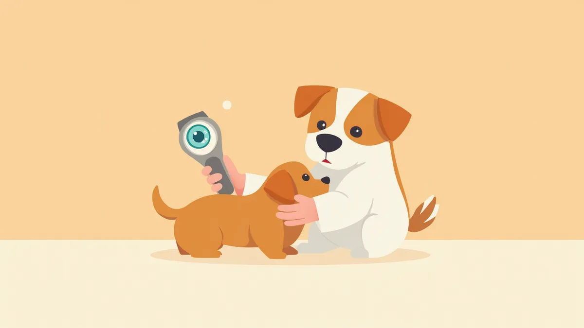
A veterinarian examining the inside of a dog’s eye with an ophthalmoscope
A fundus exam is a detailed eye test that allows direct visualization of the retina, optic disc, and retinal blood vessels inside the pupil. Early detection is key. Even if the eye appears normal on the outside, changes that could lead to blindness may already be progressing internally. If you suspect vision loss, abnormal pupillary response, or sudden blindness, your pet should undergo a fundus exam immediately.

When Should the Examination Be Performed?

Fundus examinations are performed when internal eye issues are suspected, rather than problems on the surface. According to veterinary internal medicine textbooks, this test is recommended if you notice any of the following signs: - Sudden vision loss: Bumping into furniture or showing fear of stairs - Abnormal pupil size: Pupils are unequal in size or react slowly to light - Changes in eye color: The interior appears cloudy or has a reddish reflection - Presence of systemic diseases: Regular check-ups are necessary for pets with hypertension, diabetes, or kidney disease

Diseases Detectable by Fundus Examination

Fundus exams can help detect vision-threatening diseases inside the eye at an early stage. These conditions can be broadly categorized into diseases of the retina itself, optic nerve diseases, and signs left by systemic diseases in the eye. The table below summarizes representative conditions.

Major Diseases Detected by Fundus Examination

ItemRepresentative diseaseMain findings
Retinal diseaseProgressive retinal atrophy (PRA)Reduced retinal vessels · increased reflectivity
Retinal detachmentRetinal detachment (primary · secondary)Findings of the retina lifting; hemorrhage may accompany depending on the cause
Optic nerve diseaseOptic neuritis · optic nerve atrophyOptic disc swelling or pallor
Sequelae of systemic diseaseHypertensive retinopathyVascular hemorrhage · retinal detachment
Infection · inflammationChorioretinitisRetinal lesions · exudate

Actual diagnosis is made comprehensively together with intraocular pressure testing, ultrasound, and other tests.

How Is the Examination Performed?

Fundus examinations are typically performed without anesthesia. The procedure consists of three main steps: - Pupil dilation drops: Eye drops are administered to dilate the pupils, and we wait until they are fully dilated. Commonly used tropicamide-based dilating drops usually widen the pupils relatively quickly, typically within about 15 minutes. - Observation in a dark room: In a darkened room, we directly examine the retina, optic disc, and retinal blood vessels using an ophthalmoscope. - Additional imaging: If necessary, we take photographs with a fundus camera for documentation. The examination itself is painless. The effects of the dilating drops usually last for a few hours (typically 2–3 hours) before wearing off, so your pet may experience sensitivity to bright light on the day of the exam.
Close-up photo of a cat's eye with dilated pupils after administration of atropine.

Go to the Hospital Immediately If You See These Signs

If your pet suddenly bumps into objects or their eyes become completely cloudy, it could be a medical emergency such as retinal detachment or acute glaucoma. The sooner treatment begins, the better the chance of preserving vision, so if you notice these signs, don’t wait for your next scheduled appointment—head straight to a veterinary clinic that offers after-hours care.

Preparation and Precautions Before the Examination

No special fasting is required before a fundus examination. However, since we use mydriatic eye drops to dilate the pupils, please inform us in advance if your pet has a history of glaucoma, as this can cause a rapid increase in intraocular pressure. - Current eye medications: Please let us know about any eye drops your pet has been using recently. - Systemic medications: If your pet is taking heart or blood pressure medications, please share that information with us as well. - After the exam: Keep your pet away from bright light and keep the home environment dim until the effects of the pupil-dilating drops wear off.
A dog resting in a dimly lit room after an ophthalmoscopic examination

Which Pets Are Candidates for Regular Examinations?

According to veterinary ophthalmology textbooks, regular fundus examinations are recommended for certain breeds and dogs or cats with systemic diseases, even if they show no symptoms. - Breeds prone to hereditary retinal diseases: Poodles, Cocker Spaniels, Labradors, Schnauzers - Risk of hypertension: Senior cats with chronic kidney disease or thyroid dysfunction - Diagnosis of diabetes: After confirmation, the first fundus examination should be performed as soon as possible, followed by regular follow-up exams as determined by the veterinarian - Senior dogs and cats: For older animals or those with systemic diseases, it is recommended to include a fundus examination in routine comprehensive eye checkups, even if no symptoms are present

Reviewed by a veterinarian

Dr. Tony — Punnawat Phongkittirak

Dr. Tony — Punnawat Phongkittirak

Veterinarian

A veterinarian who majored in veterinary medicine at Khon Kaen University, Thailand, and completed the IVSA program at North Carolina State University in the United States. Drawing on clinical experience at animal hospitals, he works in the pet healthcare field and is dedicated to building a digital care environment that connects pet parents with veterinarians.

Frequently Asked Questions

How much does a fundus examination typically cost?
The cost can vary significantly depending on the clinic and the equipment used, so it’s difficult to provide an exact figure. The price will differ depending on whether it’s just a basic ophthalmoscopic exam or if it also includes fundus camera imaging, so it’s a good idea to ask when you make your appointment.
Will my pet be in pain if we administer the sedative?
The eye drops may cause a slight stinging sensation, but there is little to no pain. However, light sensitivity can persist for a while, so it’s a good idea to close the curtains or dim the lights at home after the examination.
Can all eye diseases be diagnosed through a fundus examination alone?
A fundus exam allows us to look at the interior structures of the eye. However, to accurately diagnose issues with the front of the eye, such as corneal ulcers or early-stage cataracts, we need to combine it with other tests like a slit-lamp examination and an intraocular pressure measurement.
My dog is senior. Does he really need to get the vaccine every year?
For older pets or those with systemic conditions like diabetes, kidney disease, or high blood pressure, regular fundus exams are recommended even if there are no symptoms. Early detection of vision-threatening diseases is key to preserving sight for as long as possible.
Is there any point in having tests done if the pet has already gone blind?
Yes, it’s meaningful. The underlying condition could be a systemic issue (e.g., hypertension or tumors), and this information is essential for determining treatment strategies to prevent progression in the other eye or to reduce pain.

Share

Related Guides

References

[1] Maggs, D.J., Miller, P.E., Ofri, R., Slatter's Fundamentals of Veterinary Ophthalmology, 6th Ed

[2] Clinical Atlas of Canine and Feline Ophthalmic Disease, 2nd Ed — Neurophthalmic Examination

[3] Gelatt, K.N., Veterinary Ophthalmology, 6th Ed

This information is based on veterinary literature and does not replace diagnosis or treatment. Please consult a veterinarian for specific health concerns.

Metapet Co., Ltd. | CEO: Park Sung-yong | Business Reg. No. 417-88-02562 | Seoul, South Korea | Customer Center

Fundus Exam: Spotting Inner Eye Disease in Pets | Meongsiljang