Like
Share
멍실장
심전도(ECG) 검사 시기와 해석 완벽 가이드

The Complete Guide to When and How to Interpret an ECG

HeartQ&AMeongsiljang Veterinary Advisory Board

We’ve put together the essential information pet owners need to know about when an ECG is necessary and how to interpret the results.

What is an Electrocardiogram (ECG)?
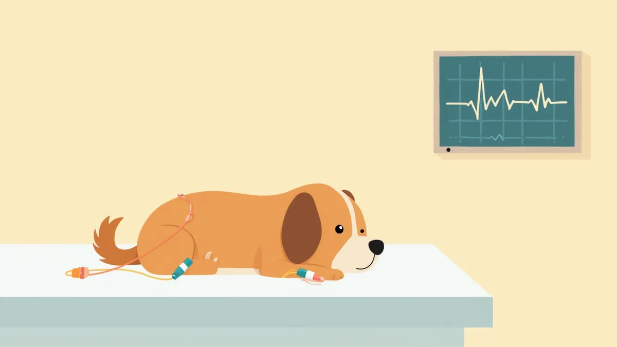
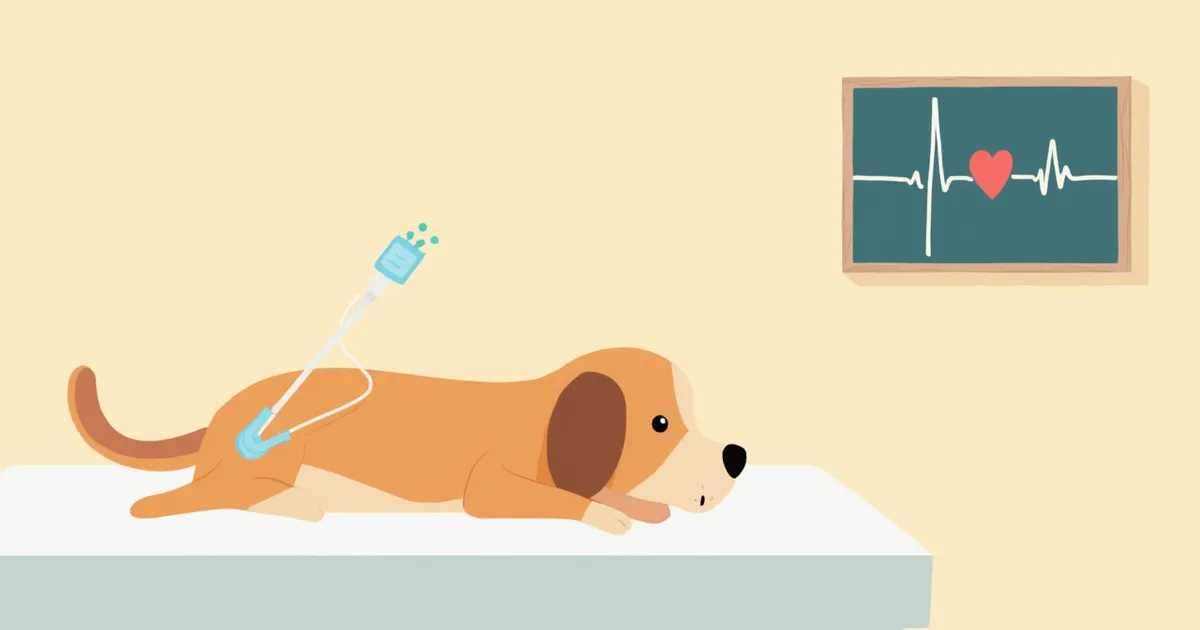
Attaching electrocardiogram electrodes to a dog's chest
An electrocardiogram (ECG) is a non-invasive test that records the tiny electrical signals generated by the heart as it beats, allowing us to assess heart rhythm and conduction. What really matters is its role in diagnosing arrhythmias. An ECG is the only way to detect irregular rhythms and electrical conduction abnormalities in real time—issues that can easily be missed with auscultation alone. However, for structural changes like atrial or ventricular enlargement, it’s more accurate to combine ECG findings with an echocardiogram. We strongly recommend an ECG before anesthesia or surgery, or if your pet has experienced fainting or collapse, to check for underlying arrhythmias.

When is an ECG Necessary?

This is the first test we recommend when heart disease is suspected or when an abnormal rhythm is detected during auscultation. It is especially necessary in the following situations: - Fainting or syncope episodes: If your pet has suddenly collapsed, it may be due to an arrhythmia. - Exercise intolerance: Becoming short of breath or collapsing after minimal exertion. - Pre-anesthesia screening: For senior pets or those with suspected heart murmurs or rhythm abnormalities. - Monitoring heart treatment progress: To assess response to medication and changes in arrhythmia. - Specific breeds: Breeds with genetic predispositions, such as Boxers, Doberman Pinschers, Maine Coons, and Ragdolls.

Comparison of Test Types and Duration

Electrocardiogram (ECG) tests are divided into three types depending on their purpose, offering a variety of options ranging from simple screening to continuous monitoring over 24 hours or more.

Comparison of ECG Test Types

ItemStandard ECGHolter monitoringEvent recorder
Measurement time3–5 minutes24–48 hours7 to 30 days
LocationIn clinicWorn at homeWorn at home
PurposeBasic rhythm checkCatching intermittent arrhythmiasTracking rare causes of syncope
Cost rangeKRW 30,000–80,000KRW 200,000–350,000KRW 400,000–600,000

Averages for general veterinary clinics as of 2025; varies by institution

How to Read ECG Results
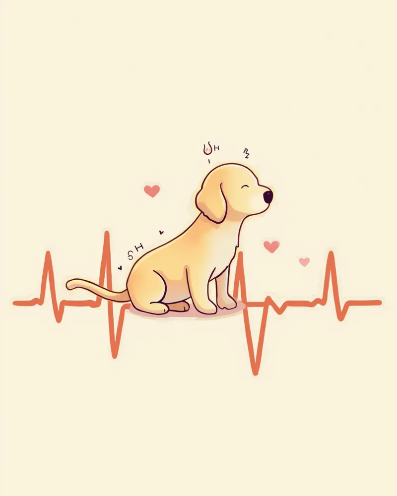
An electrocardiogram (ECG) graph consists of P waves, QRS complexes, and T waves. While pet owners don’t need to interpret every waveform, understanding these key concepts can be helpful when your veterinarian explains the results. - P wave: The signal generated when the atria contract - QRS complex: The large waveform produced when the ventricles contract — this is the most important component - T wave: The signal indicating ventricular recovery (repolarization) - Heart rate: Normal ranges are 60–160 beats per minute (bpm) for dogs and 140–220 bpm for cats - Rhythm regularity: Check whether the beats occur at consistent intervals
Explanation of the P wave, QRS complex, and T wave on an electrocardiogram (ECG) tracing

Emergency Signs Requiring Immediate Testing

If your pet shows any of the following symptoms, seek emergency veterinary care immediately. These signs require prompt evaluation and treatment, as they can be life-threatening. • Repeated episodes of collapse or loss of consciousness (syncope) • Blue or pale gums and tongue • A resting respiratory rate exceeding 40 breaths per minute • Sudden weakness and coldness in the hind legs (especially in cats, which may indicate arterial thromboembolism) • Coughing accompanied by frothy discharge These symptoms could indicate life-threatening conditions such as arrhythmias or heart failure, so do not delay seeking care.

Commonly Found Arrhythmia Types
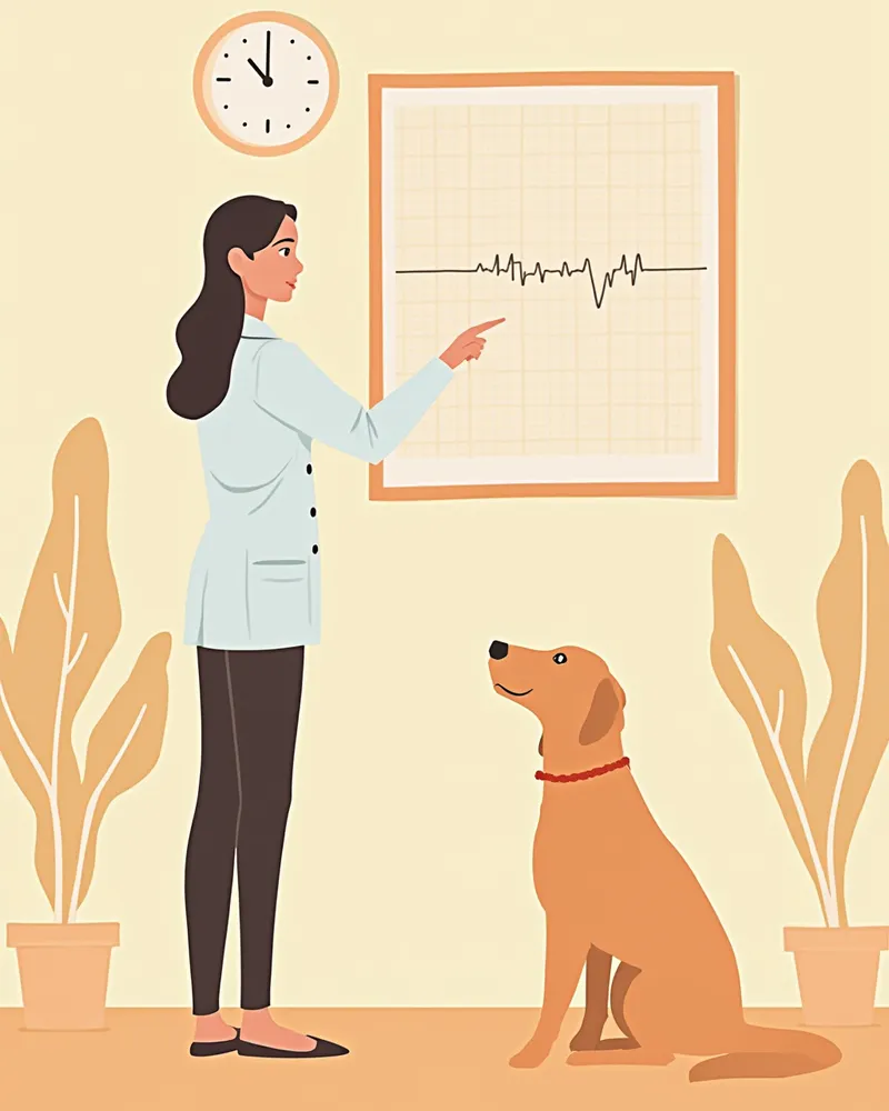
This is a classic arrhythmia diagnosed via electrocardiogram (ECG). A diagnosis doesn’t always mean danger, but the treatment approach varies depending on the type. - Atrial fibrillation: The atria quiver chaotically — common in large-breed dogs with dilated cardiomyopathy - Ventricular premature contractions (VPCs): Early beats originating from the ventricles — monitor Boxers and Doberman Pinschers closely - Atrioventricular block: Delayed or interrupted electrical conduction — third-degree block requires a pacemaker - Sinus arrhythmia: A normal variation where heart rate changes with breathing — common in puppies According to veterinary internal medicine textbooks, the prevalence of arrhythmias varies significantly by breed.
A veterinarian explaining the results of an electrocardiogram (ECG) test.

When a Holter Monitor is Needed

Even if a standard ECG appears normal, a 24-hour Holter monitor should be considered if symptoms recur. • When there is a history of syncope or fainting, but the in-clinic ECG is normal • When screening for hereditary heart conditions is needed in breeds such as Boxers and Doberman Pinschers • When monitoring medication efficacy and arrhythmia control in pets currently taking heart medication • When pre- and post-implantation assessment of a pacemaker is required Arrhythmias often occur only intermittently, so they can be missed on a short in-clinic ECG.

Test Preparation and Precautions

An electrocardiogram (ECG) is a safe test with minimal pain or side effects. However, a few preparations are necessary to ensure accurate results. - Before the test: Avoid walks or excitement and keep your pet calm for at least 30 minutes. - Positioning: Your pet is usually placed on their right side for measurement, though a standing position is possible for anxious pets. - Electrode attachment: Clip-on electrodes are attached to all four limbs, and a small amount of alcohol is applied to improve conductivity. - Current medications: Please be sure to inform us of the names and timing of any medications your pet is currently taking. - While wearing a Holter monitor: Avoid showering and excessive exercise, and keep a symptom diary.

Reviewed by a veterinarian

Dr. Tony — Punnawat Phongkittirak

Dr. Tony — Punnawat Phongkittirak

Veterinarian

A veterinarian who majored in veterinary medicine at Khon Kaen University, Thailand, and completed the IVSA program at North Carolina State University in the United States. Drawing on clinical experience at animal hospitals, he works in the pet healthcare field and is dedicated to building a digital care environment that connects pet parents with veterinarians.

Frequently Asked Questions

How often should an electrocardiogram (ECG) be performed?
For healthy pets, an annual check-up is generally sufficient. If your pet has been diagnosed with heart disease or is currently on medication, we recommend follow-up exams every three to six months. For senior pets aged seven and older, pre-anesthetic testing is essential.
Does your pet experience pain or significant stress during examinations?
An electrocardiogram (ECG) is a non-invasive test that doesn’t involve needles or radiation. You’ll only feel a slight pinch from the electrode clips, and the procedure typically takes just 3 to 5 minutes. Even sensitive pets can undergo the test with minimal stress.
How are echocardiograms and electrocardiograms different?
An electrocardiogram (ECG) assesses the heart’s electrical signals, including rhythm and arrhythmias, while an echocardiogram evaluates structural integrity and contractile function, such as valve and myocardial health. Since these two tests complement each other, they are often performed together for a comprehensive assessment of heart disease.
Can cats also undergo an electrocardiogram (ECG) test?
Yes, it’s possible. However, cats are easily stressed, and simply visiting the clinic can cause their heart rate to spike significantly. According to veterinary textbooks, if a cat’s heart rate exceeds 240 beats per minute, it’s necessary to evaluate whether treatment is required. Therefore, it’s important to measure their heart rate in a quiet environment, and if needed, use a Holter monitor at home to record their heart rhythm in a calm state.
Does a normal ECG result mean there is no heart disease?
Not exactly. An ECG primarily detects rhythm abnormalities, so issues like valve disease or weakened heart muscle contraction can only be confirmed with an echocardiogram. For an accurate diagnosis, a combination of an ECG, echocardiogram, and chest X-ray is standard.

Share

Related Guides

References

[1] Textbook of Cardiovascular Medicine in Dogs and Cats

[2] Small Animal Critical Care Medicine, 3rd Ed

[3] Advanced Monitoring for Small Animal Emergency and Critical Care, 2nd Ed

[4] Handbook of Veterinary Pharmacology

This information is based on veterinary literature and does not replace diagnosis or treatment. Please consult a veterinarian for specific health concerns.

Metapet Co., Ltd. | CEO: Park Sung-yong | Business Reg. No. 417-88-02562 | Seoul, South Korea | Customer Center

When You Need an ECG and How to Read the Results | Meongsiljang