Like
Share
멍실장
강아지 망막 변성·노화성 망막 변화 자주 묻는 질문 Q&A — 보호자가 꼭 알아야 할 핵심

What is Canine Retinal Degeneration — How to Identify Age-Related Vision Loss

Eye HealthQ&AMeongsiljang Veterinary Advisory Board

Canine retinal degeneration is a condition in which vision gradually declines due to aging or genetic factors. Here’s a summary of the key information every pet owner should know.

What is Canine Retinal Degeneration and Age-Related Retinal Changes?
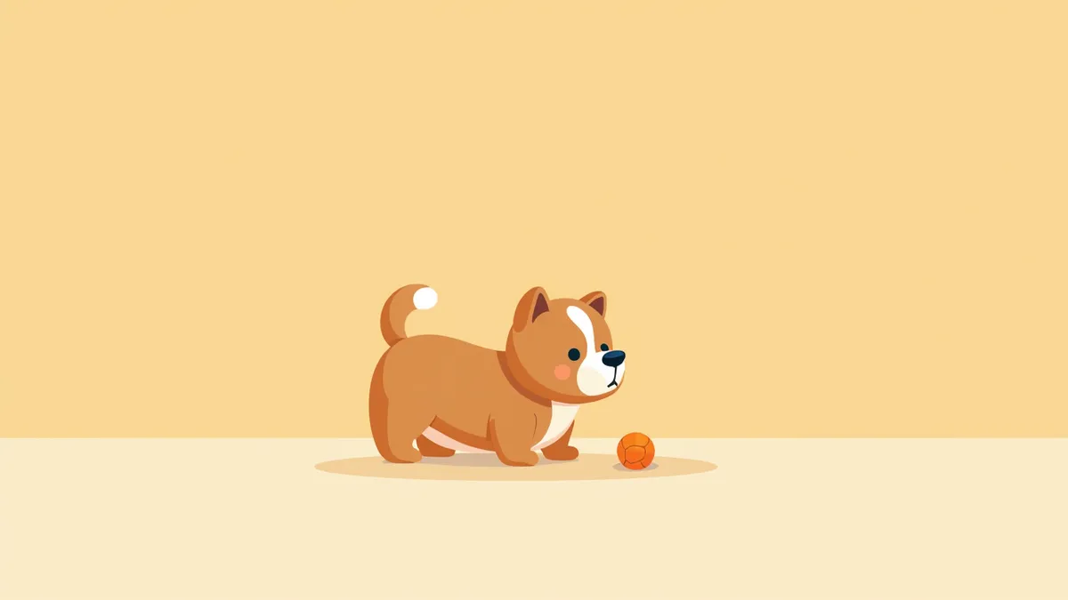
An elderly dog gazing at a toy with blurred vision.
Canine retinal degeneration, also known as retinal atrophy, is a bilateral condition in which photoreceptor cells (visual cells) gradually lose function and die, most often due to genetic factors, leading to progressive vision loss. The age of onset varies: early-onset cases typically begin between 6 weeks and 6 months of age, with blindness developing between 1 and 5 years, while late-onset cases usually start around 3 to 5 years of age, progressing to blindness between 6 and 8 years. In the early stages, the pupils may appear larger than usual (dilated), and dogs may show signs of reduced night vision, such as unsteady gait in dim light or difficulty locating doors. As the condition advances, the visual field narrows even in bright light, dogs frequently bump into obstacles, and their responses become slower. Because the disease progresses gradually, consistent monitoring by pet owners is essential. - Retinal degeneration: A condition characterized by the decline and death of photoreceptor cells, resulting in vision loss that can ultimately lead to complete blindness, making early detection crucial. - Hereditary and breed predisposition: This is a hereditary, bilateral condition that occurs more frequently and may progress more rapidly in certain breeds, including Irish Setters, Miniature Schnauzers, Collies, and Labrador Retrievers.

What are the Main Causes of This Condition?
The primary cause of canine retinal degeneration (retinal atrophy) is genetic. Textbooks classify this condition as a hereditary, bilateral retinal disease, with the causative genes varying by breed. Early-onset forms (such as rod-cone dysplasia) are seen in Irish Setters, Miniature Schnauzers, Norwegian Elkhounds, and Dachshunds; X-linked forms occur in Samoyeds, Siberian Huskies, and Collies; and late-onset forms (Progressive Rod-Cone Degeneration, PRCD) are well-documented in Labrador Retrievers and Chesapeake Bay Retrievers. While antioxidant supplements are sometimes recommended to slow disease progression, they cannot prevent blindness. - Genetic factors: Specific gene mutations that impair the survival and function of photoreceptors are the most critical cause. - Breed predisposition: Certain breeds, such as Collies, Labrador Retrievers, and Golden Retrievers, are more prone to the condition, making genetic testing helpful. - Oxidative stress: The recommendation of antioxidant products to inhibit progression suggests a link to cellular damage. - Secondary changes: Over time, secondary changes such as cataracts may develop.
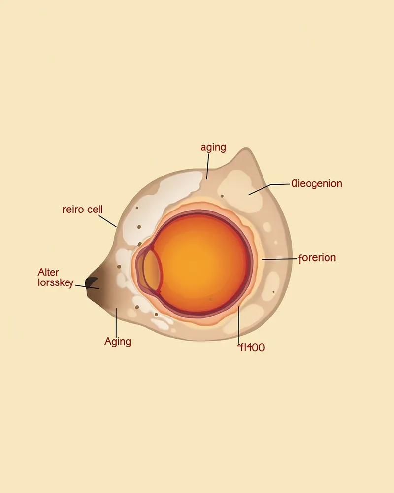
An anatomical diagram showing retinal cells damaged by aging

What Symptoms Appear?

The earliest signs of canine retinal degeneration are mydriasis (dilated pupils) and decreased night vision. Your dog may show unsteady gait in dim light, difficulty locating doors, or wandering behavior. - Mydriasis (pupil dilation): An early sign where the pupils remain more dilated than usual. - Decreased night vision (nyctalopia): An early signal of impaired mobility in low-light conditions. - Narrowed visual field: A reduced field of vision that makes your dog more prone to bumping into obstacles. - Delayed responses and unsteady movement: Slower reactions to sounds or movement, along with repeated stumbling or disorientation. Because these changes occur gradually, it is important for owners to consistently observe and record their dog’s behavior.
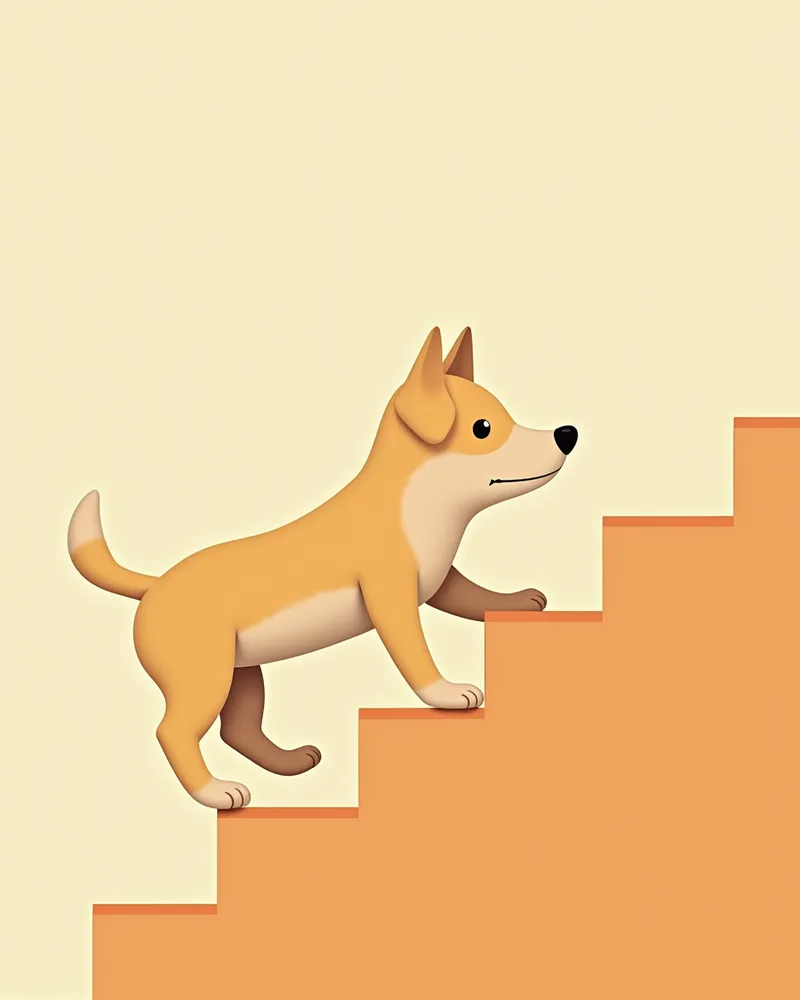
A puppy showing anxious reactions at stair edges due to retinal changes

Signs Requiring Immediate Veterinary Visit

If your pet suddenly loses vision, develops red eyes, or experiences excessive tearing, consult a veterinarian immediately. In particular, if they rapidly become blind and disoriented over the course of a few days, it could indicate an emergency condition such as sudden acquired retinal degeneration syndrome (SARDS). Since cataracts, glaucoma, and retinal detachment are also possible, prompt diagnosis is essential.

How is it Diagnosed?
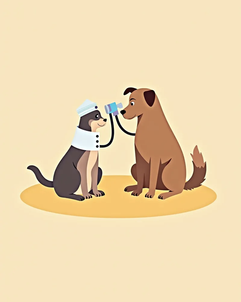
Veterinarians directly assess the condition of the retina through specialized ophthalmic examinations. Funduscopy is used to identify structural changes in the retina, such as increased tapetal reflection, vascular atrophy, and optic disc degeneration, while electroretinography (ERG) evaluates the function of photoreceptors. - Funduscopy: This examination allows for direct observation of structural abnormalities in the retina, such as vascular atrophy and increased reflection. - Electroretinography (ERG): This test measures the electrical responses of retinal cells to assess their functional status. - Genetic Testing: Genetic tests are available for breed-specific hereditary retinal atrophy, aiding in both diagnosis and breeding management. - Comprehensive Diagnosis: An accurate diagnosis is made by integrating clinical findings with results from funduscopy, ERG, and genetic testing.
A veterinarian examining a dog's eyes using ophthalmoscopy equipment

What are the Treatment Options?

Since retinal degeneration is currently difficult to cure completely, treatment focuses on slowing disease progression and alleviating symptoms rather than achieving a full recovery. While products containing antioxidants are often recommended to reduce oxidative stress and delay progression, it’s important to understand that they cannot prevent blindness entirely. The choice and use of any supplementary nutrients should always be discussed with your veterinarian to tailor the approach to your pet’s individual condition. Additionally, creating a safe home environment helps minimize accidents, while maintaining a consistent daily routine can help reduce stress. - Antioxidants: Recommended to help slow progression, but they cannot completely prevent blindness. - Supplementary Nutritional Support: The effects of additional supplements are limited, so consult your veterinarian before selecting any. - Environmental Adjustments: Ensure a safe environment by securing furniture, using auditory cues, and marking stair boundaries. - Regular Check-ups: Monitor disease progression and adjust the management plan accordingly.
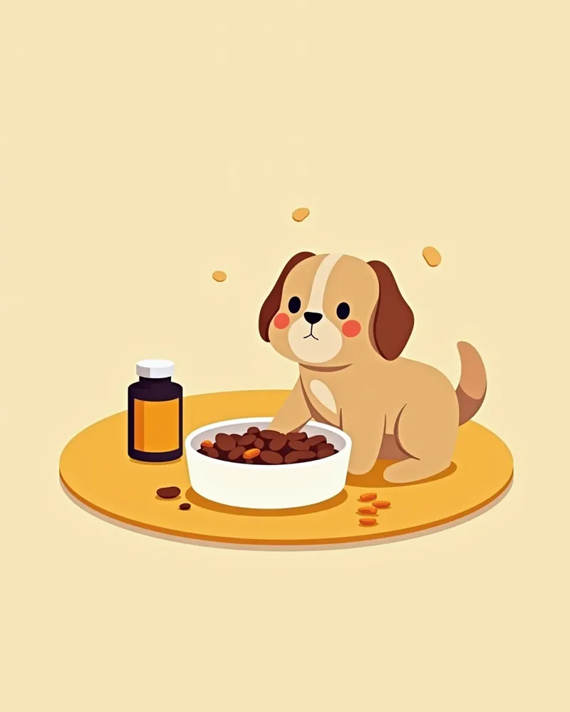
An image showing supplements and food for maintaining your dog's eye health.

Home Care Management

Creating a safe living environment for your dog is the most important step. Secure furniture in place, apply tape to stairs or thresholds to create clear boundaries, and always keep food and water bowls in the same spot. Using sound cues to indicate locations or guiding your dog with gentle touch can also be effective. Additionally, maintaining a consistent daily routine helps reduce stress and makes behavioral changes easier to anticipate. Regular veterinary check-ups are essential to monitor your dog’s condition. - Environmental Stability: Fixed furniture placement and clear boundary markers help prevent accidents. - Sound Cues: Using sound to indicate where food or water is located makes it easier for your dog to orient themselves. - Routine Maintenance: Providing meals, walks, and rest at consistent times helps reduce stress. - Regular Check-ups: Early detection of changes in condition allows for timely adjustments to the care plan.
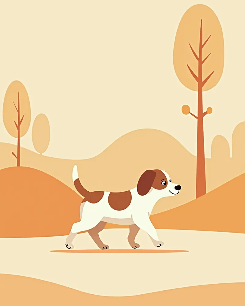
An elderly dog walking safely without obstacles

Stage-Specific Characteristics and Management of Retinal Changes in Senior Dogs

ItemMain SymptomsManagement
Early (mydriasis / reduced night vision)Pupils enlarge and gait becomes unsteady in dark placesKeep the environment consistent and provide supplemental night lighting
Middle (narrowing visual field)Frequently bumps into obstacles and cannot find doorwaysPrevent accidents by securing furniture and marking boundaries
Late (severe vision loss / blindness)Slow to respond and has difficulty perceiving surroundingsCombine sound/tactile guidance with regular checkups

Hereditary retinal degeneration can eventually progress to blindness, so stage-by-stage environmental adjustments and regular checkups are important. Antioxidant products may slow progression but cannot prevent blindness.

Reviewed by a veterinarian

Dr. Tony — Punnawat Phongkittirak

Dr. Tony — Punnawat Phongkittirak

Veterinarian

A veterinarian who majored in veterinary medicine at Khon Kaen University, Thailand, and completed the IVSA program at North Carolina State University in the United States. Drawing on clinical experience at animal hospitals, he works in the pet healthcare field and is dedicated to building a digital care environment that connects pet parents with veterinarians.

Frequently Asked Questions

Is retinal degeneration curable?
Currently, there is no cure for this condition. However, products containing antioxidants are often recommended to help slow its progression. It’s important to note that these products cannot prevent blindness itself, so early detection and proper environmental management are key.
Which breeds are at higher risk?
Hereditary retinal atrophy is relatively common in Irish Setters, Miniature Schnauzers, Collies, Labrador Retrievers, Golden Retrievers, and Samoyeds. Breed-specific genetic testing can be helpful.
When should I start giving my pet nutritional supplements?
There is limited evidence to support specific recommended timing for these supplements. Whether and when to use antioxidant products or other nutritional supplements should be decided in consultation with your veterinarian, based on your pet’s individual health status.
What can I do at home to help my pet if their vision is declining?
Keep furniture in place and use sounds to help your pet navigate, creating a safe environment.
How can you tell the difference between retinal changes and cataracts?
Cataracts cause the lens to cloud, making the eye appear hazy or with white spots, while retinal degeneration leads to dilated pupils and an initial decline in night vision and visual field. Since these two conditions can occur together, a specialized ophthalmic examination is necessary.

Share

Related Guides

References

[1] Ettinger SJ, Feldman EC, Côté E. Textbook of veterinary internal medicine: diseases of the dog and the cat. 8th ed. St. Louis, MO: Elsevier; 2017.

[2] Maggs DJ. Third eyelid. In: Maggs DJ, Miller PE, Ofri R, Slatter D. Veterinary ophthalmology. 5th ed. Oxford: Blackwell Publishing; 2017.

[3] Clinical Atlas of Canine and Feline Ophthalmic Disease, 2nd Ed. Wiley-Blackwell; 2015.

This information is based on veterinary literature and does not replace diagnosis or treatment. Please consult a veterinarian for specific health concerns.

Metapet Co., Ltd. | CEO: Park Sung-yong | Business Reg. No. 417-88-02562 | Seoul, South Korea | Customer Center

What Is Canine Retinal Degeneration? Age-Related Vision Loss | Meongsiljang