Like
Share
멍실장
강아지 MRI 판독과 필요한 경우

Canine MRI Interpretation and Indications

ImmunityQ&AMeongsiljang Veterinary Advisory Board

We’ve compiled the essential information pet owners need to know about canine MRIs, including when they’re necessary, the imaging process, how results are interpreted, and associated costs.

What is a Canine MRI?
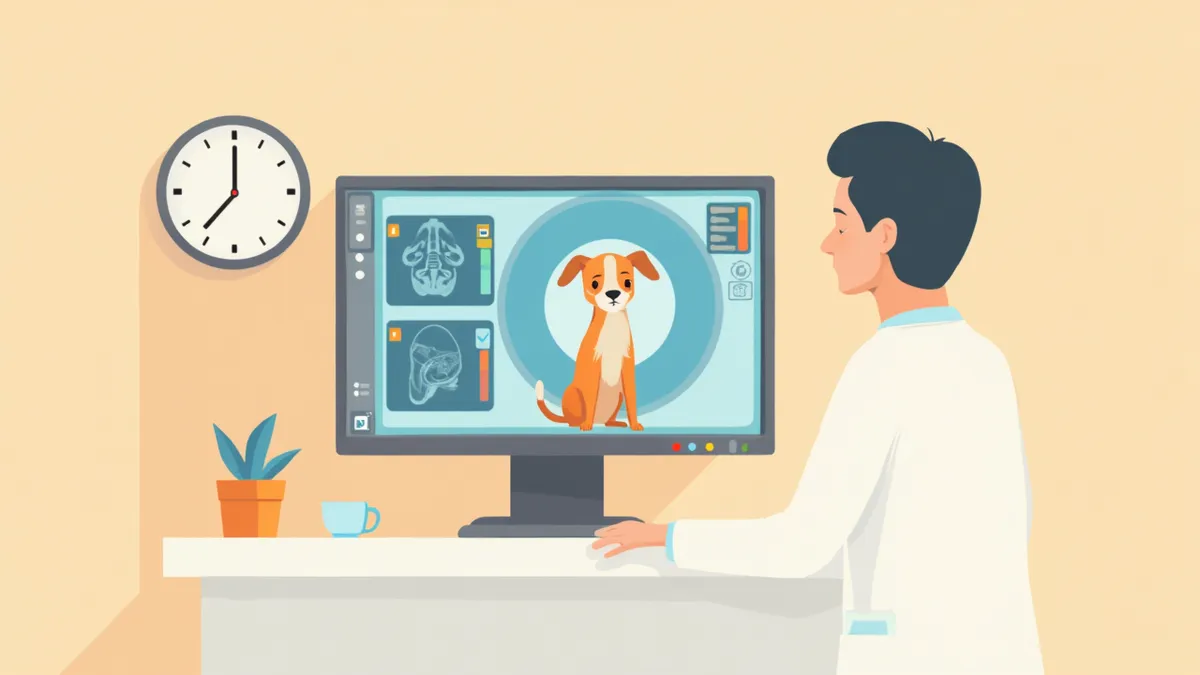
A veterinarian reviewing a puppy's brain MRI scan on a monitor.
Canine MRI (Magnetic Resonance Imaging) is an imaging test that uses magnetic fields and radio waves to produce detailed images of the brain, spinal cord, and joints. It is the most accurate method for identifying abnormalities in soft tissues and the nervous system that are difficult to detect with X-rays or ultrasound. Veterinarians often recommend MRI when neurological symptoms such as seizures, paralysis, or gait abnormalities are present, and it plays a crucial role in determining the precise location and extent of tumors or disc disease.

When is an MRI Necessary?

Here are the most common situations in which a veterinarian would recommend an MRI for your dog:
Recurrent seizures: To check for abnormalities inside the brain when seizures occur repeatedly without a clear cause.
Paralysis or gait abnormalities: To assess whether the spinal cord is being compressed if your dog shows weakness in the hind legs or appears unsteady.
Head tilt: To examine the inner ear and brainstem if your dog tilts its head to one side or moves in circles.
Chronic pain: To check for disc disease or tumors when neck or back pain persists despite showing no clear cause on X-rays.
Suspected tumors: To determine the exact location, size, and extent of invasion when a nervous system tumor is suspected.
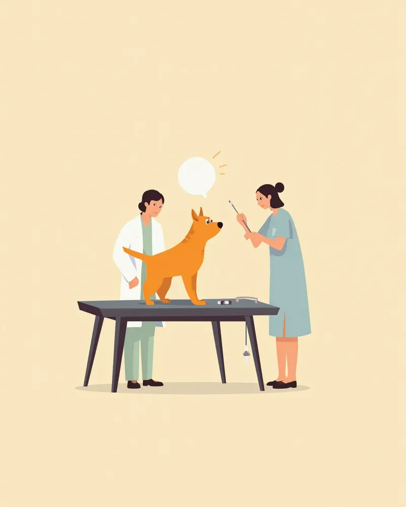
A veterinarian examining a puppy that is tilting its head on the examination table.

Comparison of MRI, CT, and X-ray

ItemMRICTX-ray
StrengthsDetailed imaging of soft tissue and the nervous systemDetailed imaging of bones and fracturesBasic imaging of bones and lungs
Areas seen wellBrain, spinal cord, and joint soft tissueFractures, skull, and thoracic structuresBasic skeleton and organ outlines
Anesthesia requiredRequiredRequiredUsually not required
Imaging time40–60 minutes10–20 minutesA few minutes
Cost range (per area)KRW 800,000–2,000,000KRW 500,000–1,000,000KRW 50,000–150,000

Costs may vary by clinic, region, and body area imaged. This is the typical range as of 2024.

The MRI Scanning Process
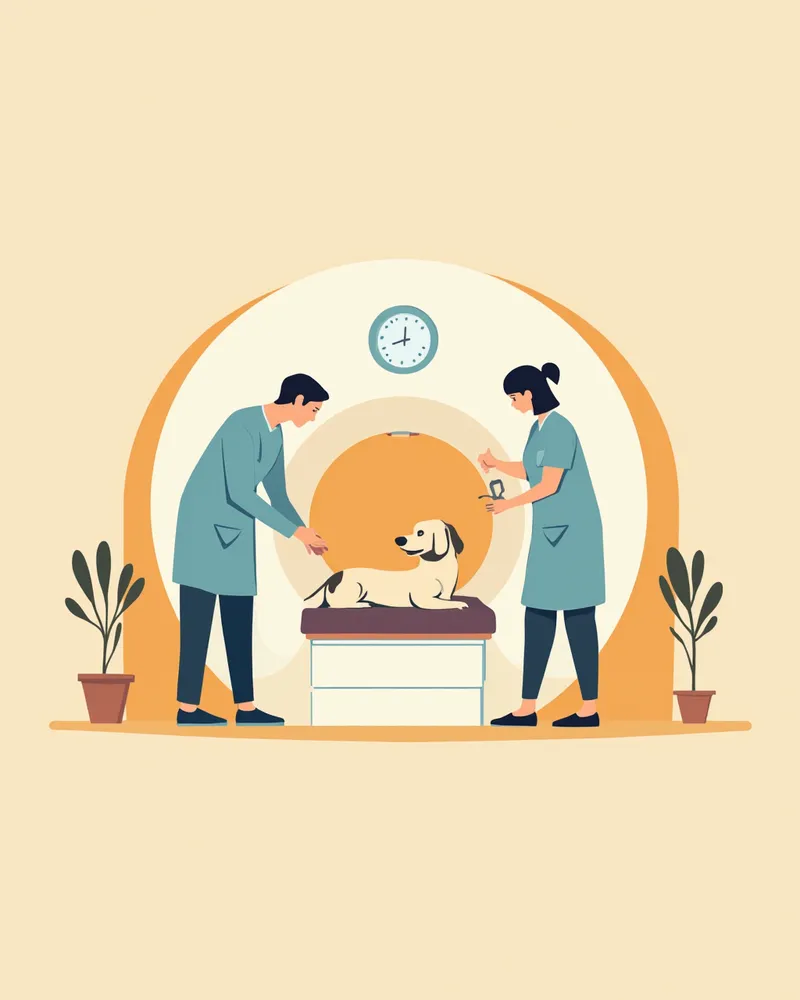
MRI scans are performed under general anesthesia. Since even slight movement can blur the images, anesthesia is essential.
Pre-scan examinations: Blood tests and cardiac evaluations are conducted first to ensure anesthesia safety.
Fasting: Fasting is required before the scan for anesthesia purposes. The exact fasting duration varies depending on the anesthesia method and your dog’s condition, so please follow your veterinarian’s instructions.
Anesthesia and scanning: After anesthesia is administered, the scan is performed inside the MRI machine. The duration may vary depending on the number of areas being scanned.
Recovery: After the scan, your dog will rest and recover at the clinic until fully awake from anesthesia.
In some cases, a contrast agent may be injected depending on the area being scanned. Using a contrast agent helps tumors or inflamed areas appear more clearly.
The veterinary medical team prepares a dog for an MRI scan.

Precautions for General Anesthesia

MRI requires general anesthesia. Senior dogs, those with heart conditions, and brachycephalic breeds (such as Bulldogs and Pugs) face a relatively higher risk of anesthesia complications. Always confirm anesthesia safety through pre-anesthetic testing, and have the scan performed at a clinic that continuously monitors heart rate, oxygen saturation, and blood pressure during the procedure.

What is Assessed in MRI Interpretation
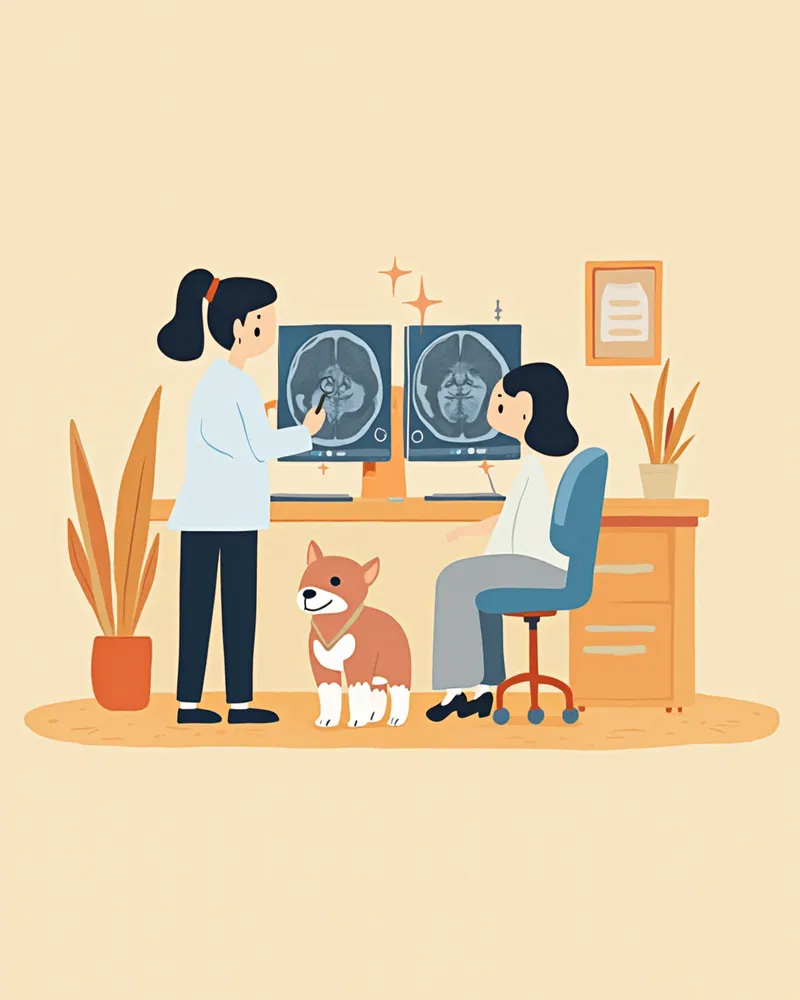
MRI scans can be categorized into several types depending on the imaging technique, and a board-certified veterinary radiologist comprehensively interprets these images.
T1-weighted images: These provide clear visualization of anatomical structures. After contrast agent administration, tumors or inflamed areas appear bright.
T2-weighted images: Areas with high water content appear bright. These images are useful for detecting edema, inflammation, and abnormalities in cerebrospinal fluid.
Additional specialized imaging: Various specialized imaging techniques may be employed depending on the nature and location of the lesion. The choice of technique is determined by the board-certified veterinary radiologist based on each patient’s clinical condition.
To accurately characterize lesions, MRI interpretation requires comparing images with different properties, such as T1 and T2, as well as signal changes before and after contrast administration. Results are typically available on the day of the scan or within two days.
A veterinary radiologist compares and interprets T1 and T2 MRI images of a dog on a dual monitor setup.

MRI Costs and Tips for Choosing a Clinic

The cost of an MRI for dogs varies depending on the body part being scanned, the clinic, and the anesthesia method. Typically, it ranges from $600 to $1,000 per area, with additional charges if multiple areas are scanned. Here are key points to consider when choosing a clinic:
Equipment specifications: High-field equipment of 1.5T (Tesla) or higher provides clearer images.
Radiology specialist: Expertise in interpreting images is as important as the scan itself. Confirm whether a specialist will personally interpret the results.
Anesthesia monitoring: Ensure that heart rate, oxygen saturation, and blood pressure are monitored in real time during the procedure.
MRIs are usually performed at university hospitals or secondary veterinary hospitals, with many cases referred from local primary clinics.

Post-MRI Care

After the MRI, your dog will be monitored at the clinic until fully awake from anesthesia. Here’s what you should keep in mind once you get home:
Same-day feeding: To reduce the risk of vomiting after anesthesia, offer small amounts of food at a time.
Activity restriction: Avoid walks or vigorous activity on the day of the procedure and allow your dog to rest comfortably.
Watch for abnormal symptoms: Contact the clinic immediately if you notice excessive vomiting, seizures, or breathing difficulties.
Most dogs return to their normal condition by the next day. Once the results are available, discuss the treatment plan with your veterinarian.

How to Understand Your MRI Report

Your MRI report may include technical terms like "high signal intensity," "low signal intensity," and "contrast enhancement." If these terms feel confusing, ask your veterinarian to walk you through the images and explain them directly. It’s important for you to fully understand the results so you can make informed decisions about your pet’s treatment plan together with your vet.

Reviewed by a veterinarian

Dr. Tony — Punnawat Phongkittirak

Dr. Tony — Punnawat Phongkittirak

Veterinarian

A veterinarian who majored in veterinary medicine at Khon Kaen University, Thailand, and completed the IVSA program at North Carolina State University in the United States. Drawing on clinical experience at animal hospitals, he works in the pet healthcare field and is dedicated to building a digital care environment that connects pet parents with veterinarians.

Frequently Asked Questions

How long does an MRI scan take for a dog?
The entire process, from anesthesia preparation to recovery, can take several hours. Since the exact duration depends on the number of areas being imaged and your dog’s condition, it’s a good idea to check with the clinic in advance for an estimated time when you make your appointment.
Should I get an MRI or a CT scan?
MRI is more accurate for examining the nervous system, such as the brain and spinal cord, as well as soft tissues. CT is better suited for detecting fractures and evaluating bone structure. Your veterinarian will recommend the most appropriate imaging test based on your pet’s symptoms.
Can my dog have an MRI without anesthesia?
It's quite challenging. MRI scans require the patient to remain completely still, so general anesthesia is essential. Even slight movement can blur the images, making accurate diagnosis impossible.
Does a normal MRI result mean there’s no problem at all?
Even if an MRI shows no abnormalities, functional issues may still be present. Additional tests, such as nerve conduction studies or cerebrospinal fluid analysis, may be necessary, so please consult with your veterinarian.
Is there anything I need to prepare before an MRI scan?
Fasting is required before anesthesia. The exact fasting duration and whether water intake should be restricted depend on the anesthetic method and your dog’s condition, so please ask your veterinarian for specific instructions during the pre-anesthetic evaluation. Pre-anesthetic blood work and a cardiac assessment are necessary beforehand, and if your dog is currently taking any medications, please inform your veterinarian in advance.

Share

Related Guides

References

[1] Schaer M, Gaschen F. Clinical Medicine of the Dog and Cat, 4th Ed. CRC Press, 2022. Chapter 14: Disorders of the Nervous System and Muscle

[2] Silverstein DC, Hopper K. Advanced Monitoring and Procedures for Small Animal Emergency and Critical Care, 2nd Ed. Wiley-Blackwell, 2023

[3] Dewey CW, Da Costa RC. Practical Guide to Canine and Feline Neurology, 3rd Ed. Wiley-Blackwell, 2016. Chapter 4: Neurodiagnostics

[4] Gavin PR, Bagley RS. Practical Small Animal MRI. Wiley-Blackwell, 2009

This information is based on veterinary literature and does not replace diagnosis or treatment. Please consult a veterinarian for specific health concerns.

Metapet Co., Ltd. | CEO: Park Sung-yong | Business Reg. No. 417-88-02562 | Seoul, South Korea | Customer Center

Dog MRI: When It's Needed, Cost & How It's Read | Meongsiljang