Like
Share
멍실장
고양이 안구 초음파 검사 자주 묻는 질문 Q&A — 보호자가 꼭 알아야 할 핵심

What is feline ocular ultrasound? — When, why, and what it reveals

Eye HealthQ&AMeongsiljang Veterinary Advisory Board

Feline ocular ultrasound is a key diagnostic tool for accurately identifying abnormalities within the eye. We’ve compiled the essential information every pet owner should know in a Q&A format.

What is feline ocular ultrasound?

A veterinarian gently holds a cat’s head while preparing for an ocular ultrasound examination.
Feline ocular ultrasound is a diagnostic method that allows non-invasive visualization of the internal structures of the eye. - Ultrasound signals: Ultrasound waves are transmitted through the transparent parts of the eye, and the signals reflected from internal tissues are analyzed to detect abnormalities. - Non-invasive nature: The procedure can be performed without anesthesia and does not cause direct damage to the eye. - Clear imaging: After pupil dilation, the detailed structures of the vitreous body and retina can be observed more clearly. - Early detection: It enables early identification of causes of vision loss, such as retinal detachment, tumors, and hemorrhage. - High safety: The test is conducted without radiation, and most cats experience minimal discomfort during the procedure.

Why is feline ocular ultrasound necessary?
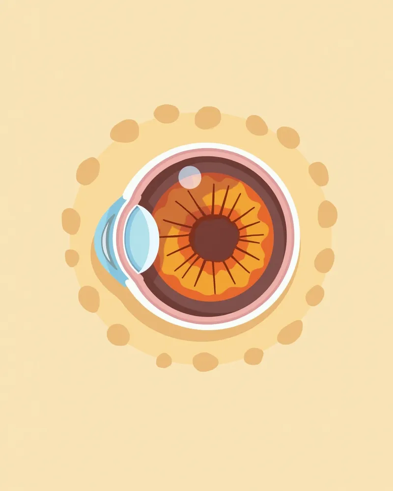
Ocular ultrasound in cats helps detect hidden abnormalities inside the eye. It is especially useful when direct visualization of the interior is difficult due to corneal or anterior segment issues. - Retinal detachment: A leading cause of vision loss, it can be diagnosed via ultrasound. Accurate assessment is crucial, as complete detachment often makes vision recovery difficult. - Vitreous hemorrhage: Blood accumulation within the eye can impair vision, and ultrasound allows for evaluation of its extent. - Intraocular tumors: Some intraocular tumors can be highly malignant, making precise identification and assessment essential. - Suspected vision changes: Consider testing if your cat suddenly bumps into objects or appears disoriented in low light. - Identifying underlying causes: Systemic hypertension is a common cause of retinal detachment and blindness in cats, so evaluating blood pressure alongside ocular findings aids diagnosis.
An ultrasound image of a cat’s eye showing the internal structures of the eyeball

How is the examination performed?

Feline ocular ultrasound is typically performed safely without general anesthesia. - Preparation: Calm your cat in a quiet environment, then apply ultrasound gel to the eye. Since cats may extend their third eyelid (nictitating membrane) if they feel pain—making the exam difficult—topical anesthetic eye drops may be used when necessary. - Procedure: A gentle probe is lightly placed on the eye to transmit ultrasound waves, allowing real-time visualization of the internal eye structures. - Duration: The procedure is relatively quick, minimizing stress for your cat. - Managing Reactions: If your cat becomes tense, take a short break before trying again. Gentle restraint and dim lighting can help facilitate the exam. - Results: Images can be analyzed immediately after the scan, enabling the veterinarian to make a prompt assessment.
A veterinarian places an ultrasound probe on the closed eyelid of a cat.

Is the examination safe?

Yes, ocular ultrasound is a very safe diagnostic method for cats. - No radiation: Ultrasound does not use radiation, making it safe even for repeated examinations. - No eye damage: The probe is gentle and exerts minimal direct pressure on the eye. - Minimal discomfort: While your cat may feel some sensation, there is no immediate pain. - Performed by experts: When conducted carefully by a veterinarian, side effects are extremely rare. - Suitable for all ages: It can be safely performed on cats of all ages, from kittens to seniors.
A cat resting comfortably after completing an ultrasound examination.

Ocular ultrasound vs. general ophthalmic examination

ItemOcular ultrasound examinationGeneral ophthalmic examination
Diagnostic scopeInternal eye structures such as the vitreous and retinaThe surface and anterior structures of the eye, such as the cornea and anterior chamber
Non-invasivenessHigh (probe contact)High (eye drops, light illumination, etc.)
Key strengthCan evaluate the interior when the inside of the eye is hard to see due to anterior structural abnormalitiesDirectly observes the eye surface and anterior structures
Example usesChecking internal abnormalities such as retinal detachment and intraocular tumorsEvaluating lesions of the cornea, anterior chamber, etc.

The two examinations are complementary, and ultrasound is especially useful when the internal structures cannot be seen directly.

How to interpret the test results
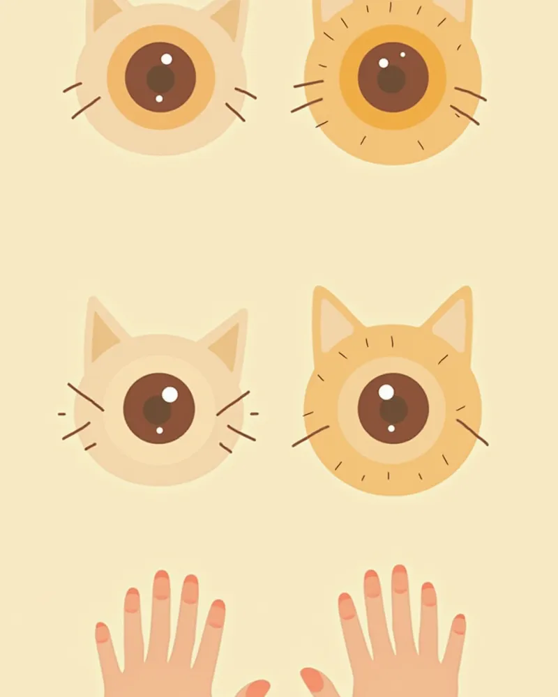
The test results visually reveal structural changes inside the eye. - Vitreous hemorrhage: Appears as hazy shadows or blurred areas, allowing us to assess the extent of bleeding. - Retinal detachment: Looks like a separated membrane or a broken line. - Tumors: Appear as solid masses, enabling us to evaluate their size, location, and border clarity. - Edema or inflammation: Manifests as thickened tissue or uneven density. - Veterinarian’s assessment criteria: Based on these images, the veterinarian determines the treatment plan and recommends additional tests if necessary.
Comparison of normal and abnormal feline eye ultrasound images

Immediate examination required in these cases

If your cat has red eyes, a sudden drop in vision, frequent blinking, or keeps its eyes closed, you should seek immediate veterinary care. These symptoms could indicate an emergency such as retinal detachment or intraocular hemorrhage.

Precautions after the examination

After the examination, your cat may feel tense or anxious. - Provide a quiet environment: Prepare a calm space where your cat can rest comfortably. - Eye care: Gently wipe away any gel that may have gotten into the eyes during the exam using a soft cloth. - Monitor behavior: If your cat rubs its eyes excessively, shows increased redness, or produces more tears than usual, contact your veterinarian immediately. - Return to normal routine: Your cat can resume regular activities without any special restrictions. - Next appointment: Depending on the results, schedule any additional tests or treatments as needed.
A cat resting quietly in a calm environment after completing an ultrasound examination.

Reviewed by a veterinarian

Dr. Tony — Punnawat Phongkittirak

Dr. Tony — Punnawat Phongkittirak

Veterinarian

A veterinarian who majored in veterinary medicine at Khon Kaen University, Thailand, and completed the IVSA program at North Carolina State University in the United States. Drawing on clinical experience at animal hospitals, he works in the pet healthcare field and is dedicated to building a digital care environment that connects pet parents with veterinarians.

Frequently asked questions

Is an ocular ultrasound painful for cats?
It doesn’t hurt. The probe only lightly glides over the eye, so most cats don’t experience significant discomfort. If needed, we may use topical anesthetic eye drops.
Is anesthesia necessary?
In most cases, the procedure can be performed without general anesthesia. However, if your cat is overly stressed or unable to cooperate due to pain, we may use a topical anesthetic eye drop or consider additional sedation.
When will the test results be ready?
After the tests, the veterinarian will explain the results right away. If needed, they’ll also guide you through any additional tests or treatment plans.
What conditions is ocular ultrasound effective in diagnosing?
It is especially useful for detecting abnormalities inside the eye, such as retinal detachment, vitreous hemorrhage, and intraocular tumors. It is also very helpful when it is difficult to directly visualize the interior of the eye due to problems with the cornea or anterior structures.
How is the frequency of examinations determined?
Testing is recommended if you notice any concerning symptoms or if your veterinarian suggests it, and conditions with a high risk of recurrence may require regular follow-up exams.

Share

Related Guides

References

[1] Smith, J. et al. (2021) Ocular Ultrasound in Feline Patients: Clinical Applications and Diagnostic Accuracy. Journal of Feline Medicine and Surgery, 23(4), 345–352.

[2] Johnson, L. & Brown, R. (2020) Advanced Imaging Techniques in Veterinary Ophthalmology. Veterinary Clinics of North America: Small Animal Practice, 50(3), 567–589.

[3] American College of Veterinary Ophthalmologists (2022) Guidelines for Diagnostic Imaging in Feline Ocular Disease. ACVO Practice Standards, 12(1), 1–24.

This information is based on veterinary literature and does not replace diagnosis or treatment. Please consult a veterinarian for specific health concerns.

Metapet Co., Ltd. | CEO: Park Sung-yong | Business Reg. No. 417-88-02562 | Seoul, South Korea | Customer Center

What Is Feline Ocular Ultrasound? When, Why, and What It Reveals | Meongsiljang