Like
Share
멍실장
고양이 안구 돌출·프로프토시스 자주 묻는 질문 Q&A — 보호자가 꼭 알아야 할 핵심

Feline Proptosis FAQ — Essential Information for Pet Owners

Eye HealthQ&AMeongsiljang Veterinary Advisory Board

Feline exophthalmos is an acute ophthalmic emergency. We’ve compiled essential information that pet owners need to know, including causes, symptoms, immediate response steps, and prognosis.

Feline Proptosis Is a Life-Threatening Emergency
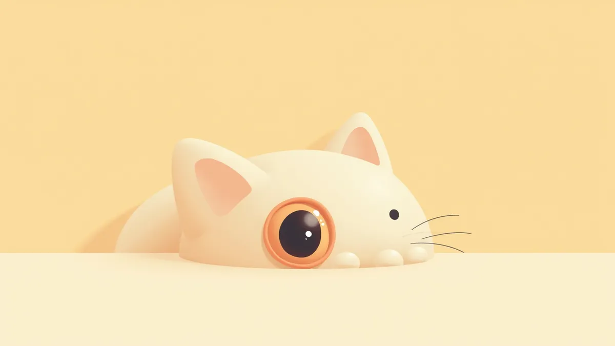
An emergency situation image showing a cat's bulging eyes
Feline proptosis, or eyeball protrusion, is an acute ophthalmic emergency in which the eye bulges out of the orbit, requiring immediate veterinary attention. It most commonly results from trauma; because cats require significant blunt force to dislocate the eye, concurrent head injuries are frequent. Therefore, it is crucial to assess not only the eye but also the cat’s overall systemic condition for any life-threatening injuries. - Proptosis: The eye protrudes from the orbit, often accompanied by hemorrhage in the conjunctiva or sclera, anterior chamber hemorrhage, and damage to the exposed cornea. - Immediate First Aid: Do not touch or pull on the eye. Instead, keep the ocular surface moist with sterile saline and gently cover it with a clean cloth for protection. - Prognosis: The prognosis for proptosis in cats is generally poorer than in dogs, with a significant risk of vision loss or the need for enucleation (eye removal). - Owner’s Role: Early detection and rapid transport to a veterinary clinic are decisive factors in preserving the eye. Do not delay; seek immediate veterinary care.

What Are the Main Causes of Proptosis?

The most common cause of ocular proptosis in cats is trauma. It occurs when strong impact is delivered to the eye socket, such as from fights with other animals, vehicle accidents, or falls from heights. Unlike dogs, cats require significant blunt force to cause the eye to protrude, so ocular proptosis is often accompanied by concurrent head injuries. - Trauma: Strong impact increases pressure inside the eye socket and damages the surrounding structures, forcing the eye outward. - Concurrent injuries: Due to the nature of blunt force trauma, brain or skull damage may also be present, necessitating a comprehensive systemic evaluation. - Breed factors: This condition is reported in brachycephalic breeds with shallow eye sockets and prominent eyes, such as Persians. However, the brachycephalic structure itself is known to be a relatively favorable prognostic factor. - Prevention: A key role for pet owners is to reduce the risk of trauma from falls, traffic accidents, and fights by providing a safe environment.
An image showing the eye structure of brachycephalic cats

Main Symptoms of Proptosis

When a cat’s eye becomes proptosed, it slips out of the socket and shifts from its normal position. You may also notice bleeding in the conjunctiva and sclera, hyphema (bleeding inside the front chamber of the eye), and severe swelling of the eyelids. The exposed cornea can dry out and become damaged. Due to pain, the cat may refuse to eat, remain immobile, or exhibit avoidance behaviors. - Vision loss: Proptosis causes significant vision impairment, and cats often suffer permanent blindness. - Pain response: Touching the area around the eye may trigger screaming or attempts to flee. - Signs of accompanying injuries: Because this condition usually results from blunt force trauma, head injuries may also be present. Watch for additional signs such as decreased consciousness or lethargy. - Immediate action required: If you observe these symptoms, seek veterinary care immediately. Prompt treatment significantly affects the likelihood of saving the eye.
A cat experiencing pain due to proptosis (eye bulging)

Emergency Situations Requiring Immediate Veterinary Visit

If the eyeball protrudes from the socket, seek emergency veterinary care immediately. The longer you wait, the more the cornea dries out and damage progresses, reducing the chances of saving the eye. Do not touch or pull on the eye. Instead, keep the surface moist with sterile saline or clean water, then gently cover it with a clean, soft cloth for protection.

Diagnosis and Treatment of Proptosis
The veterinarian first stabilizes the pet’s overall condition to rule out any life-threatening concurrent injuries, then directly examines the eye and surrounding structures. If head or orbital trauma is suspected due to blunt force, imaging tests may be performed to check for additional injuries. For eyes that can be saved, the surface is moistened under general anesthesia, the eye is repositioned, and the eyelids are temporarily sutured (temporary tarsorrhaphy) to protect it. - Surgical Goal: The procedure is performed as quickly as possible to preserve the eye and minimize damage. However, cats generally have a poorer prognosis than dogs, and vision recovery is often difficult. - Post-Operative Care: Eye drops are used to protect the ocular surface, and medications are prescribed to reduce pain and inflammation. - Severe Damage: If the eye cannot be recovered, enucleation (surgical removal of the eye) may be necessary. - Temporary Suture Maintenance: The temporary tarsorrhaphy is typically maintained for about 10 to 14 days. - Regular Check-ups: Ongoing monitoring by the veterinarian is essential even after treatment.
A surgical procedure in which a veterinarian repositions the eyeball under general anesthesia

Home Care and Recovery Process

After surgery, an eye mask is used to protect the eyes. Regular use of eye drops and strict adherence to medication are essential. Limiting activity and supporting recovery in a quiet environment are important. - Eye mask: Must be worn to protect the eyes and reduce irritation. - Medication: All prescribed medications must be taken as directed to prevent recurrence or complications. - Environment management: Avoid bright light and dusty areas; allow rest in a quiet space. - Observation points: Contact the clinic immediately if redness, increased discharge, or signs of pain occur. - Recovery period: Typically takes 2–4 weeks, with a gradual return to normal activities under veterinary guidance.
A cat recovering while wearing an eye mask

Comparison of Responses Based on Severity of Proptosis

ItemMildModerateSevere
Eyeball PositionSlightly protrudedHalf protrudedCompletely protruded
Pain LevelMildModerateSevere
Prognostic FactorsLittle displacement, pupil constriction and light reflex present (favorable)ModerateAccompanied by muscle rupture, scleral rupture, hyphema (poor)
Need for Emergency ResponseImmediate veterinary visitImmediate veterinary visitImmediate veterinary visit

Regardless of degree, ocular proptosis is always an emergency, and cats have a poorer prognosis than dogs, with vision recovery often difficult, so immediate veterinary visit is necessary.

Brachycephalic Breeds Are at Higher Risk for Proptosis

Globe protrusion has been reported in brachycephalic cat breeds with prominent eyes, such as Persians. However, unlike dogs, cats require significant blunt force to dislodge an eye, so this condition most often occurs alongside trauma from incidents like traffic accidents, falls, or fights, and is frequently accompanied by head injuries. To prevent such injuries, minimize household hazards like fall risks and sharp objects, and take care to avoid exposing your cat to outdoor accident risks.

Reviewed by a veterinarian

Dr. Tony — Punnawat Phongkittirak

Dr. Tony — Punnawat Phongkittirak

Veterinarian

A veterinarian who majored in veterinary medicine at Khon Kaen University, Thailand, and completed the IVSA program at North Carolina State University in the United States. Drawing on clinical experience at animal hospitals, he works in the pet healthcare field and is dedicated to building a digital care environment that connects pet parents with veterinarians.

Frequently Asked Questions

Can vision return after a prolapsed eyeball?
Prognosis for ocular proptosis in cats is generally poorer than in dogs. In one retrospective study, all cats with proptosis experienced permanent blindness, indicating that vision recovery is often difficult. However, if the displacement is minimal and pupillary constriction or light reflexes remain, the prognosis is relatively better, making prompt treatment essential.
Can eye protrusion be life-threatening in cats?
It’s not the eye bulging itself that’s life-threatening, but rather the accompanying head or brain injuries and bleeding. Since a cat’s eye only pops out when significant blunt force is applied, head trauma is often present, making an immediate vet visit essential.
Can the eye be saved after it has been displaced from the socket?
Surgery can sometimes be used to reposition and preserve the eye, but because cats often suffer severe damage, enucleation (surgical removal of the eye) is frequently necessary. In one study, 12 out of 18 cats with proptosis (eye displacement) required enucleation.
Can you prevent proptosis?
Most cases of exophthalmos (bulging eyes) in cats are caused by trauma, so the most important thing is to reduce the risk of accidents such as traffic incidents, falls, and fights. Please ensure both indoor and outdoor environments are safe.
Can a cat become active again after experiencing eye bulging?
After the recovery period, many cats return to their normal daily lives. However, if a cat has had an eye removed or has lost its vision, it is important to adjust the home environment to suit the cat's needs.

Share

Related Guides

References

[1] Ettinger SJ, Feldman EC, Côté E. Textbook of veterinary internal medicine: diseases of the dog and the cat. 8th ed. St. Louis, MO: Elsevier; 2017.

[2] The Cat, Clinical Medicine and Management, 2nd Edition. Elsevier; 2008.

[3] Clinical Atlas of Canine and Feline Ophthalmic Disease, 2nd Ed. Elsevier; 2015.

This information is based on veterinary literature and does not replace diagnosis or treatment. Please consult a veterinarian for specific health concerns.

Metapet Co., Ltd. | CEO: Park Sung-yong | Business Reg. No. 417-88-02562 | Seoul, South Korea | Customer Center

Cat Eye Protrusion: Causes, Treatment, and Emergency Signs | Meongsiljang